 History: A 42-year-old woman with a one month history of dry cough was radiographically thought to have a left cardiac myxoma and bilateral hydrothorax. At surgery, however, she was found to have a 7.0 x 6.0 x 4.5 cm gray fleshy solid mass in the left pulmonary vein which extended into the left atrium.
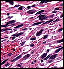
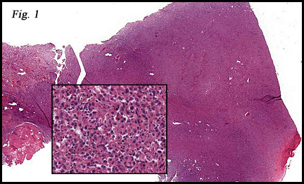
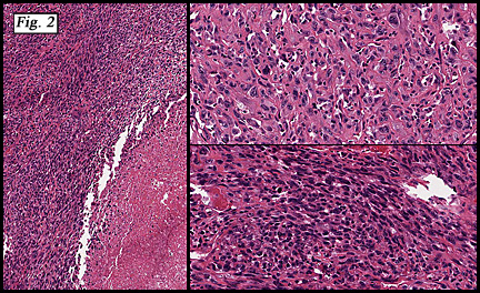
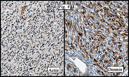
Microscopically, the tumor grew in broad sheets and fascicles and was composed of spindled and epithelioid cells (Fig. 1). Pleomorphic cells had blunt-ended nuclei and bizarre atypical nuclei with conspicuous nucleoli and a high mitotic rate. The cytoplasm was eosinophilic. Areas of tumor necrosis and dilated vessels with hemorrhage were also seen (Fig. 2, 3). Immunohistologically, the lesional cells were strongly and diffusely positive for desmin and focally marked for smooth muscle actin (Fig. 4). The tumor was negative for S100.
Diagnosis: “Pulmonary vein leiomyosarcomaâ€
Jin Guo, MD and Donald R. Chase, MD
Department of Pathology and Human Anatomy
Loma Linda University and Medical Center, Loma Linda, California
California Tumor Tissue Registry, Loma Linda, California
Discussion: Although leiomyosarcoma of the pulmonary vein is extremely rare, it is the most common of primary pulmonary vein neoplasms. Including the present case, there are only 20 cases of pulmonary vein leiomyosarcoma reported worldwide. Of the non-leiomyosarcoma tumors of pulmonary vein origin, only single cases of angiosarcoma, lipomyxosarcoma, malignant fibrous histiocytoma, alveolar soft part sarcoma and leiomyoma have been reported.
Cardiac leiomyosarcoma is very rare occurring in less than 0.2% of all cardiac tumors and generally favoring the left atrium. It may secondarily invade the pulmonary veins, mitral valve, or even the lung. The average reported patient age of pulmonary vein leiomyosarcoma is 48.4 years old (27-74 years). It occurs more commonly in females (13 females, 7 males), and in the right pulmonary vein.
Leiomyosarcomas from either the pulmonary vein or the left atrium have similar morphological features consisting of highly cellular proliferations of spindle cells varying in size and shape, usually with bizarre pleomorphism. Proliferative indices are high including mitotic rates ranging from 1-25 per 10 high power fields. Regional necrosis is common. The tumors tend to show immunoreactivity to desmin and actin, thereby confirming myogenous differentiation. They fail to show epithelial, neural or vascular components by uniformly being negative for EMA, S100, CD34 and factor VIII.
The symptoms of pulmonary vein or left atrial leiomyosarcoma are non-specific and may include dyspnea, hemoptysis/bloody sputum, cough, chest pain, weight loss, ipsilateral pleural effusion, palpitations, paroxymal tachycardia and/or orthopnea. Severe hemoptysis may necessitate blood transfusion. Sometimes, occlusion of the right superior pulmonary vein may occur.
Since cardiac leiomyosarcomas preferentially arise in the left atrium as myxoid neoplasms, distinction from atrial myxoma may be difficult on a limited, pre-surgical biopsy. It is therefore recommended to resect all atrial myxoid tumors with a wide (at least 1 cm) margin.
The diagnosis of pulmonary or cardiac leiomyosarcoma is frequently delayed because of the non-specific nature of the symptoms. By the time of diagnosis, the tumor is usually large, showing invasion and/or metastasis. The prognosis is generally poor with postoperative survivals of 6 months (75%), 1 year (73%), 2 years (50%) and 3 years (33%), respectively. The current therapy of choice is early, complete surgical extirpation with post-operative chemotherapy being reserved for tumors which are incompletely removed.
Suggested Reading:
Gyhra AS, Santander CK, Alarcon EC, et al: Leiomyosarcoma of the pulmonary veins with extension to the left atrium. Ann Thorac Surg. 61:1840-1841, 1996.
Ko TM, Mayer DA, Tsapogas MJ Forbes A and Marchioro TL. Leiomyosarcoma of the pulmonary vein. J Cardiovasc Surg. 37(4):421-3, 1996.
Oliai BR, Taxelarr HD, Lloyd RV, et al: Leiomyosarcoma of the pulmonary veins. Am J Surg Pathol 23:1082-1088, 1999.
Laroia ST, Potti A, Rabbani M, Mehdi SA, Koch M. Unusual pulmonary lesions: case 3. Pulmonary vein leiomyosarcoma presenting as a left atrial mass. J Clin Oncol. 1;20(11):2749-51, 2002.
Gürbüz A, Yetkin U, Yilik L, Ozdemir T, Türk F. A case of leiomyosarcoma originating from pulmonary vein, occluding mitral inflow. Heart Lung. Heart lung. 32(3):210-4, 2003.
Khemichian S, Bemanian S. Pulmonary vein leiomyosarcoma. Intern Med J 36(12):793-4, 2006.