October 2014: A 69 year old man with dry skin hyperpigmentation and fatigue

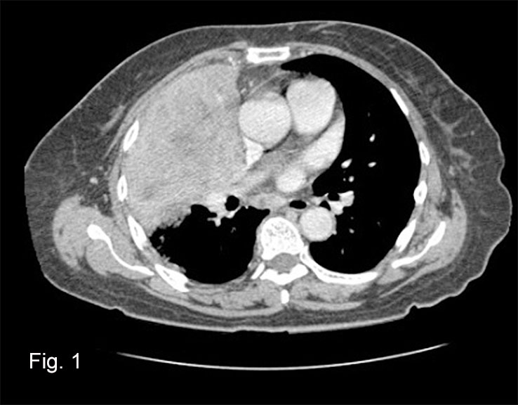
History: A 69-year-old man was hospitalized for 10 days with complaints of easy fatigability. He was found to have dry skin with hyperpigmentation over the lower legs and some cracking of the skin of the lower abdomen. Lab studies showed a sodium level of 129 mEq and a potassium level of 4.5 mEq. The work up discovered chronic active hepatitis, arteriosclerotic heart disease, chronic renal disease, and early COPD. He had a femoral arteriogram to evaluate lower extremity vascular insufficiency, but died 2 months later of apparent cardiac arrest.

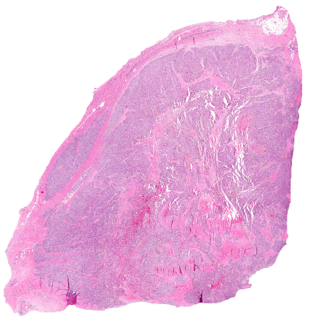
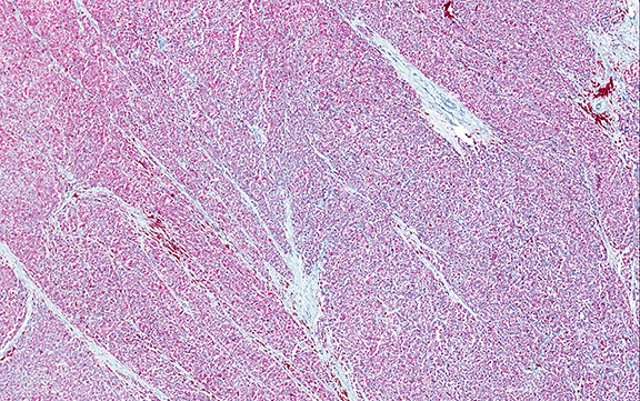
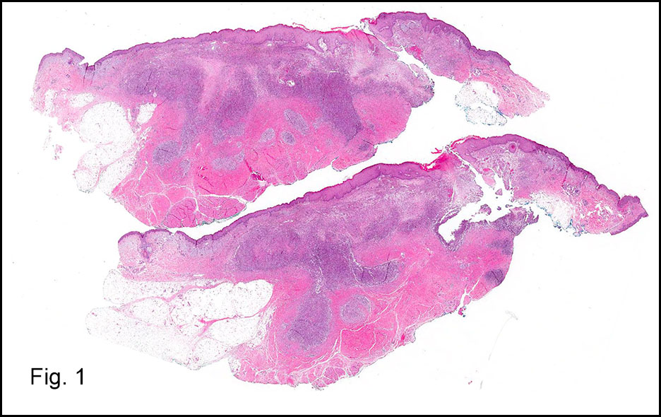
At autopsy, both adrenal glands were enlarged to 5 cm and appeared completely replaced by tumor. Each weighed 30 grams. There was no infiltration of surrounding tissue. Metastases were not seen.

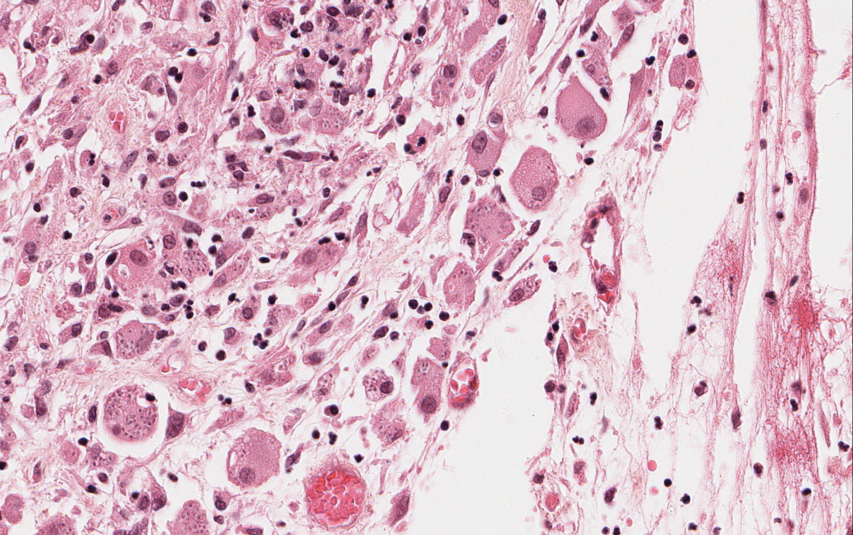
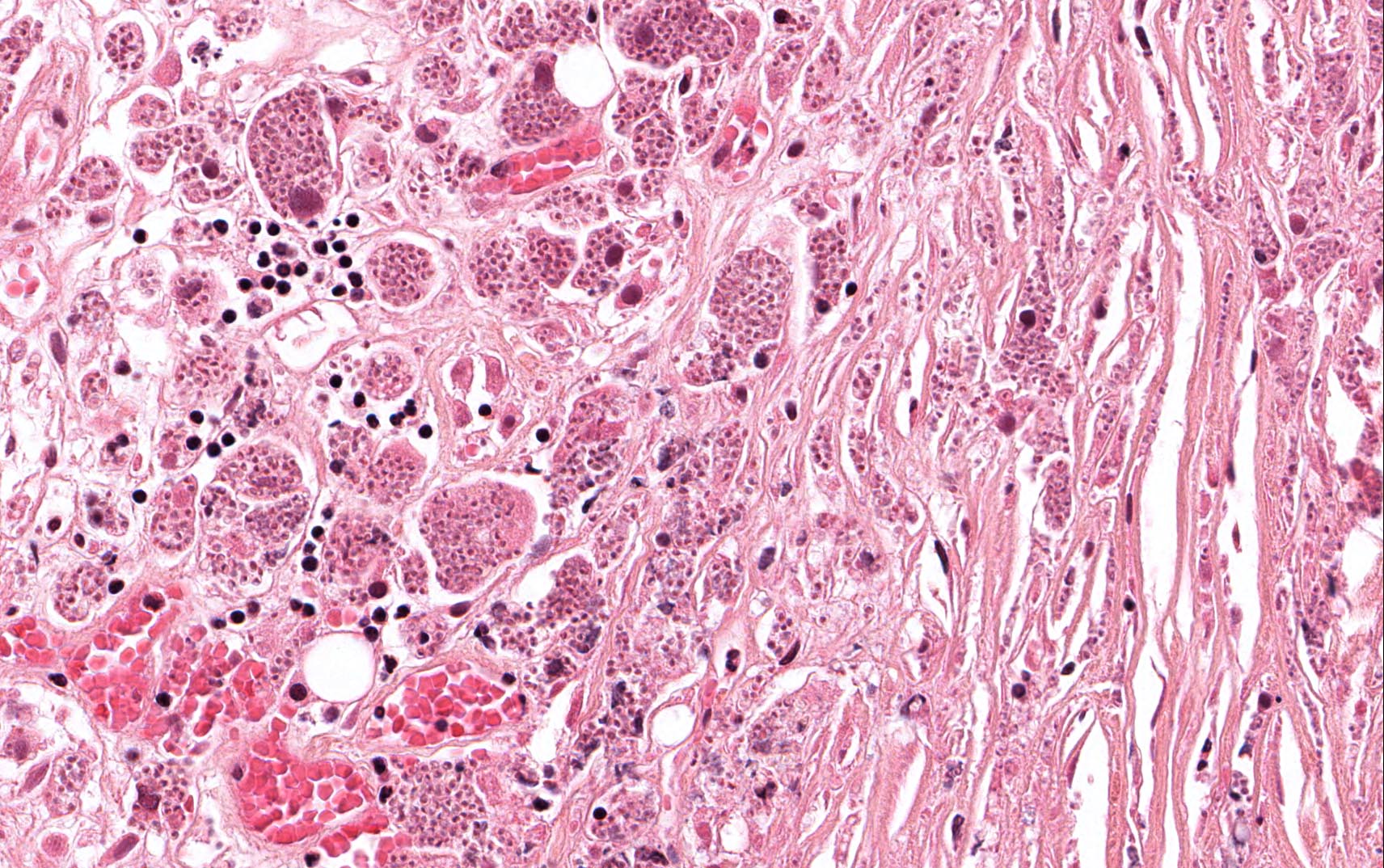
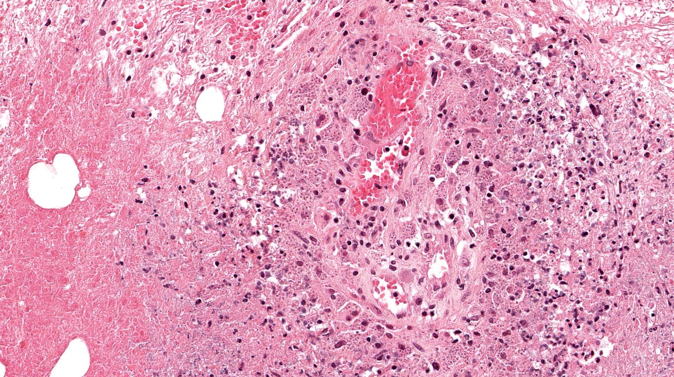
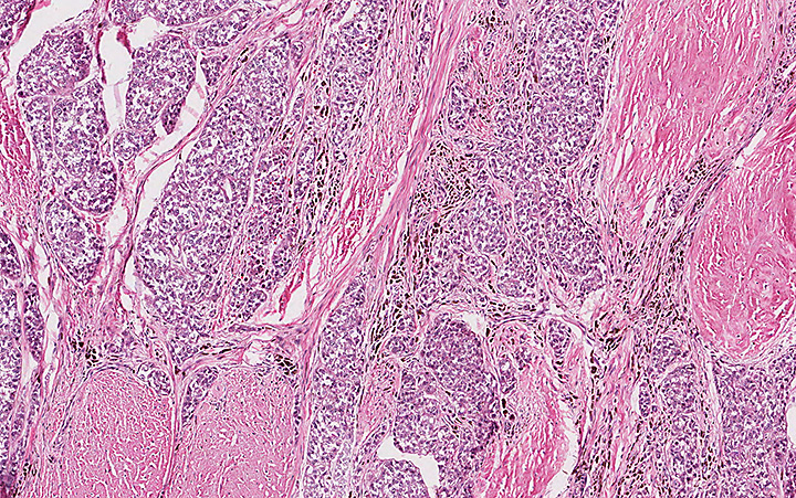
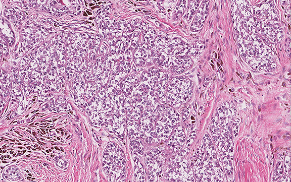
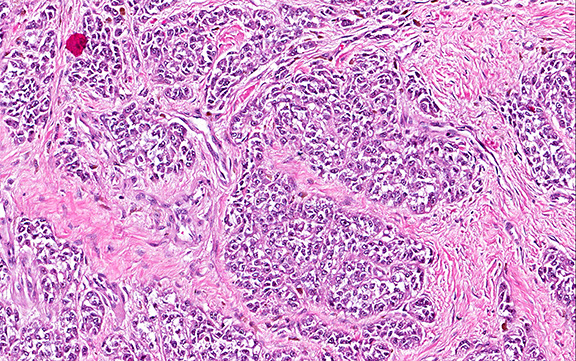
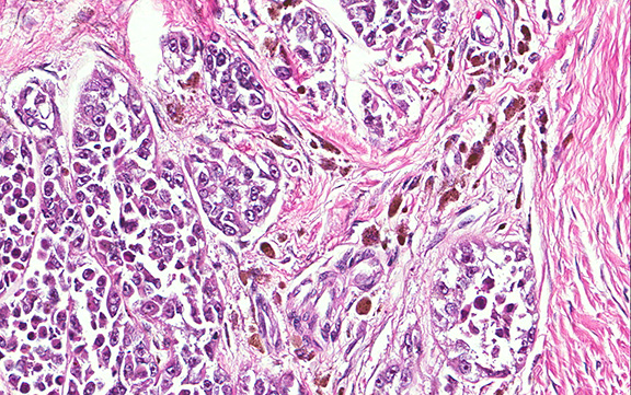
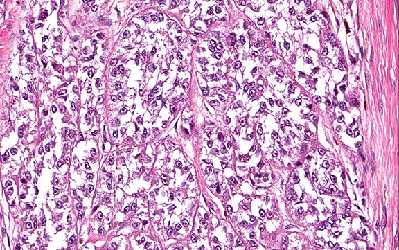
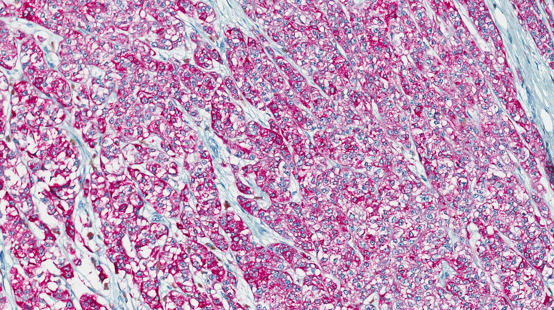
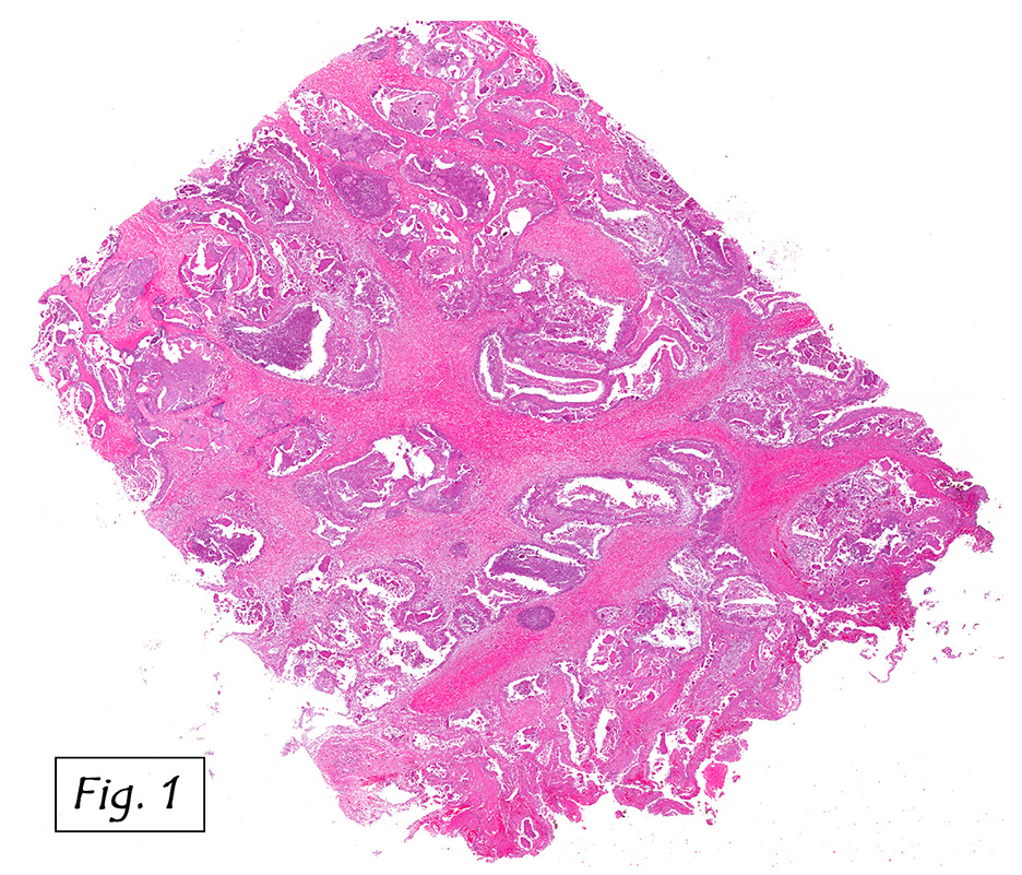
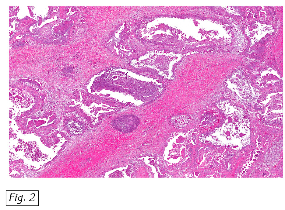
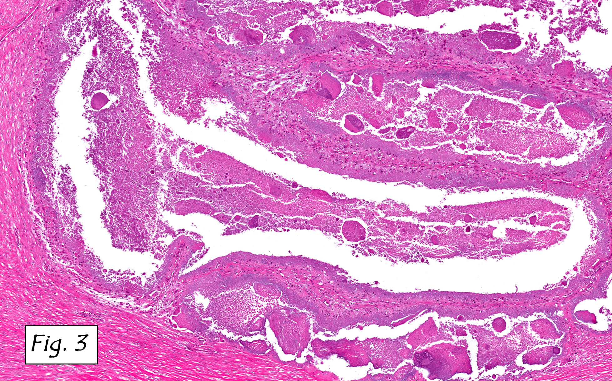
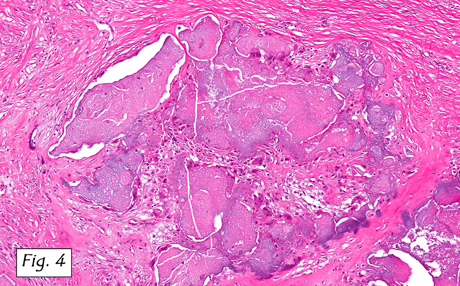
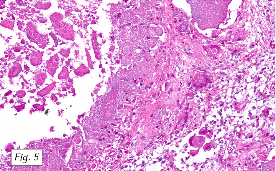
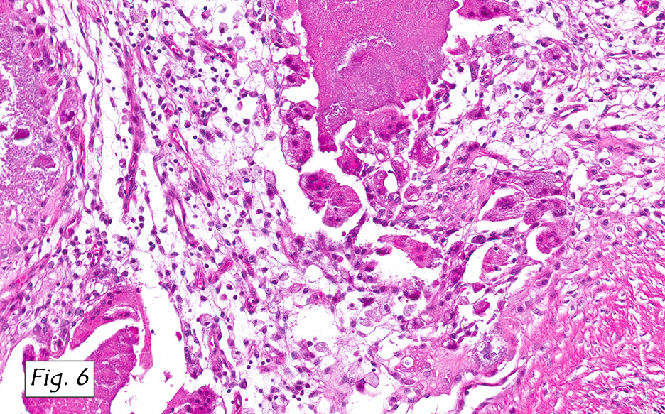
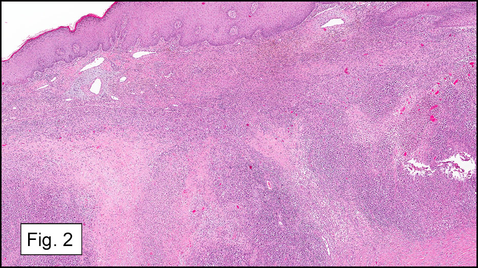
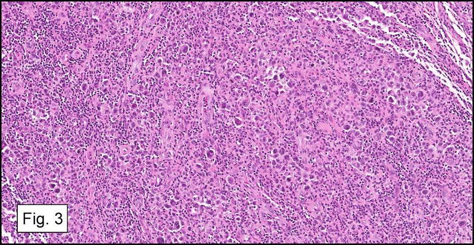
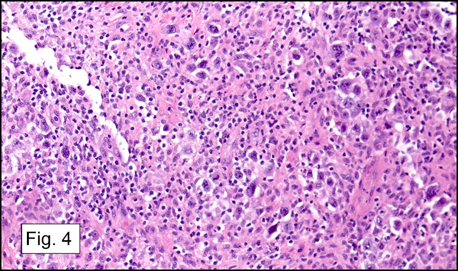
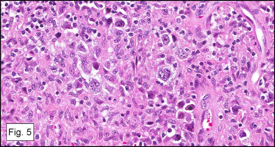
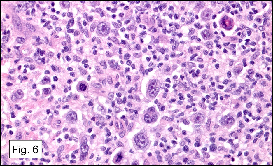
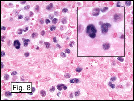
Microscopically, the adrenal glands were necrotic and focally hemorrhagic (Fig. 1). Higher magnification revealed multiple organisms, including intracellular organisms within macrophages (Figs. 2, 3), some of which were adjacent to the areas of necrosis (Fig. 4).

Diagnosis: Adrenal Histoplasmosis

Dennis Aaron Reinke, M.D. and Donald R. Chase, M. D.
Department of Pathology and Human Anatomy,
Loma Linda University Medical Center, Loma Linda, California
California Tumor Tissue Registry, Loma Linda, California

Discussion: Histoplasmosis is caused by the fungus Histoplasma capsulatum. It can be seen in many countries including the United States where it usually occurs in the eastern and central regions. It is transmitted by spore inhalation which can result in a pulmonary infection. It can disseminate to other organs via the blood to affect the spleen, liver, lymph nodes, bone marrow and adrenal glands.

There are a number of causes of adrenal insufficiency which can be categorized as either primary or secondary. Primary causes to consider include infectious, autoimmune, bilateral adrenal hemorrhage (resulting from sepsis, etc.), drug-induced, certain genetic disorders, adrenal infiltration (resulting from metastases, amyloidosis, etc.), or bilateral adrenalectomy. Secondary causes would include isolated adrenocorticotropic hormone deficiency, prior chronic glucocorticoid excess (which may be due to Cushing’s syndrome or steroid therapy), hypothalamic-pituitary region tumors, or pituitary apoplexy.

The clinical presentation of adrenal histoplasmosis may include weight loss, chronic fatigue, fever and anorexia. Primary adrenal insufficiency/Addison’s disease may also manifest in 5-71% of cases, and it is the most frequent cause of fatality. In these cases symptoms may include malaise, fever, nausea, vomiting and orthostatic hypotension. Lab values may show eosinophilia, hyponatremia and hyperkalemia. Imaging studies can reveal symmetrical adrenal enlargement with areas of hemorrhage and necrosis. These findings would suggest a differential diagnosis of aspergillosis, tuberculosis, or other infections, but a definitive diagnosis of adrenal histoplasmosis is not made by imaging studies.

In immunocompromised individuals, the fungus develops in phagocytic cells and macrophages. In patients that are immunocompetent, granulomas may develop that appear similar to noncaseous tuberculosis.

Microscopically, intracellular Histoplasma can be seen on H&E-staining. However, in necrotic and epithelioid cell granulomatous lesions, special stains (i.e. GMS) may be necessary.

Yeast in these tissue sections are around 3 µm in diameter, may be grouped, and have a single bud connected by a narrow base. These organisms have a firm cell wall, and during fixation the protoplasm withdraws from this firm wall. This leaves a resulting clear space that presents the false effect of an unstained capsule in these organisms.

The differential may include Leishmania, Toxoplasma, Blastomyces and Cryptococcus. Leishmania may look very similar on low power, but higher magnification can show the kinetoplast of these organisms. Toxoplasma is generally smaller and not identified within macrophages. Blastomyces have two or more nuclei, while Histoplasma has a single nucleus. Cryptococcus can be differentiated from Histoplasma, as the former’s walls should stain with carmine dyes.

Suggested Reading:

Arlt W, Allolio B. Adrenal insufficiency. The Lancet. 2003;361(9372):1881-1893.

Binford, CH, Dooley, J. Histoplasmosis. In: Binford, CH, ed., Conner, DH, ed. Pathology of Tropical and Extraordinary Diseases. Volume 2. Washington, D.C.: Armed Forces Institute of Pathology; 1976:578-580.

Larbcharoensub N, Boonsakan P, Aroonroch R, et al. Adrenal histoplasmosis: a case series and review of the literature. Southeast Asian J Trop Med Public Health. 2011;42(4):920-5.

Vyas S, Kalra N, Das PJ, et al. Adrenal histoplasmosis: An unusual cause of adrenomegaly. Indian J Nephrol. 2011;21(4):283-5.

September 2014: A 38 year old woman with a knee mass

History: A 38 year old woman presented with a several month history of an enlarging painful mass of the right knee. Physical examination showed a firm, tender mass over the medial knee region. An MRI revealed a 6.9 x 4.7 x 3.4 cm, heterogeneously enhancing soft tissue mass with areas of hemorrhage and probable
Grossly, the mass was firm, variegated, white-tan to dark-brown, and well-circumscribed (Fig. 1).
Microscopically, the tumor infiltrated into tendon (Fig. 2). Tumor cells showed a nesting pattern and clear cells with focal pigment (Figs. 3-4). Prominent nucleoli were present (Figs. 5-6). The tumor was positive for a melanoma cocktail (Fig. 7) and S100 (Fig. 8).

Immunohistochemical studies showed the tumor to be positive for S100 and Melanoma cocktail, but were negative for synaptophysin and NFP. FISH was performed and showed 97% tumor cells were positive for fusion transcript of EWSR1(22q12) and ATF1 (12q13.13) loci.

Diagnosis: Clear Cell Sarcoma of Soft Parts

Huina Zhang, M.D., Ph.D., Ravi Raghavan, M.D.
and Donald R. Chase, M.D.
Department of Pathology & Human Anatomy
Loma Linda University Medical center, Loma Linda, California

Discussion: Clear cell sarcoma (CCS) is a rare soft tissue sarcoma with melanocytic differentiation, which is also known as malignant melanoma of soft parts. Although it has certain morphologic similarities to malignant melanoma, CCS is a clinicopathologically and genetically distinct from the conventional malignant melanoma. It mainly affects young adults between the ages of 10 to 40 years with a median age of approximately 30 years. CCS typically presents as a slowly growing, firm, deep-seated nodule in the distal extremities, most often located around the ankle or foot, followed by the knee, wrist, and hands. It is usually associated with tendons, tendon sheaths, or aponeuroses, with rare involvement of the subcutaneous tissue or dermis. CCS usually follows a protracted clinical course, with frequent local recurrences and late metastases to regional lymph nodes, lungs, and bone. The long-term prognosis of CCS is poor and one study showed the survival rates at 5, 10, and 20 years were 67%, 33% and 10%, respectively. Early diagnosis and initial wide excision are essential for local control and a more favorable outcome. Unfavorable prognostic factors include large tumor size (>5 cm), presence of necrosis, early local recurrence, and positive resection margins.
Grossly, CCS is usually a lobulated or multi-nodular, firm, tan-grey mass and the size ranges from 0.4 cm up to 14.5 cm in greatest diameter. It may show infiltration into the tendons and aponeuroses. The cut surface may show focal haemorrhage, necrosis or cystic changes and often is gritty.
Microscopically, CCS is highly infiltrative and is characterized by a nested, bundled or fascicular growth of uniform fusiform or spindled-to-epithelioid cells, separated by prominent fine collagenous framework of various thicknesses. The tumor cells contain abundant clear or pale eosinophilic cytoplasm and centrally located, round to ovoid vesicular nuclei with prominent basophilic nucleoli. Scattered multinucleated giant cells with 10 to 15 peripherally distributed nuclei in a wreathlike pattern are commonly present. In about half to two thirds of cases finely granular melanin pigment may be identified. The stroma may be barely visible, fibrotic or hyalinized. Focal areas of necrosis may be also noted. The mitotic rate is usually low, and pleomorphism is absent.

Immunohistochemical studies of CCS reveal that the tumor cells express antigens associated with melanin synthesis including diffuse immunoreactivity with HMB-45, nuclear and cytoplasmic immunoreactivity to S-100 protein, and reactivity with the microphthalmia transcription factor (MITF). There may be focally reactivity with Melan-A, neuron-specific enolase and vimentin, but the tumor is typically negative for EMA, keratins, and desmin.

Genetically, CCS has a distinct genetic background including a reciprocal chromosome translocation t(12;22)(q13;q12), which can be detected by conventional chromosomal analysis, FISH or reverse transcriptase polymerase chain reaction in 70-90% of CCS. This translocation results in fusion of the 3’ portion of the Ewing sarcoma (EWSR1) oncogene on chromosome 22q12 with the 3’ portion of the activating transcription factor 1 (ATF1) oncogene on chromosome 12q13, giving rise to the EWSR1/ATF1 chimeric transcript. EWSR1/ATF1 functions as a transcriptional regulator that induces the expression of MITF in CCS and plays a crucial role in melanocytic differentiation and growth or survival of tumor cells.

Differential Diagnoses:

• Metastatic melanoma is the most important mimic of CCS in the soft tissue and can be extremely difficult to distinguish from CCS because the significant overlapping features at morphologic, immunophenotypic and ultrastructural levels. The clinical context including a deep-seated infiltrative mass in the distal extremities, no proceeding history of cutaneous, mucosal or ocular tumor, the absence of junctional activity, and the uniform cytomorphology are helpful clues to the diagnosis of CCS, but in some cases detection of the translocation or its gene product is needed.

• Paraganglioma-like dermal melanocytic tumor is a recent-described, rare, benign neoplasm derived from melanocytes. It is primarily a non-pigmented, well-demarcated skin nodule at the extremities, most common in adult females. It is composed of clear to amphophilic epithelioid cells in an organoid or nest-like pattern separated by delicate fibrous strands and blood vessels, which is a reminiscent of those of paraganglioma. The tumor cells may also express S-100 protein, Melan-A, HMB-45, and microphthalmia transcription factor but lacks pancytokeratin or smooth muscle actin. FISH analysis of 5 cases revealed an intact EWS gene locus, supporting absence of the clear cell sarcoma 12;22 translocation.

• Malignant peripheral nerve sheath tumor (MPNST) is usually associated with a large peripheral nerve, and usually has manifestations of neurofibromatosis. It has significant myxoid stroma, long fascicles of spindle cells with slender, tapering or wavy nuclei and indistinct cytoplasm. Although it expresses S-100 protein, MPNST is negative for HMB-45 and Melan-A.

Summary: CCS is an extremely rare soft tissue malignancy with melanocytic differentiation, which is characterized by nests and short fascicles of uniform spindled-to-epithelioid cells with pale, eosinophilic cytoplasm and prominent nucleoli, separated by dense fibrous septa. Immunohistochemically, expression of HMB-45 is usually stronger and more diffuse than S-100 protein. Furthermore, t(12;22) translocation with EWSR1-ATF1 fusion is a typical genetic feature in CCS.

Suggested Reading:

Hornick JL. Practical Soft Tissue Pathology: A Diagnostic Approach. Elsevier.
Dim DC, Cooley LD, Miranda RN. Clear cell sarcoma of tendons and aponeuroses: a review. Arch Pathol Lab Med. 2007, 131(1):152-6.
Deyrup AT1, Althof P, Zhou M, Morgan M, Solomon AR, Bridge JA, Weiss SW. Paraganglioma-like dermal melanocytic tumor: a unique entity distinct from cellular blue nevus, clear cell sarcoma, and cutaneous melanoma. Am J Surg Pathol. 2004, 28(12):1579-86.
Goldblum JR, Folpe AL and Weiss SW. Enzinger & Weiss’s Soft Tissue Tumors: 6th ed. Elsevier.

August 2014: A 21 year old man with a mass in the inguinal region

History:  A 21 year old man presented with an 11 month history of a slow growing, painless subcutaneous mass in the left inguinal area. A pelvic X-ray showed a 7 x 4 x 2.8 cm well circumscribed periarticular calcified mass within the soft tissue of the hip. It consisted of multiple small calcified nodules. Serum calcium, phosphate and alkaline phosphatase levels were normal.
The mass was composed of white, granular material surrounded by a fibrous tissue capsule. The cut surface was hard, calcified, light tan and chalky. Microscopically, it showed multiple, solitary and confluent nodules of calcification in a matrix of granular debris (Figs. 1, 2, 3, 4 ). Associated were histiocytes, multinucleated giant cells and fibroblasts (Figs. 5, 6).

“August 2014: A 21 year old man with a mass in the inguinal region”Continue reading

July 2014: A large lung mass in a 69 year old woman from South Asia

History:A 69 year-old woman from Laos presented with hemoptysis and a 9 cm mass in the right middle and lower lung lobes (Fig. 1). It obliterated the right middle lobe bronchus, obstructed the inferior pulmonary vena cava and extended to the chest wall. Ten years prior, the tumor was recognized by CT scan, but therapy had not been given. Metastatic workup at the current presentation failed to show a different site of origin. “July 2014: A large lung mass in a 69 year old woman from South Asia”Continue reading

June 2014: A non-healing, skin ulcer in a woman with dermatomyositis

History: A 58 year-old woman with a 6 year history of dermatomyositis presented with a 7 month history of a painless 1.5 x 1.0 cm skin ulcer near her right elbow on the dorsal surface of the proximal forearm. She had a 6 year history of methotrexate (MTX) treatment during which time other cutaneous wounds had arisen, but unlike this one, had healed. Physical examination also showed decreased muscle strength in her shoulder and hip flexors, but neither lymphadenopathy nor hepatosplenomegaly were identified.

Microscopically the mass was centered in the dermis (Figs. 1,2) and consisted of hypercellular, sheet-like dermal infiltrates (Figs. 3,4). The infiltrate was particularly noteworthy for scattered atypical cells with large bi- or mono-lobed nuclei. These cells also had pale vesicular chromatin, amphophilic cytoplasm and were felt to be Reed-Sternberg (RS) cells and variants (Figs. 5,6). Admixed with them were small lymphocytes, plasma cells, neutrophils and rare eosinophils. The RS cells and variants stained positive for CD15, CD30, and PAX-5, but negative for CD3, CD20 and CD45 (Fig. 7). An Epstein-Barr virus (EBV) staining by EBER in-situ hybridization was positive in the RS cells/variants (Figs. 8).

After withdrawing MTX therapy the site healed well, and the patient was spared additional chemotherapy and radiation. However after 13 months the tumor recurred locally at the same location.

Diagnosis: Primary Cutaneous Hodgkin Lymphoma Presenting as an Epstein-Barr Virus Positive Methotrexate-associated Lymphoproliferative Disorder

Ying Zhao, MD; Ravi Raghavan, MD; Jun Wang, MD; Donald R Chase, MD
Department of Pathology and Human Anatomy,
Loma Linda University Medical Center, Loma Linda, California
California Tumor Tissue Registry, Loma Linda, California

Discussion:
There are only a few reported cases of Hodgkin lymphoma (HL) presenting as a primary cutaneous lesion (Mukesh et al). Patients are usually in the terminal phase of the disease and have already sustained extensive systemic and lymph node involvement.

According to the 2008 WHO classification of tumors of hematopoietic and lymphoid origin, our case presentation tumor falls into an iatrogenic subgroup of immunodeficiency-associated lymphoproliferative disorders (LPD). In fact, MTX was the first reported agent associated with LPD, noticed when it occurred in patients being treated for rheumatoid arthritis (RA). The frequency of this relationship is not well known. In one large survey, 478 lymphoma cases were recorded among 101,589 patients treated with methotrexate and/or anti-TNF therapy for RA (Wolfe et al.). Among the MTX-LPD, 40-50% were extranodal, including gastrointestinal tract, skin, liver, spleen, lung, kidney, thyroid gland, bone marrow and soft tissue (Hoshida et al). According to the WHO classification, MTX-LPD mainly consists of diffuse large B-cell lymphoma (35-60%) and classical Hodgkin lymphoma (12-25%). Overall, 40% MTX-LPD were EBV positive, with EBV detected more frequently in Hodgkin lymphoma (~80%) than in DLBCL (~25%) or other B-cell lymphoma types. The hypothesis of pathogenesis of MTX-LPD is that the immunosuppressive state induced by MTX administration might contribute to development of LPD. It’s also noteworthy that patients with RA may have a T-lymphocyte defect that allows EBV-infected B-lymphocytes to survive.

In this case report, the tumor cell morphology and immunophenotype demonstrate classical Hodgkin lymphoma, which is associated with MTX treatment for the dermatomyositis and has EBV positivity. The differential diagnosis mainly includes cutaneous CD30+ lymphoproliferative disorders as listed below:

• Hodgkin lymphoma-like post-transplantation lymphoproliferative disorder (PTLD), is mainly seen in immunosuppressed recipients following solid organ or bone marrow transplantation. HL-like PTLD cases are almost always positive for EBV, with the EBER present not only in the large atypical cells but also in a substantial subset of the associated bystander small lymphocytes (Wang et al). HL-like PTLD may be either nodal or extranodal. The large atypical RS-like cells express CD45 and B-cell markers (CD20 and/or CD79a) with variable expression of CD30 but not CD15. This entity shows an aggressive clinical course.

• Primary cutaneous anaplastic large cell lymphoma, is a CD30+ T-cell lymphoma presenting with skin nodules or ulceration with diffuse, non-epidermotropic infiltrates. The large CD30+ anaplastic tumor cells have the characteristic morphology of anaplastic cells, showing round, oval or irregularly-shaped nuclei, prominent eosinophilic nucleoli and abundant cytoplasm. The neoplastic cells express T-cell markers such as CD3, CD4, granzymeB and TIA, but are negative for CD15 and EBV.

• Mycosis fungoides with large cell transformation, is a tumor stage of MF and defined by presence of over 25% large lymphoid cells in the dermal infiltrates. With progression, the dermal infiltrates become more diffuse and epidermotropism may be lost. The large CD30+ transformed cells present as large cribriform cells or blast cells with prominent nucleoli. They generally express a T-cell phenotype as CD2+, CD3+, CD4+, but CD15 -.

• Lymphomatoid papulosis is a chronic, recurrent, self-healing papular or nodular skin lesion. The atypical cells can present as anaplastic, immunoblastic or Hodgkin-like cells, which are CD30+, CD15-, CD4+, and CD20- . Although this CD30 positive T-cell lymphoproliferative disorder has a malignant morphology, it is self-healing.

Spontaneous complete remission after cessation of MTX has been reported in DLBCL (up to 40%) and classical Hodgkin lymphoma (up to 30%) (Hoshida et al., Rita Rizzi et al.). The majority of responses occurred in EBV-positive cases and remission generally occurred within 4 weeks. In our case, despite positive margins, the patient went into remission for 13 months. Its recurrence, however, suggests the advisability to continue to monitor the patient for many months. Little is known about the interval for the recurrent tumor after initial spontaneous remission. It will be important to accumulate and evaluate many more cases of MTX-LPD and examine their clinical correlates.

Suggested Reading:
Mukesh M et al, Primary cutaneous Hodgkin’s lymphoma. Clin Exp Dermatol. 2009 Dec;34(8):e673-5.
Hoshida et al, Lymphoproliferative disorders in rheumatoid arthritis: Clinicopathological analysis of 76 cases in relation to methotrexate medication. .J Rheumatol. 2007 Feb;34(2):322-31.
Rita Rizzi et al, Spontaneous remission of ‘‘methotrexate-associated lymphoproliferative disorders’’ after discontinuation of immunosuppressive treatment for autoimmune disease.Review of the literature. Med Oncol (2009) 26:1–9.
Jun Wang et al. Hodgkin lymphoma-like post-transplantation lymphoproliferative disorder. Arch Pathol Lab Med. Vol130: 559-560
Wolfe F et al. The effect of methotrexate and anti-tumor necrosis factor therapy on the risk of lymphoma in rheumatoid arthritis in 19,562 patients during 89,710 person-years of observation. Arthritis Rheum 56:1433-1439.
Takemori N et al. Complete Remission of Methotrexate-Related Epstein-Barr-Virus-Associated Hodgkin-Like Lymphoma following Withdrawal of MTX Coupled with Clarithromycin Administration. Case Rep Hematol. 2012: 658745.
Satoh K et al. Reversible methotrexate-associated lymphoproliferative disorder resembling advanced gastric cancer in a patient with rheumatoid arthritis. Am K Med Sci. 2009 Oct: 338(4): 334-5.

Scroll to top