March 2010: A 50 year old woman with an upper abdominal/chest wall mass

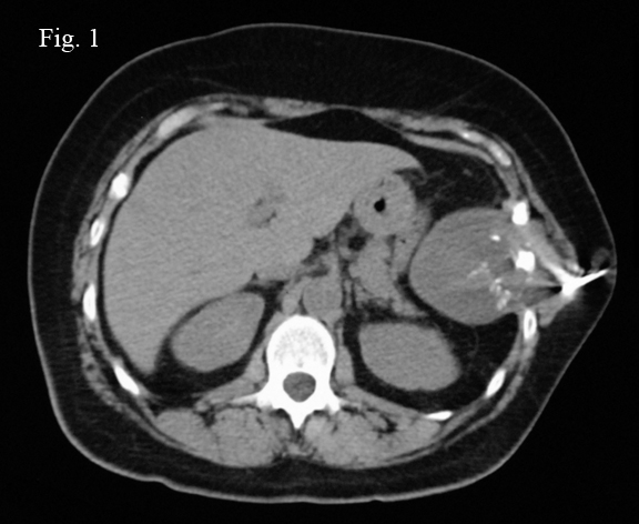
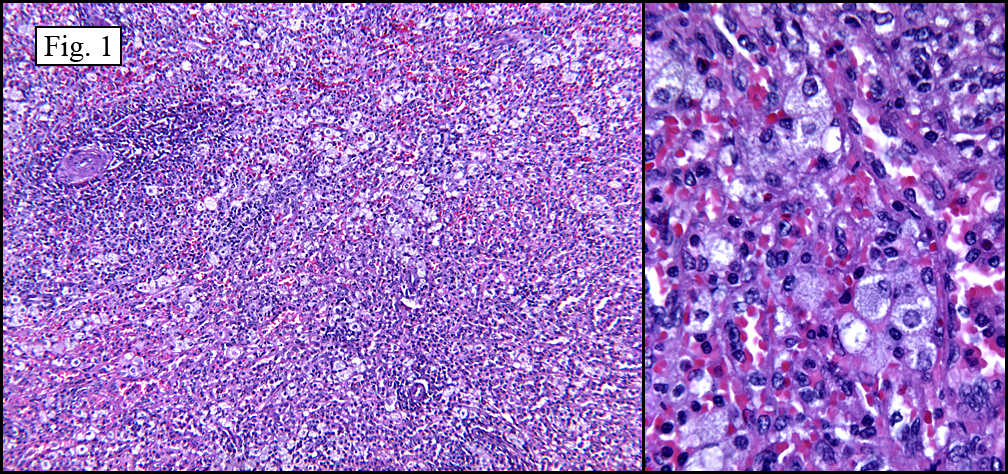
History: A 50 year-old woman presented with a 9 x 7 x 6 cm slowly enlarging, painless upper abdominal mass extending to the flank. A CT scan demonstrated a focally calcified, well-demarcated tumor which completely encompassed and obliterated one rib with involvement of adjacent ribs, as well as extending peripherally into the soft tissues of the thoracic wall (Fig. 1). She subsequently underwent needle biopsy resulting in a small aggregate of gray-tan semi-translucent tissue fragments.

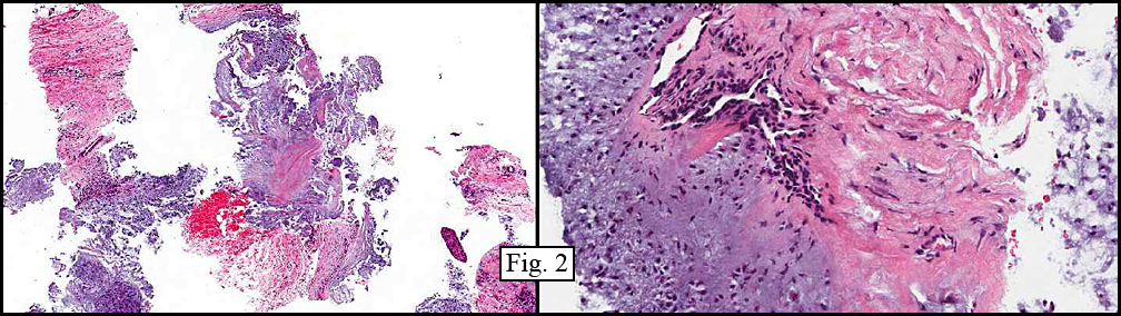
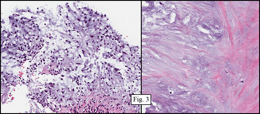
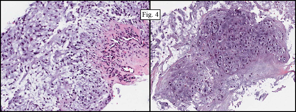
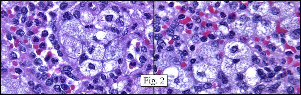
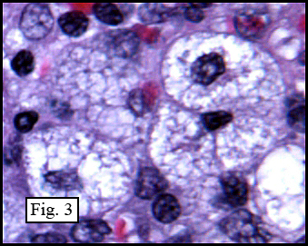
Microscopically, the material varied greatly in cellularity, including fibrous regions, prominent supporting blood vessels, and abundant myxomatous regions incorporating bland round to oval cells with occasional stellate cells (Fig. 2). Mitotic figures were not encountered. The predominant pattern was myxomatous and had no particular growth pattern (Fig. 3 left). There were also regions of necrosis and fibrosis in which were scattered hyphal mycotic organisms (Fig. 3 right). In addition to haphazard growth (Fig. 4 left), several areas showed nodular patterns with groupings of small, hyperchromatic cells residing in lacunae (Fig. 4 right.). Physaliferous cells were not encountered.

Diagnosis: “Myxoid Chondrosarcoma”

Laura Denham MS3, Donald R. Chase, M.D.
Department of Pathology, Loma Linda University and Medical Center
California Tumor Tissue Registry, Loma Linda, California

Discussion: Myxoid Chondrosarcoma (MC) is a rare soft tissue tumor that comprises less than three percent of all soft tissue tumors. It was first described by Stout and Verner in 1953 but not until 1972 did Enzinger and Shiraki elaborate on the clinicopathologic features. MCs have traditionally been divided into skeletal and extraskeletal tumors based on their location of origin, which at times may be difficult to elucidate. It is believed that extraskeletal myxoid chondrosarcoma is a distinct entity whereas skeletal myxoid chondrosarcoma is a separate variant of conventional chondrosarcoma. In 1985 it was discovered that the majority of extraskeletal myxoid chondrosarcomas showed a genetic translocation which was lacking in neoplasms arising from bone. This finding, along with certain histologic and clinical findings, suggests that they are separate entities.

The tumor is grossly characterized as being well circumscribed, lobulated to nodular, and having a gelatinous gray to tan cut surface. There is often hemorrhage, which may be quite prominent. Microscopically, it is usually composed of fairly uniform small round to oval cells with hyperchromatic nuclei edged by eosinophilic cytoplasm. The cells are often arranged in cords, strands and/or nests in a myxoid matrix which may be separated into lobules by fibrous tissue. Occasionally the cells may be arranged in a more diffuse pattern, which is reportedly more common in the skeletal version of myxoid chondrosarcoma. Although somewhat unusual, it is possible to find foci of well-differentiated chondrocytes with typical lacunae in a specimen that has been well sampled, however calcification is rare. Mitoses and fibroblastic cells may also occasionally be found. A cellular variant of myxoid chondrosarcoma is described, characterized by sheets of large cells with prominent nucleoli and a more open, vesicular chromatin pattern and with less myxoid change between cells. Rarely, areas of blue cells reminiscent of Ewings sarcoma/PNET, as well as rhabdoid-like cells with peri-nuclear inclusions have been reported.

Neoplastic cells stain positively for S100 protein in 20-40% of the cases. The decoration is generally weak, but diffuse. Because many of the differential diagnoses also stain strongly for S100 protein, Enzinger and Weiss argue that the usual lack of staining in MC is a useful diagnostic feature. Curiously, MCs occasionally stain positively for cytokeratins, such as EMA and may also stain positively for chromogranin and neuron-specific enolase, suggesting a possible neuroendocrine lineage. Neurosecretory granules seen via electron microscopy also lend support this possibility. Other ultrastructual and histochemical findings such as dilated rough endoplasmic reticulum, prominent mitochondria, diastase sensitive PAS positive intracytoplasmic material and hyaluronidase resistance tend to support chondroid lineage.

Genetic studies show approximately 70% of extraskeletal myxoid chondrosarcomas have a t(9; 22) (q22; q12) translocation. This balanced translocation combines the EWS gene with the CHN (known as NOR1 in rodents). Other translocations have also been described, however, to date similar translocations have not been found in the skeletal myxoid chondrosarcoma.

Differential Diagnosis:

• Chondromyxoid fibroma – Typically the tumor cells are arranged in fused lobules, with a fibrous band surrounding the lesion. The periphery of a lobule tends to be more cellular, with more pleomorphic and fibroblastic cells, while the center of the lobule is usually more myxomatous and may show lacunae formation. It is not unusual to find multinucleated giant cells, especially in areas with bland cells. Occasional foci of calcification or ossification may also be present. An area of well-vascularized connective tissue separates lobules from each other, as well the neoplasm from adjacent bone.

• Ossifying Fibromyxoid Tumor of Soft Parts (OFMT) – Grossly the tumor is surrounded by a thick fibrous capsule and is separated into lobules by fibrous tissue with an underlying shell of lamellar bone in the majority of cases. The uniform round to spindled cells are rimmed by pale cytoplasm and are found in nests, cords or sheets within a myxoid to collagenous matrix. There may be areas of cartilage formation, as well as intricate vasculature often with perivascular hyalinization, both of which are rare in myxoid chondrosarcomas. Furthermore, the majority of OFMTs are S100 positive.

• Myxoid liposarcoma – Grossly these tumors are gelatinous and may be multinodular. The round to spindled cells are characteristically denser around the periphery and are suspended in a myxoid matrix, which is sensitive to hyaluronidase. Lipoblasts are normally present in varying numbers, usually at the periphery of a tumor lobule. A distinct plexiform “chicken-wire” vasculature is a common feature, which helps to differentiate this tumor from myxoid chondrosarcomas. There may also be foci of high cellularity which may or may not have round cell differentiation and little myxoid stroma.

• Myoepithelioma – This mixed tumor may be composed of a mixture of epithelioid, clear, spindled or physaliferous cells found in cords, nests or sheets. The stromal component is also variable and may be myxoid, chondroid or focally osteoid. If ductal differentiation is not present, it may be difficult to differentiate between this entity and myxoid chondrosarcoma. However, in contrast to myxoid chondrosarcomas, these tumors typically co-express S100 and epithelial markers such as EMA and cytokeratin, and may express smooth muscle actin as well.

• Myxoma – A fairly common neoplasm, myxomas are essentially avascular, consisting of widely-spaced, bland spindled to stellate cells with round to oval nuclei in an abundant myxoid matrix with varying amounts of reticulin fibers. There may also be infiltration of the neoplasm into the surrounding skeletal muscle. Myxomas are much less cellular than myxoid chondrosarcomas and tend to have a haphazard spindled pattern rather than a lobular chondrocyte-type pattern.

• Myxoid variant of Extraskeletal (Soft Part) Chondroma – These multinodular lesions are composed of chondrocytes, which tend to be more differentiated toward the periphery of the lesion. The cells may be surrounded by stippled calcification and may occasionally have foci of multinucleated giant cells. Although they are usually less cellular than myxoid chondrosarcoma, the two entities may be difficult to differentiate. However, myxochondromas may be distinguished from myxoid chondrosarcomas based on their predilection for hands and feet, their propensity to form mature hyaline cartilage, and their unique peri-cellular granular calcification pattern.

• Chondroid Lipoma – This rare tumor is generally smooth, well circumscribed and consists of hibernoma-like cells with vacuolated, often granular cytoplasm. The cells are in cords or nests with surrounding myxoid stroma. Although some cells may resemble chondroblasts, this benign neoplasm can be distinguished from myxoid chondrosarcomas by the intracytoplasmic vacuoles, rounded, less lobular appearance, greater vascularity and more consistent positive staining of S100 protein.

Clinically, myxoid chondrosarcomas are mostly found in the deep tissues of the lower extremities, especially buttock and thighs, as well as in the proximal upper extremities such as shoulders or neck. Both skeletal and extraskeletal forms of MC are characterized by slow growth. However, despite its bland morphology and slow growth pattern extraskeletal myxoid chondrosarcoma has a propensity for metastases, most commonly to the lung. Furthermore, recurrences are very common in this entity and may often be multiple. Both chemotherapy and radiation have little role in the eradication of disease, and early total surgical extirpation is the preferred treatment.

Suggested Reading:

1. Enzinger FM, Skiraki M. Extraskeletal myxoid chondrosarcoma: an analysis of 34 cases. Hum Pathol. 3:421-435, 1972.

2. Meis-Kindblom JM, Bergh P, Gunterberg B, Kindblom LG. Extraskeletal myxoid chondrosarcoma: a reappraisal of its morphologic spectrum and prognostic factors based on 117 cases. Am J Surg Pathol 23:636-50, 1999.

3. Atonescu CR, Argani P, Erlandson RA, healey JH, Ladanyi M, Huvos AG. Skeletal and extraskeletal myxoid chondrosarcoma: a comparative clinicopathologic, ultrastructural and molecular study. Cancer 83:1504-21, 1998.

4. Rubin BP, Fletcher JA. Skeletal and extraskeletal myxoid chondrosarcoma: related or distinct tumors? Adv Anat Pathol 6:204-212, 1999.

5. Meis JM, Enzinger FM. Chondroid lipoma: a unique tumor simulating liposarcoma and myxoid chondrosarcoma. Am J Surg Pathol 17(11): 1103-1112, 1993.

6. Rahimi A, Beabout JW, Ivins JC, Dahlin DC. Chondromyxoid fibroma: a clinicopathologic study of 76 cases. Cancer 30:726-736, 1972.

7. Kilpatrick SE, Hitchcock MG, Kraus MD, Calonje, E, Fletcher C. Mixed tumors and myoepitheliomas of soft tissue: a clinicopathologic study of 19 cases with a unifying concept. Am J Surg Pathol 21(1):13-22, 1997.

8. Mackenzie DH. The myxoid tumors of somatic soft tissues. Am J Surg Pathol 5(5): 443-458, 1981.

9. Smith TA, Easley KA, Goldblum JR. Am J Surg Pathol 20(20):171-180, 1996.

10. Enzinger FM, Weiss SW, Liang CY. Ossifying fibromyxoid tumor of soft parts: a clinicopathological analysis of 59 cases. Am J Surg Pathol 13(10) 817-827, 1989.

11. Enzinger and Weiss, Soft Tissue Tumors, 5th edition.

December 2010 Seminar

Diagnostic Identifiers and Biologic Behavior Contrasted in Adult Versus Pediatric Neoplasms
Drs. Pedram Argani and Juan Rosai

Sunday, December 5, 2010
8:30AM to 4:30 PM

Hyatt Regency Hotel
5 Embarcadero Center
San Francisco, CA
(415) 788-1234‎


View Larger Map

Pedram (“Pete”) Argani, MD is Professor of Pathology and Oncology and Director of the Breast Pathology Progam at John Hopkins University and Hospital in Baltimore, MD. After graduating Summa Cum Laude in molecular biology at Princeton, Dr. Argani graduated medical school at the University of Pennsylvania where he also did pathology residency followed by two fellowships at Memorial-Sloan Kettering Cancer Center in New York. He has over 130 published papers in peer-reviewed journals and has established himself as an international leader in surgical pathology, molecular studies breast and GU pathology. He has been on over 25 manuscript review boards and has written 14 book chapters. Honors have included numerous teaching awards and the 2007 Arthur Purdy Stout Prize.

Over the past several decades, Juan Rosai, MD, has been perhaps the most influential leader of surgical pathology. He is currently head of the Pathology Consultation Centre at the Centro Diagnostico Italiano, Milan, Italy and Senior Diagnostic Pathologist at Genzyme Corp., New York. He received his MD degree from the University of Buenos Aires, Argentina and in 1967, completed his postgraduate training in Pathology at Barnes Hospital and Washington University (St. Louis). Affiliations/professorships have included Washington University (St. Louis), the University of Minnesota, Yale University, Memorial Sloan-Kettering Cancer Center, adjunct professor at Cornell University, and professor at the National Cancer Institute in Milan. His book editorships have included several AFIP fascicles, the WHO book on Histologic Classification of Thymic Tumors, and Rosai and Ackerman’s Surgical Pathology, currently in its ninth edition. In addition, he is the author of approximately 300 publications which include original articles, reviews and essays. He has received Doctor Honoris Causa Degrees from the University of Bologna (Italy), the University of Santiago de Compostela (Spain), the University of Ioannina (Greece) and the University of Cordoba (Argentina). He has also been awarded the Honorary Fellowship of the Royal College of Pathologists (England) and the Maude Abbott Lectureship of the US and Canadian Academy of Pathology. Additionally, Dr. Rosai has described several new tumor entities and other diseases, one of which bears his name (Rosai-Dorfman disease).

Objectives: At the conclusion of the seminar the participants will have been updated on the behavioral differences of pediatric and adult neoplasms, and will have a better grasp of new diagnostic modalities and how they augment traditional diagnostic methods.

February 2010: A 31 year old man with traumatic laceration of the spleen

History: A 31 year-old trauma patient underwent a splenectomy to remove a lacerated spleen. The organ was enlarged, weighing 500 grams and had overall diameters of 19.5 x 10.5 x 3.5 cm. The parenchyma was uniformly firm and red with indistinct follicular and trabecular markings. No focal lesions were identified.

Microscopically, the red pulp was expanded, largely by a macrophage infiltration (Fig. 1). These cells had bubbly, vacuolated cytoplasm giving the cytoplasm a “clear cell” appearance (Figs. 2,3). Special stains included:

CD68 Positive
PAS (+/- diastase) Faintly positive
Giemsa Rare ceroid-containing (‘sea blue’) histiocytes
Iron Negative
AFB Negative

Diagnosis: Lipid Storage Disease, Consistent With Niemann-Pick Disease

Melissa Skaugset, MSIV and Donald R Chase MD
Department of Pathology, Loma Linda University and Medical Center, and
California Tumor Tissue Registry, Loma Linda, California

Discussion: Niemann-Pick disease is a disorder of lipid metabolism and storage that causes accumulation of sphingomyelin in tissue macrophages (histiocytes) due to a deficiency of acid sphingomyelinase. Patients can be classified both by clinical features and by genetic mutation. Four types have been identified. Types A and B are associated with a defect in the SMPD1 (acid sphingomyelinase) gene with type A being the neuronopathic form and B the non-neuronopathic form. Mutations in both the NPC1 and NPC2 genes cause so-called type C disease. All forms have autosomal recessive inheritance.

Clinically, presentation varies with type of disease, with Type A showing the most severe impairment characterized by hepatomegaly, jaundice, failure to thrive, seizures, and mental retardation in infancy. Children with Type A disease rarely survive past 18 months. Type B disease has no neural involvement, so patients typically present later, most commonly in late childhood/early teens. These children present with poor growth, hepatosplenomegaly, recurrent lung infections, and may have lab abnormalities, including elevated lipids and cholesterol and thrombocytopenia.

Type C disease differs significantly from A or B disease in both pathologic mechanism and clinical presentation. The defect in the NPC gene causes failure of a transporter in the endosomal-lysosomal system of cells, causing a buildup of cholesterol and glycolipids in lysosomes. The clinical presentation of Type C disease varies widely. In children who manifest disease, progressive neurological deficits develop which may or may not be associated with hepatosplenomegaly or jaundice. Neurologic disease is diffuse and has been documented to include dysphagia, dysarthria, ataxia, seizures, gaze palsies, ptosis, dystonia, hypotonia, psychiatric disorders, and dementia. Types A, B, and C are increased in Ashkenazi Jews.

A fourth form exists, occurring mostly in a population from Yarmouth County, Nova Scotia. This Type D or “Nova Scotian” form has been demonstrated to also have a defect in the NPC1 gene. Interestingly, it seems that those with Type D disease share common ancestry with Joseph Muise (born in Nova Scotia in 1679) and his wife Marie Amirault (born in Nova Scotia in 1684), making one or both of them the likely carrier of original mutation causing disease in this population. It is unclear why what may be a new mutation in either Joseph or Marie resulted in such a high carrier rate, but close communities (this is seen in the Acadian population of Nova Scotia) with the resultant limitation in genetic diversity may have played a role. Once a high carrier rate was achieved, it became possible for this autosomal recessive disease to manifest as children of dual carrier parents who were homozygotic for the disease causing gene, therefore having clinical disease. Similar patterns of emergence of rarer genetic diseases have been seen in populations such as the Old Order Amish (Ellis-Van-Crevald syndrome), French Canadian Chicoutimi (hereditary tyrosinemia). More recently there has been an emergence of fumarase deficiency noted in the Fundamentalist Church of Jesus Christ of Latter Day Saints particularly at the Arizona-Utah border, that shows similar patterns. This is also and autosomal recessive disorder and in a community is able to definitively trace its ancestry to a limited group of church/community leaders, however it has not yet been elucidated when the defective gene was found in the founding members or introduced to the community at a later date.

Pathologic specimens from patients with Niemann-Pick disease may be limited to bone marrow biopsies if the diagnosis is suspected clinically. These are used to confirm disease and to help guide additional biochemical and genetic testing. When surgical specimens are available, gross findings include enlarged spleens and livers, with diffuse involvement of the liver and spleen evident on cut sections. Expansion of the cordal macrophages gives a firm texture to the cut surface of the spleen. The tissue is frequently pale and homogeneous on cross section. The normal markings of the spleen are made less distinct by the proliferation of histiocytes. Later findings in hepatic involvement include nodular fibrosis of the liver.

Histologically, Niemann-Pick cells are enlarged with accumulation of small vacuoles containing sphingomyelin (Figs 2,3). These give the cells a foamy or bubbly appearance and make them lighter in color than the similar appearing Gaucher cells. Niemann-Pick cells are CD-68 positive histiocytes. PAS staining is only faintly positive, but Sudan Black B and Oil Red O are positive, indicating that neutral fat contained in the vacuoles. These lipid deposits are birefringent and have yellow-green fluorescence in UV light. Electron microscopy shows lamellated structures in the lysosomes (similar to myelin figures) and may also demonstrate “zebra bodies”, parallel lamellated structures in the cytoplasm. Giemsa staining can highlight “sea blue” histiocytes containing ceroid, most common in Type C disease.

Many of the congenital enzyme deficiencies cause accumulation of material within the histiocytes of the spleen, liver, and CNS, causing enlargement of the parenchyma and the clinical manifestations of the disease. Many cases are diagnosed using tests for enzyme activity, but histologic diagnosis is often the mainstay of initial evaluation. Histology allows for guidance of enzymatic and/or genetic testing by helping differentiate the cells noted in different metabolic diseases. The cells of Gaucher disease are identified by their “crinkled tissue paper” cytoplasm, seen best on touch imprints. Niemann-Pick histiocytes have bubbly, multi-vacuolated cytoplasm, as do the gangliosidoses and mucopolysaccharidoses (i.e. Tay-Sachs, Hunter’s, or Hurler’s disease), however the latter with ballooned cells makes it distinct from the former. Fabrys, Wolmans, and von Gierkes disease all have foamy appearing histiocytes. Hermansky-Pudlak syndrome causes ceroid accumulation, leading to sea blue colors in the cells, though it should be noted that ceroid containing histiocytes are also seen on other, more common diseases (and in fact were noted in this case) and should therefore be considered somewhat non-specific. Evaluation by of enzyme activity is required for more precise classification of disease.

Because treatment regimens are very limited, patient prognosis depends more on the type of disease, rather than on the treatment. Type A disease almost invariably ends in infantile death. Type B patients may live significantly longer, but have significant morbidity, particularly due to lung involvement. Bone marrow transplantation has been used in Type B patients with some success. It is thought that Types C and D may benefit from a low cholesterol diet, though this has not been demonstrated in clinical studies. The prognosis for Types C and D is widely variable, with some childhood deaths. The less severely affected patients may live into adulthood. An ongoing clinical trial of enzyme replacement therapy uses a recombinant human acid aphingomyelinase, and to date, this protocol shows the greatest promise for improving outcomes in the most severe forms of the disease.

Suggested reading:

Hopkin, R.J., Grabowski, G.A. Lysosomal Storage Diseases (Ch. 341). In: Kasper, D., Fauci, A., Longo, D., Braunwald, E., Hauser, S., and Jameson, J. Harrison’s Principles of Internal Medicine, 16th Ed. New York: McGraw-Hill (2005). pp2315-2319.

Neiman, R., Orazi, A. Disorders of the Spleen, 2nd ed. Philadelphia: W.B. Saunders Company (1999). pp167-175.

Vanier, M.T., Kinuko, S. (1998). Recent Advances in Elucidating Niemann-Pick C Disease. Brain Pathology. 8: 163-174.

Winsor, E.J.T., Welch, J.P. (1978). Genetic and Demographic Aspects of Nova Scotia Niemann-Pick Disease (Type D). Am J Genet. 30: 530-538.

Scroll to top