August 2009: An 8 year old boy with a scrotal mass

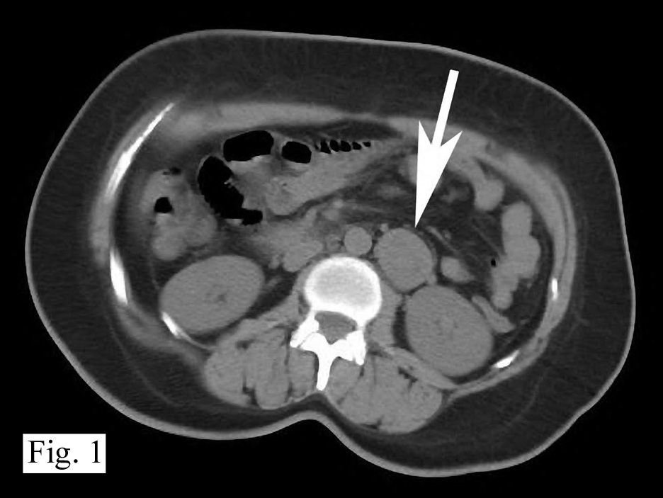
History: An 8 year old otherwise healthy boy was found to have a left scrotal mass. An ultrasound showed the left testis to be “inhomogeneous” and surrounded by a tumor confined to the scrotum. The right testis was normal. Regional inguinal lymph nodes were not enlarged and an abdominal CT scan was unremarkable.

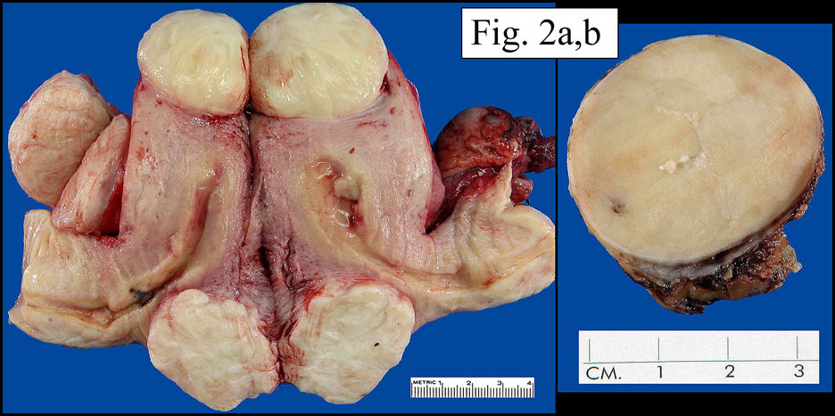
A left orchiectomy specimen included a 7.0 x 3.5 x 2.5 cm grey-tan solid tumor that surrounded and compressed an uninvolved 1.5 cm testis.

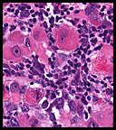
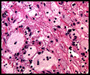
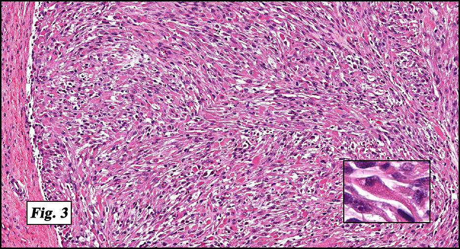
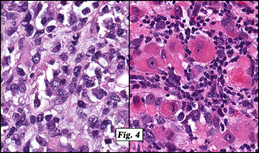
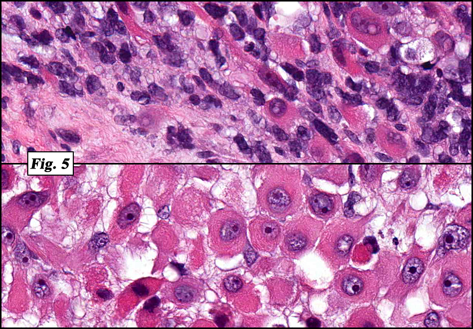
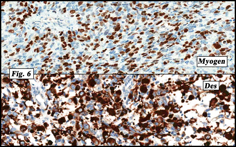
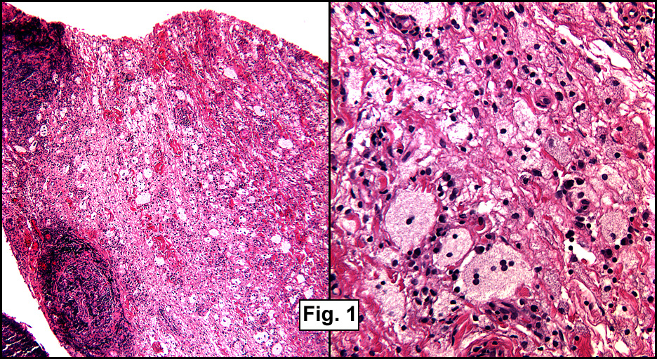
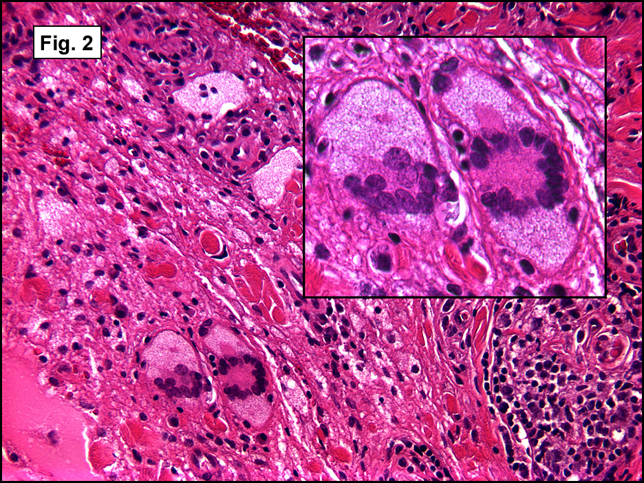
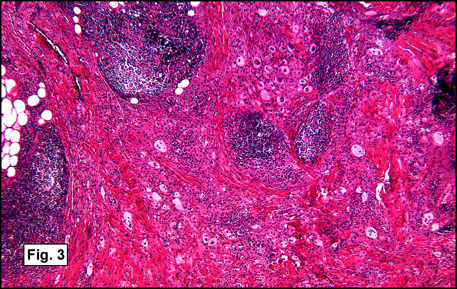
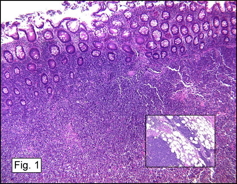
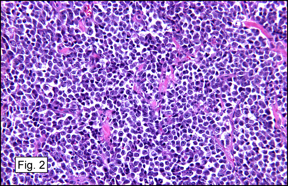
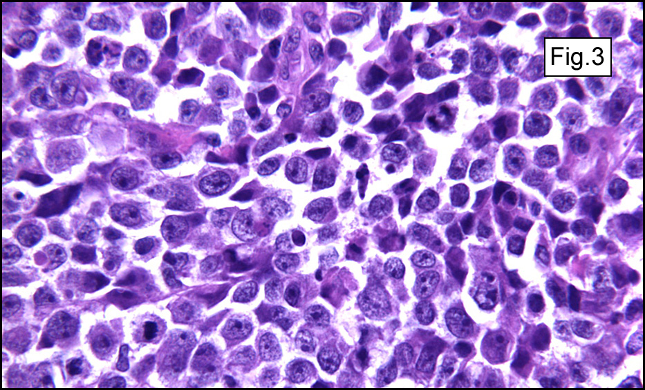
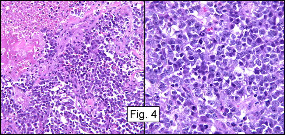
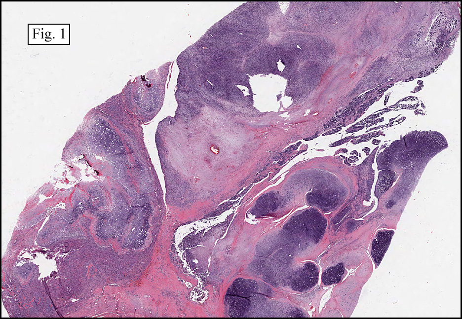
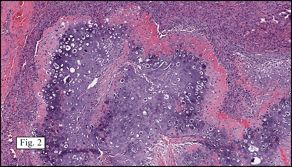
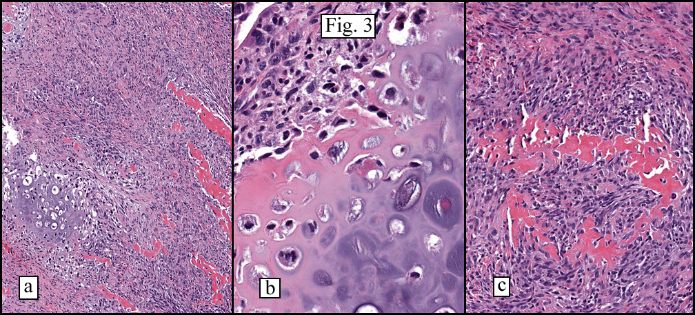
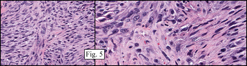
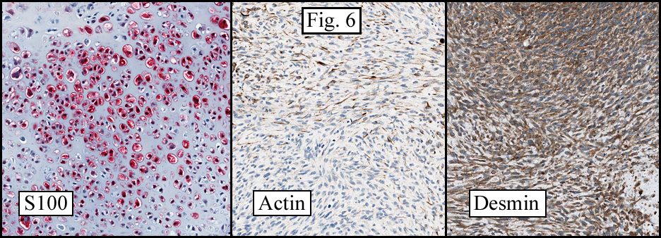
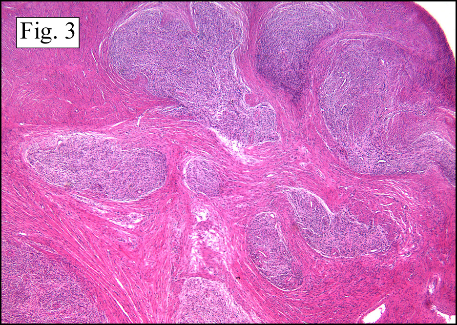
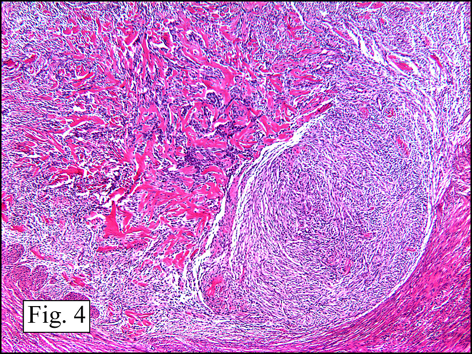
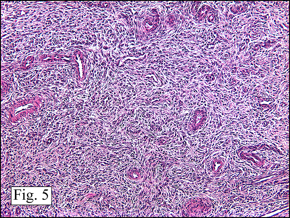
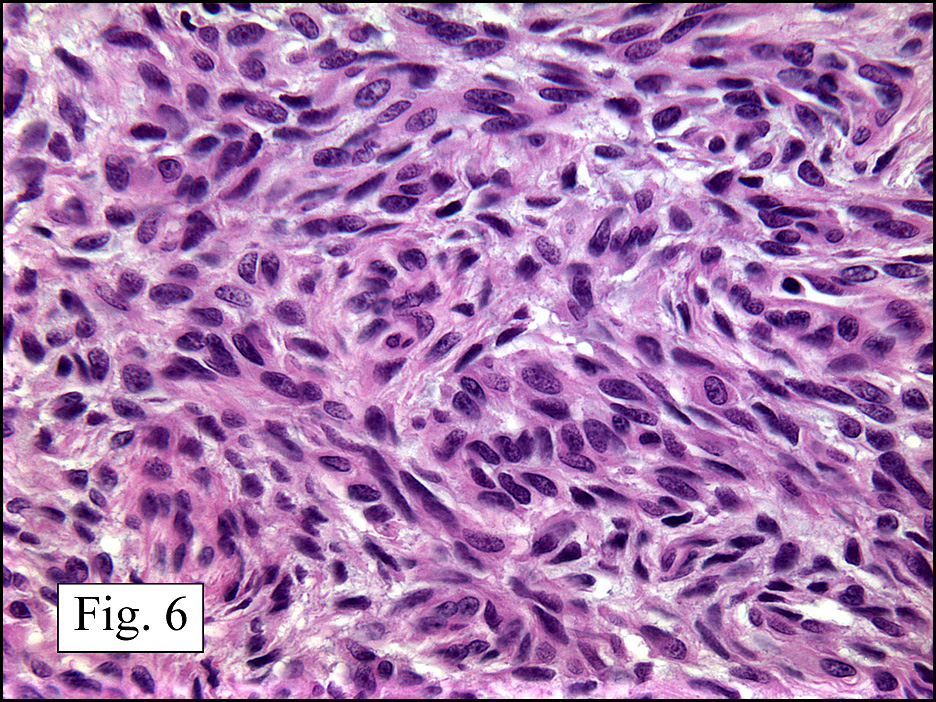
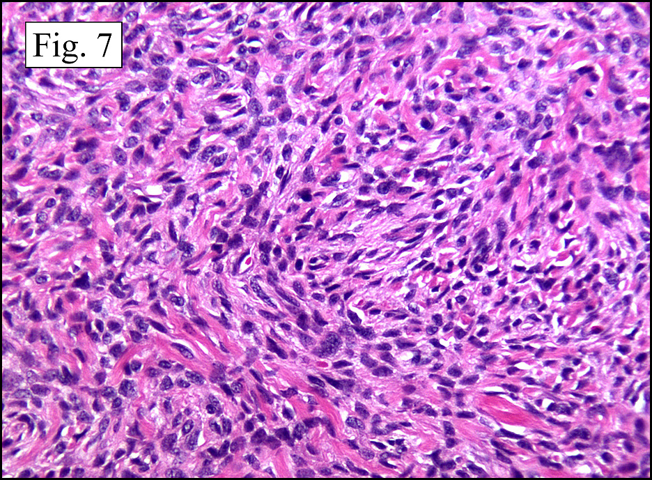
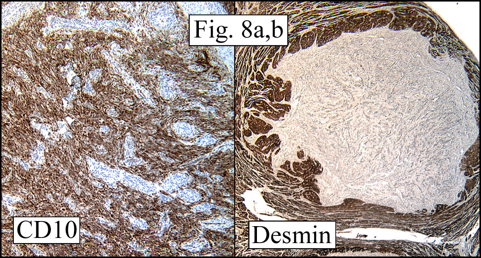
Microscopically the tumor was solid, partly necrotic and highly cellular (Fig. 1). It was partially compartmentalized by fibrous bands separating two populations of cells which varied in amounts of cytoplasm (Fig. 2). Some regions showed spindling with occasional fusiform cells displaying cross striations (Fig. 3). Most of the tumor, however, consisted of areas of primitive small rounded hyperchromatic cells with limited cytoplasm, and other regions having an admixture of primitive cells with larger, more differentiated cells containing abundant eosinophilic cytoplasm and concentric cytoplasmic striations (Fig. 4,Fig. 5). Immunohistochemical stains showed the tumor to have moderate nuclear staining for myogenin (“myogen”) and strong cytoplasmic decoration for desmin (“des”) (Fig. 6).

Diagnosis: “Paratesticular Embryonal Rhabdomyosarcoma”

Krishna Ahuja MD, Donald R. Chase MD, Anwar Raza MD, Craig Zuppan, MD
Department of Pathology, Loma Linda University and Medical Center, Loma Linda, California
California Tumor Tissue Registry, Loma Linda, California

Discussion: Rhabdomyosarcoma (RMS) is a malignant mesenchymal tumor, usually of childhood, showing striated muscle differentiation. It generally arises in body regions with skeletal muscle, though it may also occur in locations lacking these elements such as the prostate or nasopharynx. As a result, RMS is currently felt to devolve not from myocytes, but from primitive mesenchyme having myogenic tendencies. Although the etiology is not known, it is associated with:

Genetic Disorders:
Neurofibromatosis type 1
Adenomatous polyposis coli
Beckwith-Wiedemann syndrome
Gorlin nevoid basal cell carcinoma syndrome

Developmental Disorders:
Congenital lung cyst
Central nervous system malformations
Genitourinary malformations
Fetal alcohol syndrome

Traditionally the morphological classification of RMS includes three main types, each having slightly different ages of occurrence:

• Embryonal rhabdomyosarcoma (infants, children, adolescents, young adults)
• Alveolar rhabdomyosarcoma (adolescents, young adults)
• Pleomorphic rhabdomyosarcoma (adults)

The WHO, however, currently recommends a classification by subtype which emphasizes prognosis.

Prognostic Group and Type % of Cases 5-year Survival
Superior:
Botryoid embryonal RMS 6% 95%
Spindle cell embryonal RMS 3% 88%
Intermediate:
Embryonal RMS, NOS 49% 66%
Poor:
Alveolar RMS 31% 54%
Undifferentiated sarcoma 3% 40%

*(From Newton, 1995 and CTTR seminar of June, 2008 by Cheryl Coffin)

Embryonal RMS

ERMS accounts for approximately 50 % of all RMS mostly affecting children younger than 10 years of age, it also occurs in adolescents and young adults and is uncommon in patients older than 40 years of age. The tumor seems to display various stages of myogenesis and its patterns are variable, ranging from poorly differentiated tumors that are difficult to diagnose without immunohistochemical or electron microscopic examination, to well-differentiated neoplasms that resemble fetal or even adult muscle. The percentage of differentiated rhabdomyoblasts serves as a rough estimate of differentiation. Tumors with mostly primitive small cells tend to do worse than those dominated by larger, rounded rhabdomyoblasts. Two important sub-types with more favorable prognoses are:

Spindle cell RMS: Usually involves paratesticular soft tissue, followed by the head and neck area. Histologically it is composed almost exclusively of elongated fusiform cells with cigar-shaped nuclei and prominent nucleoli. Cross striations are more readily discernible in this subtype than in others.

Botryoid RMS (BRMS): Most commonly found in mucosa-lined hollow organs such as nasal cavity, nasopharynx, bile duct, urinary bladder and vagina. According to ICR criteria, a “cambium layer”, characterized by a subepithelial condensation of tumor cells separated from an intact surface epithelium by a zone of loose stroma, must be present to recognize this variant.

Alveolar RMS

This tumor occurs more often in limb girdles than does BRMS and usually arises in adolescents. The classic alveolar pattern consists of anastomosing fibrovascular septa lined by discohesive tumor sometimes accompanied by multinucleated cells. The neoplasm is aggressive and may present with distant metastasis. Curiously, in addition to lung, it may metastasize to breast or the bone marrow where it can cause a leukemic-like presentation.

Pleomorphic RMS

Accounting for no more than 10% of the tumors, the “adult form” of RMS is rare. It usually arises in the limbs and appears to be less chemosensitive than the other variants. The histology is that of a pleomorphic sarcoma (nos) showing positivity for muscle markers such as myogenin, desmin, etc. The expected 5-year survival is only around 20%. Of interest is that it may be associated with prior radiation to or near the primary site.

With combination therapy, especially improved chemotherapy, the prognosis of RMS has greatly improved through the years. Despite the dramatic improvement in treatment of RMS, early diagnosis and accurate classification remains vital. Continued therapeutic success is expected as we enter the molecular era with the potential for targeted therapies.

Suggested Reading:

Stout AP. Rhabdomyosarcoma of the skeletal muscles. Ann Surg 123:447, 1946.

Newton WA Jr, et al. Classification of rhabdomyosarcomas and related sarcomas. Pathologic aspects and proposal for a new classification – an Intergroup Rhabdomyosarcoma Study. Cancer 76:1073, 1995.

Parham DM: Pediatric Neoplasia: Morphology and Biology, Chapter 5, pp 87-100, Lippincoot-Raven Publishers, Philadelphia 1996.

Cheryl M. Coffin: “The New International Rhabdomyosarcoma Classification, Its Progenitors, and Considerations beyond Morphology.” Advan Anat Pathol, Vol 4, No 1, pp1-16, 1997.

Enzinger and Weiss’: Soft Tissue Tumors 5th Ed. Editors SW Weiss, JB Goldblum. Mosby, Inc. pp 595-632, 2008.

July 2009: A 55 year old man with ten year history of swollen eyes, asthma, and free kappa light chains

History: A 55-year-old man with a ten year history of swollen eyes presented with asthmatic symptoms. He was found to have enlarged upper and lower eyelids with ocular secretions that prevented him from seeing properly. The clinical differential included inflammatory pseudotumor of the orbits, proptosis from Grave’s disease and multiple myeloma. The workup showed an elevation of free kappa light chains. A periocular biopsy was performed.

The examined tissue consisted of a diffuse infiltration of lymphocytes admixed with lipid-laden histiocytes (xanthoma cells). They had small rounded nuclei and abundant clear or vacuolated cytoplasm (Fig. 1). There were also scattered multi-nucleated giant cells of the Touton type (Fig. 2). Within the lymphoid component were prominent follicles with parafollicular fibrosis (Fig. 3). Immunohistochemically, the foamy histiocytes were strongly positive for CD68 and negative for S100. The lymphoid infiltration had a reactive profile with the germinal cells being CD20 positive and negative for Bcl-2. The parafollicular T cells were CD3 positive (Fig. 4).

Diagnosis: “Adult-Onset Asthma and Periocular Xanthogranuloma”

Jin Guo MD, Jun Wang MD and Donald R. Chase MD
Department of Pathology, Loma Linda University and Medical Center, Loma Linda, California
California Tumor Tissue Registry, Loma Linda, California

Discussion: Adult-onset asthma with periocular xanthogranuoloma (AAPOX) was first described by Jakobiec et al in 1993 when they reported 6 patients with periocular xanthogranulomatous disease associated with adult onset asthma, but lacking necrobiosis. The entity has now been placed into a heterogeneous group of syndromes called adult xanthogranulomatous disease (AXGD) that are rare and poorly understood. AXGD has variable clinical features which have resulted in subclassification into:
• Adult onset xanthogranuloma
• Necrobiotic xanthogranuloma
• Erdheim-Chester disease, and
• AAPOX

Histopathologically, each of these four entities is characterized by infiltration of “hallmark cells” specifically foamy histiocytes and Touton-type giant cells, both of which are negative for S100 and CD1. The infiltration along with accompanying lymphocytes can replace the normal lacrimal gland architecture, causing mass effects and loss of tear production.

Adult onset xanthogranuloma is an isolated xanthogranulomatous lesion without significant systemic involvement.

Necrobiotic xanthogranuloma is characterized by subcutaneous skin lesions that tend to ulcerate and become fibrotic. Associated systemic findings frequently include paraproteinaemia and multiple myeloma.

Erdheim-Chester Disease is an idiopathic condition of lymphohistiocytic infiltration of the heart, lungs, retroperitoneum, bones, and other tissues. The elements are xanthogranulomatous with Touton giant cells and are accompanied by regional fibrosis.. The condition is often fatal, with death due to cardiomyopathy, severe lung disease, or chronic renal failure.

AAPOX often presents with bilateral yellow-orange, elevated, indurated and non-ulcerated xanthomatous eyelids and/or orbital masses. It typically extends into the anterior orbital fat, and sometimes involves the extraocular muscles and/or the lacrimal gland(s). Most patients experience adult-onset asthma within a few months to a few years of onset. Even when asthmatic symptoms are severe enough to require systemic corticosteroids and/or inhalation therapy, a chest X-ray may be negative.

AAPOX is rare with only 21 previous reported cases. Like other AXGDs, it usually has sheets of mononucleated foamy histiocytes, with lymphocytes, plasma cells and Touton giant cells. The cells characteristically infiltrate the orbicularis muscles and the anteriorbital tissues. Their Touton cells have a ring of nuclei about a central eosinophilic zone which is surrounded by a zone of pallor extending to the periphery of the cell. Lymphoid follicles are commonly scattered throughout. Unlike Erdheim-Chester Disease, fibrosis is usually minimal to moderate and unlike necrobiotic xanthogranuloma there is no necrobiosis of collagen. Because the eyelids remain intact and the process does not reach the deep orbital and perioptic connective tissues, visual acuity may not be affected and ocular motility is generally well preserved unless the extraocular muscles are involved. Only rarely are the corneas exposed to the infiltrate, but in rare cases where lacrimal gland involvement causes punctuate corneal epitheliopathy the patient may suffer from a dry eye condition. CT scanning may reveal preseptal and anterior orbital involvement with occasional posterior tracking along or within the orbital muscles and fat, usually sparing the perioptic nerve connective tissues. Facial bones are usually not involved.

Systemic evaluations are usually normal except for rare reports of elevated alpha high-density cholesterol and/or M-protein (IgG) on serum immunoelectrophoresis. The latter suggests that the inflammatory infiltrates have stimulated B cell populations.

The main differential diagnoses of AAPOX include other AXGDs including the aforementioned adult onset xanthogranuloma, necrobiotic xanthogranuloma, and Erdheim-Chester Disease. These conditions all share similar lymphogranulomatous infiltrates but have subtle differences in the amount of lymphoid follicles, number of Touton giant cells, degree of fibrosis and presence of necrosis. Curiously, these features may vary within the same specimen and change with time and treatment. Based upon morphology alone, therefore, the AXGDs are difficult to sub-classify on histomorphologic grounds alone and usually require correlation with patterns of systemic involvement.

Necrobiosis with palisading epithelioid histiocytes is most often seen in necrobiotic xanthogranuloma whereas large lymphoid aggregates with germinal centers are often found in cases of AAPOX. Whereas orbit/adnexal xanthogranulomas tend to be anterior in adult-onset xanthogranuloma, AAPOX, and in necrobiotic xanthogranuloma, in Erdheim-Chester disease the disease is often diffuse (and posterior) leading to visual loss. Bone involvement is common and death may occur despite aggressive therapies.

While the majority of cases can be classified as one of the four syndromes, there are cases that fall in between. These patients have varying combinations of periocular xanthogranuloma and other blood dyscrasias including thrombocytopenia, paraproteinaemia and/or monoclonal gammopathy of undetermined significance (MGUS).

In addition to AXGDs, AAPOX should also be differentiated from other non-Langerhans disorders of histiocytes, including juvenile xanthogranuloma (JXG). This tumor is usually manifested as a self-limited, corticosteroid-sensitive skin tumor that rarely has systemic manifestations. Infants and small children are mainly affected with head and neck lesions. Many extracutaneous sites, however, have been reported, particularly the eye where JXG may cause spontaneous hyphema and result in secondary glaucoma and eventual blindness. Approximately one half of patients with ocular involvement also have skin lesions.

Other entities involving ocular adnexal or orbital tissue that may require distinction from adult xanthogranulomatous disease include:
• Langerhans histiocytosis
• Rosai-Dorfman syndrome
• Inflammatory MFH
• Inflammatory myofibroblastic tumor (inflammatory pseudotumor)
• Graves disease
• Multiple myeloma, and
• Lymphoma.

Despite surgical debulking, AAPOX often recurs within 6-12 months. Although systemic prednisone treatment may cause temporal shrinkage, it alone is usually not successful in causing a lasting resolution. Combined prednisone and systemic chemotherapy for paraproteinaemia may result in more complete resolution of the xanthogranulomatous eyelid deposits. Radiotherapy has also been successfully employed, but the results are more anecdotal than statistical. The eyelid lesions in necrobiotic xanthogranuloma can be successfully be treated with radiotherapy or systemic prednisalone and chlorambucil unless extensive destruction has occurred. Patients without detectable systemic disease at the time ocular adnexal lesions appear should be spared surgical debulking because of possible scarring. In this setting high doses of systemic corticosteroids and low doses of periobital radiotherapy may be given with hope of protection of the globe. If this approach fails, systemic corticosteroids and light chemotherapy may be tried. In patients with MGUS, follow-up should include serial serum protein immunoelectrophoresis and bone biopsy as needed.

A recent study evaluated the efficacy of methotrexate in the treatment of the periorbital changes in adult-onset xanthogranuloma with or without asthma. With follow-up of over 3 years, 10-20 mg/weeks of methotrexate with folate supplementation and a course of corticosteroids showed promising results in significantly reducing inflammation and ptosis.

In summary, AAPOX as well as other entities in the adult xanthogranulomatous disease are rare and pose challenges in daily clinical practice. Recognizing their clinical and histopathological features will facilitate early diagnosis and appropriate therapeutic management.

Suggested reading:

Sivak-Callcott JA, Rootman J, Rasmussen SL, Nugent RA, White VA, Paridaens D, Currie Z, Rose G, Clark B, McNab AA, Buffam FV, Neigel JM, Kazim M. Adult xanthogranulomatous disease of the orbit and ocular adnexa: new immunohistochemical findings and clinical review. Br J Ophthalmol 90(5):602-8, 2006.

Jakobiec FA, Mills MD, Hidayat AA, Dallow RL, Townsend DJ, Brinker EA, Charles NC. Periocular xanthogranulomas associated with severe adult-onset asthma. Trans Am Ophthalmol Soc 91:99-125, 1993.

Hammond MD, Niemi EW, Ward TP, Eiseman AS. Adult orbital xanthogranuloma with associated adult-onset asthma. Ophthal Plast Reconstr Surg 20(4):329-32, 2004.

Hayden A, Wilson DJ, Rosenbaum JT. Management of orbital xanthogranuloma with methotrexate. Br J Ophthalmol 91(4):434-6, 2007.

Vick VL, Wilson MW, Fleming JC, Haik BG. Orbital and eyelid manifestations of xanthogranulomatous diseases. Orbit 25(3):221-5, 2006.

Rayner SA, Duncombe AS, Keefe M, Theaker J, Manners RM. Necrobiotic xanthogranuloma occurring in an eyelid scar Orbit 27(3):191-4, 2008.

Chaudhry IA, Al-Jishi Z, Shamsi FA, Riley F. Juvenile xanthogranuloma of the corneoscleral limbus: case report and review of the literature. Surv Ophthalmol 49(6):608-14. Review, 2004

Elner VM, Mintz R, Demirci H, Hassan AS. Local corticosteroid treatment of eyelid and orbital xanthogranuloma. Trans Am Ophthalmol Soc 103:69-73, 2005

June 2009: A 53 year old woman with acute abdominal pain

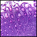
History: A 53 year old woman presented with acute abdominal pain assumed to be diverticulitis. A right hemicolectomy was performed.

The resected colon showed extensive serosal fibrinopurulent exudate. There was also an 8 x 7 x 4 cm mass in the cecum and ascending colon which narrowed the lumen by approximately 70%. It ulcerated the overlying mucosal and perforated into the underlying fat. The attached portion of ileum was uninvolved.

Microscopically, the tumor involved the submucosa with upward extension into the mucosa and more deeply into the muscularis and serosa (Fig. 1). The tumor was composed of sheets of fairly uniform large cells with a faint organoid growth pattern (Fig. 2). The cells were round to oval with moderate amounts of cytoplasm, vesicular nuclei and prominent nucleoli (Fig. 3). Numerous mitotic figures (up to 30/10 HPFs) were accompanied by apoptotic bodies as well as large areas of necrosis (Fig. 4). Immunohistochemically, the tumor cells were strongly positive for synaptophysin and CD56 and moderately positive for chromogranin (Fig. 5). They were negative for cytokeratins, CDX2, TTF1, CD45 and CD117.

Diagnosis: Large Cell Neuroendocrine Carcinoma of the Cecum
Yvonne Noronha MD, Donald R Chase, MD
Department of Pathology and Human Anatomy,
Loma Linda University and Medical Center, Loma Linda, CA
California Tumor Tissue Registry, Loma Linda, CA.

Discussion: Originally described in the lung, large cell neuroendocrine carcinoma (LCNEC) has subsequently been reported in other organs. Because of its rarity, clear-cut criteria were originally lacking and the tumor was graded as if it was of pulmonary origin. But it is now recognized that unlike carcinoid tumors/atypical carcinoids, LCNEC is high grade, supported by their high mitotic rates, generally >10/10 HPF and aggressive clinical behavior.

Recently, in a study of 65 high grade neuroendocrine carcinomas of the GI tract, Shia et al used the WHO/ International Association for the Study of Lung Cancer criteria for pulmonary neuroendocrine tumors as a guideline to classify neuroendocrine carcinoma of the GI tract. To be classified as high grade neuroendocrine carcinoma, the tumor had to have >10 mitoses/10 HPF and had to fulfill criteria from one of the following three sub-groups:

1) Small cell carcinoma:
• Tumors with typical small cell morphology: markedly high nuclear to cytoplasmic ratio, hyperchromatic nuclei, finely granular chromatin with absent or faint nucleoli and frequent nuclear molding.
• Immunohistochemical evidence of epithelial differentiation: Positive staining for AE1:AE3.
• Positive staining for neuroendocrine markers helps but was not a requirement for diagnosis.

2) Large cell neuroendocrine carcinoma:
• Tumor with large cell neuroendocrine morphology: diffuse growth pattern/ neuroendocrine architecture (organoid growth pattern, trabeculae, and rosette formation); monotonous round to oval cells with moderate amounts of cytoplasm, granular or vesicular nuclei, with or without prominent nucleoli. Focal intracytoplasmic mucin or lumen formations were not criteria for exclusion.
• Positive immunohistochemical staining for chromogranin or synaptophysin in > 20% of tumor cells.

3) Mixed neuroendocrine carcinoma:
• Tumors showing histologic features in between small and large cell neuroendocrine carcinoma with immunohistochemical evidence of neuroendocrine differentiation.

The differential diagnosis of LCNEC is broad and should include (apart from carcinoids and small cell neuroendocrine carcinomas) lymphomas, plasma cell myelomas, poorly differentiated carcinomas and sarcomas. The correct diagnosis is based on the recognition of the typical morphology on H&E staining and is supported by the use of a limited immunohistochemical panel (synaptophysin and chromogranin). CD56 can be used but may not clarify the diagnosis since it can be positive with lymphomas, plasma cell myelomas, synovial sarcomas and rhabdomyosarcomas which are part of the differential diagnoses of LCNEC on H&E.

LCNECs are aggressive tumors. In one study of 83 cases of LCNEC of lung, the median survival was 17 months. The clinical significance of these tumors has yet to be fully evaluated. Their recognition, however, is of potential therapeutic significance since their neuroendocrine nature links them to classic small cell carcinomas which are sensitive to chemotherapy. Reports available to date regarding the chemosensitivity of LCNECs are contradictory. Although there are no prospectively controlled data, surgical resection when feasible, followed by combination chemotherapy similar to that employed for small cell carcinoma, appear to offer the best chance for improved survival in patients with early stage disease.

Suggested Reading:

1. Hamilton S.R and Aaltonen L.A. World Health Organization Classification of Tumors. Pathology and Genetics of Tumors of the Digestive System. Lyon: IARC Press; 2000.
2. Travis WD, Colby TV, Shimosato Y, et al. in association with Sobin LH and pathologists from 14 other countries. WHO/IASLC Classification of Lung and Pleural Tumors. Berlin: Springer; 1999.
3. Shia J, Tang LH, Weiser MR et al. Is Nonsmall Cell Type High-Grade Neuroendocrine Carcinoma of the Tubular Gastrointestinal Tract a Distinct Disease Entity? Am J Surg Pathol 32(5): 719-731, 2008.
4. Bernick PE, Klimstra DS, Shia J et al. Neuroendocrine Carcinomas of the Colon and Rectum. Dis Colon Rectum 47: 163-169, 2004.
5. Gaffey MJ, Mills Se, Lack EE. Neuroendocrine Carcinoma of the Colon and Rectum. A clinicopathologic, ultrastructural and immunohistochemical study of 24 cases. Am J Surg Pathol; 14: 1010-1023, 1990.

May 2009: A 32 year old male with a short history of exertional dyspnea

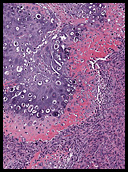
History: A 32 year old previously healthy Hispanic male presented with a 3-week history of dyspnea upon exertion.  A CT-scan showed a mass in the left cardiac atrium which extended into the pulmonary veins.  The tumor was well encapsulated and attached to an atrial appendage.  It measured 6 x 5 x 4 cm and was solid, pale pink-tan with gelatinous regions.  Foci of necrosis and hemorrhage were present.  The surgeon was not able to completely excise the mass.

The neoplasm was microscopically remarkable for having a large number of sarcoma patterns (Fig. 1).  There was a blending of spindle-cell sarcoma with osteosarcoma and chondrosarcoma (Fig. 2,3) Other areas consisted of sheets of uniform fusiform cells growing in long sweeping fascicles with a faint herringbone pattern (Fig. 4).  Yet other regions showed cells with more pleomorphism and abundant eosinophilic cytoplasm with faint longitudinal and cross striations (Fig. 5).

The cartilaginous, but not the spindle cell areas marked for S100 protein. An actin stain was weakly positive and a desmin stain was strongly positive, especially in the more pleomorphic spindle cell areas (Fig. 6)

Diagnosis: “Chondrosarcoma, Osteosarcoma, Myosarcoma and Fibrosarcoma of the atrium (‘Malignant Mesenchymoma’)”

Timothy R. Smith, MS3 and Donald R. Chase, MD
Department of Pathology and Human Anatomy 
Loma Linda University and Medical Center, Loma Linda, California 
California Tumor Tissue Registry, Loma Linda, California

Discussion: Primary tumors of the heart are very rare.  They are mostly benign with relative frequencies of:

myxomas (78%)
sarcomas of any type (10%)
fibroma (3%)
lipoma (3%)
vascular tumors (2%), and 
rhabdomyoma (2%).   

Most cardiac tumors present with non-specific symptomology reflecting disruption of the conduction system or blockage of blood flow.  In addition, high grade sarcomas may also present with distant metastasis, especially when they involve the great vessels of the heart.  When malignant, cardiac tumors (virtually all of which are sarcomas) are usually of a “pure” histologic type and the combination of sarcoma subtypes in our presentation case is exceedingly rare.  Although it is common practice to identify the specific type of sarcoma by morphologic and immunohistochemical patterns, the exercise of doing so is of little clinical value.  In addition to location of a sarcoma in the great vessels, low resectibility, a high mitotic rate and the presence of necrosis portend an accelerated aggressive behavior.

There is still debate about the classification of soft tissue tumors that show more than one tumor type.  In soft tissue, the uterus, and other in other organs, when both an epithelial and sarcomatous component are present the term, “carcinosarcoma” is still in use.  In soft tissue the term, “malignant mesenchymoma” has been used since it was first coined by Stout in 1948.  He defined these complex neoplasms as “non-epithelial malignant tumors showing two or more unrelated tissue types in addition to a fibrosarcomatous element”  Stout and Lattes later changed this definition to mean sarcomas composed of two or more unrelated tumor elements, with or without a fibrosarcomatous component. More recently, the World Health Organization decided that “malignant mesenchymoma” was a vague term and did not reveal the various morphologies of the mixed sarcomas.  The current thought, expounded by Weiss, Goldblum, and Fletcher is that the term “malignant mesenchymoma” should be deleted from the classification scheme of soft tissue tumors. 

Differential Diagnosis of Cardiac Neoplasms:

• Cardiac Rhabdomyomas usually arise in the myocardium of children, generally below the age of five years and is sometimes associated with tuberous sclerosis. The cells are usually “bubbly”, stain for desmin, and may have striations.  Mitotic figures are virtually non-existent.  Occasional rhabdomyomas may spontaneous regress, usually by maturation into more normal-appearing myocytes. 
 
• Cardiac Fibromas also arise mostly in children and may be associated with adenoma sebaceum of the skin and kidney tumors.  They consist of a uniform proliferation of fibroblasts without significant mitotic activity.  They lack either a cartilaginous or an osseous matrix.

• Myxomas have varied gross appearances and are smooth or lobular, firm or gelatinous, and contain a myxoid stroma. They account for the majority of primary cardiac tumors. Seventy-five percent are found in left atria.  Curiously, benign myxomas usually are found in the left side of the heart, while sarcomas are usually found in the right side.

• Vascular neoplasms may be benign (hemangioma) or malignant (angiosarcoma) and when large, may interrupt blood flow through the heart.  Angiosarcoma is particularly prone to early metastases.  Both tumors stain for vascular markers such as CD31, CD34 and HFVIII.

• Lipomas usually originate in the endocardium or epicardium and have a broad pedunculated base.  They consist of mature, adult-type fat.

• Pericardial cysts may mimic a cardiac tumor or pericardial effusion on chest x-ray.  They are usually asymptomatic, although some cause compressive symptoms (eg, chest pain, dyspnea, cough).  The center of the process is of blood-tinged fluid which is bounded by pericardial-type cells. 

• Sarcomas are usually vimentin-positive and are composed of spindled cells of varying pleomorphism.  A negative desmin stain tends to exclude myogenous origin and a negative S100 stain helps in the exclusion of neural tumors and melanoma.  A negative epithelial stain helps rule out spindled carcinomas and synovial sarcoma.  Osseous and/or cartilaginous matrix formation rules out the previously mentioned benign cardiac tumors.

• Teratomas usually occur in midline structures such as the anterior mediastinum but rarely arise in the heart.  By definition, these germ cell tumors contain tissue types representing all three germ layers.

A high index of suspicion is necessary for early diagnosis of these cardiac neoplasms.  Patients with signs of cardiac obstruction, chest pain or discomfort, or dyspnea should have appropriate radiologic and laboratory work-ups.  But even with early diagnosis, sarcomas do poorly, and even with positive indicators such as low mitotic rate, low levels of necrosis, presence in the left side of the heart, and good resectibility, the survival rate of cardiac sarcomas is under five years.  Cardiac transplant is a last measure for patients with Unmanageable primary cardiac tumors. 

Suggested Reading:

Weiss SW, Goldblum JR. Enzinger & Weiss’ Soft Tissue Tumors 5th ed: 1213-1214, 2008.

Stout AP. Mesenchymoma: the mixed tumor of mesenchymal derivatives. Annu Surg: 127:278-90, 1948.

Stout AP, Lattes R. Tumors of the soft tissues. In: Firminger GI, ed. Atlas of Tumor Pathology. 2nd series. Fascicle 1. Washington, DC: Armed Forces Institute of Pathology;: 172-5, 1967

Virmani, Burke, Farb, Atkinson. Tumors and tumor-like lesions of the heart and great vessels. Cardiovascular Pathology 2nd ed;: 424-458, 2001.

Merck online manual section cardiovascular disorders, subject Cardiac tumors, Topics Cardiac tumors, 2008.

April 2009: A 43 y.o. woman with multiple uterine masses and left periaortic lymphadenopathy

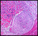
History: A 43-year-old gravida 0 woman with a history of menorrhagia was found to have a 3.5 cm left retroperitoneal, periaortic mass on abdominal CT six years previously (
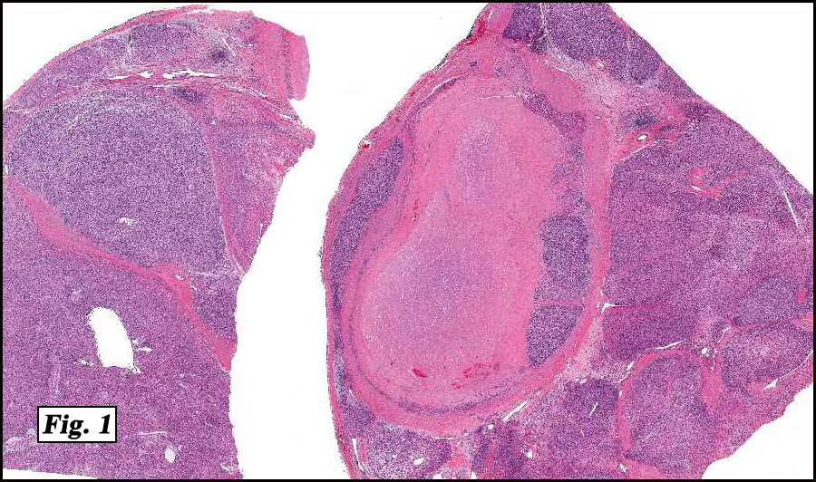
Fig. 1).  She underwent CT-guided biopsy of the left periarotic mass and was diagnosed to have an epithelioid and spindle cell neoplasm consistent with epithelioid leiomyosarcoma.  Despite chemotherapy the retroperitoneal lesion persisted, though no progression was seen by CT follow-up.  A current pelvic examination demonstrated a mobile anteverted enlarged uterus without adnexal masses. A vaginal hysterectomy with bilateral salpingo-oophorectomy was performed.

The 920 gm, 19.0 x 18.0 x 8.0 cm uterus contained multiple serosal, intramural and submucosal nodules with pale-tan whorled cut surfaces measuring up to 8.0 cm in diameter (Fig. 2a). Additionally, the anterior lower uterine segment was distorted by numerous firm, poorly-circumscribed nodules ranging from 0.2 to 1.0 cm in diameter.  The excised 6.0 cm left periaortic mass proved to be an enlarged lymph node with fleshy-pink, homogeneous cut surfaces (Fig. 2b).

Microscopically, the lower uterine segment was extensively infiltrated by numerous small nodules with irregular tongues invading the adjacent myometrium (Fig. 3).  The nodules were densely cellular with a “blue” appearance at low magnification and areas with prominent stromal hyalinization (Fig. 4).  Composed of a spindle cell proliferation, the neoplasm was punctuated by a plexiform capillary network with edematous change (Fig. 5).  Spindle cells featured uniform oval to round nuclei, vesicular chromatin, small nucleoli and elongated inconspicuous cytoplasm (Fig. 6).  No striking pleomorphism and relatively few mitotic figures were seen ranging from 2 to 5 mitoses per 10 hpf with occasional atypical forms.  No hemorrhagic necrosis, definitive vascular invasion, sex cord-like arrangements, or epithelioid differentiation was identified.  The periaortic lymph node was predominately replaced by neoplastic cells with identical histology as the lower uterine segment nodules (Fig. 7).  Immunohistochemical studies performed on both the uterine and lymph node tumors exhibited identical patterns: strong positivity for CD10, focally positivity for SMA, and negative staining for desmin (Fig. 8a & 8b).  A Ki67 proliferative index demonstrated low activity (~ 10%).

Diagnosis: “Low-grade endometrial stromal sarcoma with retroperitoneal periarotic lymph nodes metastasis”

Mingyi Chen M.D., Evelyn Choo M.D., Amita T. Mistry, M.D., Donald R. Chase, M.D.
Department of Pathology, Loma Linda University and Medical Center
California Tumor Tissue Registry, Loma Linda, California

Discussion: Endometrial stromal tumors are the second most common category of mesenchymal tumors of the uterus; however, they only account for approximately 10% of all uterine sarcomas.  Low-grade endometrial stromal sarcoma (LGESS) occurs in younger women (median age ~40 years) compared to other uterine neoplasms such as carcinosarcoma (malignant mullerian mixed tumor), leiomyosarcoma, or endometrial carcinoma.  Patients typically present with abnormal vaginal bleeding, pelvic pain, and a clinical impression of uterine leiomyomata due to an enlarged uterus with an irregular contour.

Endometrial stromal tumors are divided into two classes based on the presence of circumscribed margins (stromal nodule), or infiltrating margins with or without vascular intrusion (stromal sarcoma).  The two lesions are otherwise indistinguishable histologically.  Infiltration is seen as irregular, jagged islands or tongues of neoplastic stromal cells between the smooth muscle bundles of the surrounding normal myometrium.   Determining myometrial infiltration can be problematic, especially when there is subtle interdigitation of stromal and smooth muscle cells at a compressive margin examined at high magnification, which should not be counted as an infiltrative margin.  A diagnosis of stromal sarcoma is rarely possible in curettage and biopsy specimens since margin evaluation is questionable.  For vascular involvement, clumps of tumor cells must be present in spaces within the myometrium.

LGESS is comprised of uniform, oval to spindle-shaped cells with inconspicuous cytoplasm and round to oblong nuclei that are morphologically, ultrastructurally and immunohistochemically identical to endometrial stromal cells found in proliferative endometria.  This monotonous cellular proliferation is interrupted by a characteristic regular network of delicate ramifying small vessels resembling spiral arteries and capillaries.  The presence of fragments of proliferative phase endometrial stroma without glands in curettage or biopsy specimens raises the possibility of an endometrial stromal tumor.  Significant cytological atypia and pleomorphism are absent as well as stainable glycogen and mucin.  Occasionally, an abundance of distinctive hyalinized osteoid-like collagen is present, and may be so extensive as to reduce the cellular component to attenuated chains of cells, resembling a hyalinized leiomyoma. This pattern of hyalinization may be preserved in pelvic recurrences and pulmonary metastases.  Foam cells may be present in stromal neoplasms and are morphologically identical to the foam cells seen in endometrial hyperplasia and carcinoma.  Neoplastic cells are positive for CD10, almost always estrogen and progesterone receptor positive and focally positive for actin.

The most common cytogenetic alteration observed in low-grade endometrial stromal tumors is the t(7;17)(p15;q21) translocation, resulting in the fusion of the JAZF1 and JJAZ1 genes.

Differential Diagnosis:

•    Benign smooth muscle tumors: In a uterine tumor composed of uniform spindle cells with circumscribed margins, the distinction between smooth muscle and endometrial stromal tumors is less critical since both leiomyoma and endometrial stromal nodules are benign. With infiltrating margins, the distinction between either a leiomyoma or a LGESS is clinically significant.  Cellular leiomyomata can be especially problematic.  As in the current case, the concomitant presence of extensive leiomyomas can cause difficulties in the diagnosis of endometrial stromal sarcoma.  Small tissue sampling can also add to the complexity.  Positive staining of desmin, smooth muscle actin, and/or CD10 immunostains can be helpful in distinguishing LGESS from smooth muscle neoplasms.  If the cellular lineage remains ambiguous, it may be prudent to assign the tumor to the low-grade stromal category, particularly if there are tongue-like islands infiltrating within the myometrium or vascular invasion is present.  Though unequivocal smooth muscle elements may be seen within otherwise typical endometrial stromal neoplasms, such tumors behave more like LGESS than their smooth muscle counterparts (such as intravascular leiomyomatosis, leiomyomatosis, or leiomyomas).  Intravascular leiomyomatosis is far less common than endometrial stromal sarcoma; and should not be diagnosed unless tumor cells show unambiguous smooth muscle differentiation.

•    Leiomyosarcoma: LGESS lacks significant cytological atypia and is less mitotically active when compared to leiomyosarcoma.  Mitotic indices more than 10 mitotic figures/10 hpf are unusual in endometrial stromal sarcoma but do not exclude the diagnosis.  In addition, the tumor cells in leiomyosarcoma have more abundant cytoplasm, and can often be focally arranged in long fascicles.  Angioinvasive leiomyosarcoma is distinguished from endometrial stromal sarcoma by its greater cytologic atypia, and lack of plexiform vasculature.

•    Adenomyosis:  Occasionally, adenomyotic foci do not contain glands, raising the possibility of LGESS.  Stromal nodules and sarcomas produce mass lesions, whereas small foci of gland-free endometrial stroma in the absence of a mass within the myometrium are almost always a manifestation of adenomyosis.  Looking for the characteristic smooth muscle hypertrophy that often accompanies adenomyosis as well as gland-containing lesions elsewhere in the myometrium usually establishes the correct diagnosis.  Very rarely, fragments of benign proliferative phase stroma without glands are found within myometrial vessels.  As long as no mass lesion is identified and the intravascular fragments are microscopic, this phenomenon is undoubtedly a manifestation of adenomyosis.

•    Hemangiopericytoma:  Vasculature resembling endometrial spiral arterioles is uniformly distributed throughout LGESS potentially leading to a mistaken diagnosis of hemangiopericytoma.  Immunohistochemical studies can establish the diagnosis with CD10 staining the endometrial stromal cells in LGESS, and the weak and patchy staining of CD34 in hemangiopericytoma.

•    Adenosarcoma and malignant mullerian mixed tumor (MMMT): LGESS with foci of glandular differentiation raises the differential diagnostic consideration of adenosarcoma which contains endometrial stromal cells as the mesenchymal component.  Glands in endometrial stromal sarcoma are usually sparse and small without surrounding stromal condensation, whereas those of adenosarcoma often feature periglandular stromal condensation and tend to be large and dilated.  However, when glands are numerous and stromal condensation is questionable, considerable overlap exists between these two neoplasms with malignant mesenchymal elements.  LGESS typically expresses estrogen and progesterone receptors, but MMMT shows marked pleomorphism and a high mitotic rate.  Prognostically, both LGESS with glands and adenosarcoma with a low-grade stromal proliferation are low-grade neoplasms, though the prognosis worsens when the primaries occur in extra-uterine sites.

LGESS is characterized by indolent growth with up to half of all patients developing one or more pelvic or abdominal recurrences, some as much as 20 years later.  Median interval for recurrence is 3-5 years.  Women with stage I disease have >80% survival, while survival is less favorable (~50%) when advanced stage disease is present.  Pulmonary metastases occur in 10% of stage I tumors.  The single most important prognostic factor is the stage at presentation, and risk of recurrence is determined by both stage and the type of surgical procedure.  Adnexal spread may not be appreciated during the operation and pelvic recurrence occurs more frequently with ovary conservation.  Both recurrent and metastatic disease can remain localized for long periods and are amenable to successful treatment by resection, radiation therapy, progestin therapy or a combination thereof.  Curative therapy is not guaranteed by bland histology and low mitotic indices.  Although progestin therapy may be effective in cases with progesterone receptor expression, chemotherapy alone is usually ineffective.

Suggested Reading:

1.    Kempson RL, Hendrickson MR. Smooth muscle, endometrial stromal, and mixed Mullerian tumors of the uterus. Modern Pathology. 13(3):328-42, 2000.
2.    Clement PB, Young, RH. Atlas of gynecologic surgical pathology. 1st Edition, 177-210, 2000.
3.    Kurman RJ. Blaustein’s pathology of female genital tract. 5th Edition, 583-605, 2002.
4.    Robboy SJ, Anderson MC, Russell P. Pathology of the female reproductive tract. 1st Edition, 370-80, 2002.
5.    Kurihara S, Oda Y, Ohishi Y, Iwasa A, Takahira T, Kaneki E, Kobayashi H, Wake N, Tsuneyoshi M. Endometrial stromal sarcomas and related high-grade sarcomas: immunohistochemical and molecular genetic study of 31 cases. Am J Surg Pathol. 32(8):1228-38, 2008.

Scroll to top