April 2014: An adult woman with a painful mass in her back

History: An adult woman experienced a painful mass in her back. The mass, excised for symptomatic relief, was in the left paraspinal region at the inferior angle of scapula. Clinically, it was 9 x 4 cm, soft and movable. It had been gradually enlarging for about 3 years.

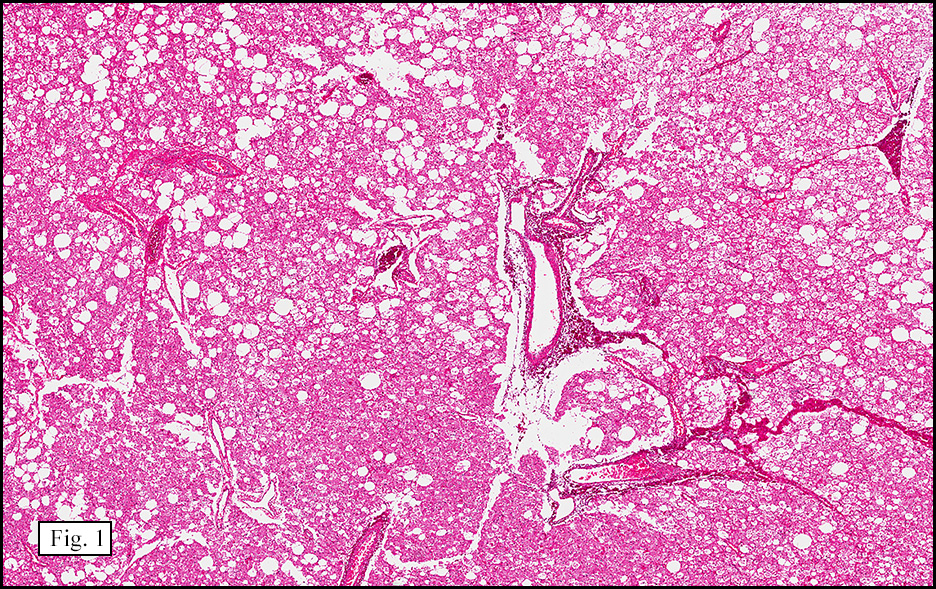
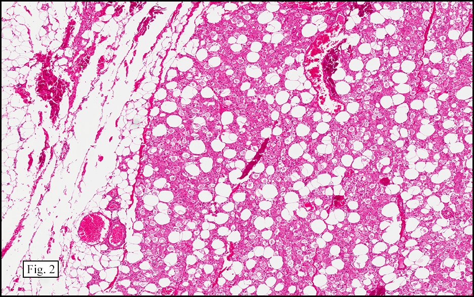
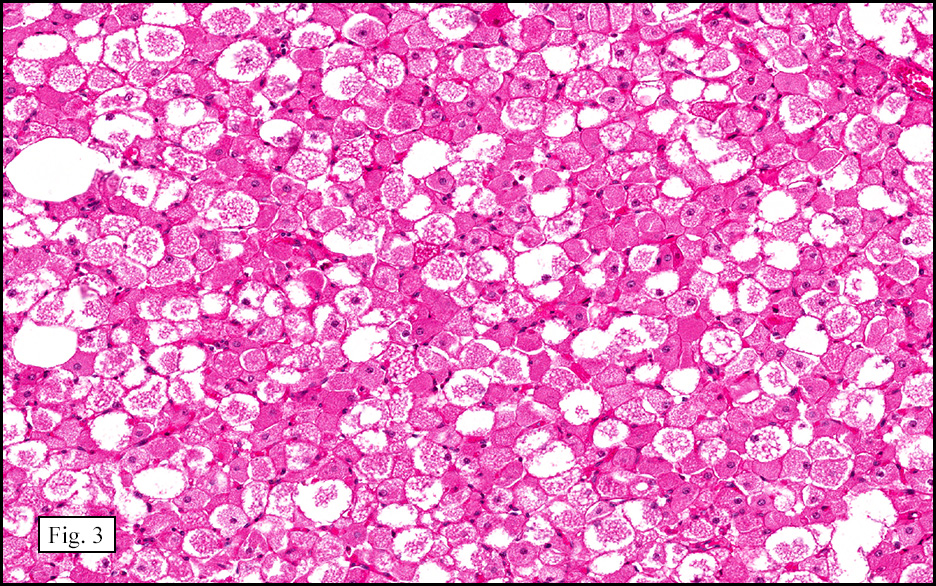
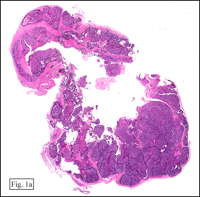
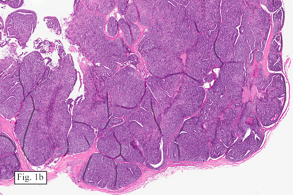
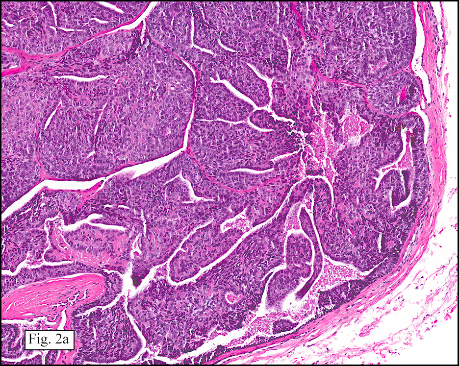
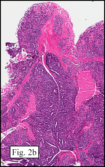
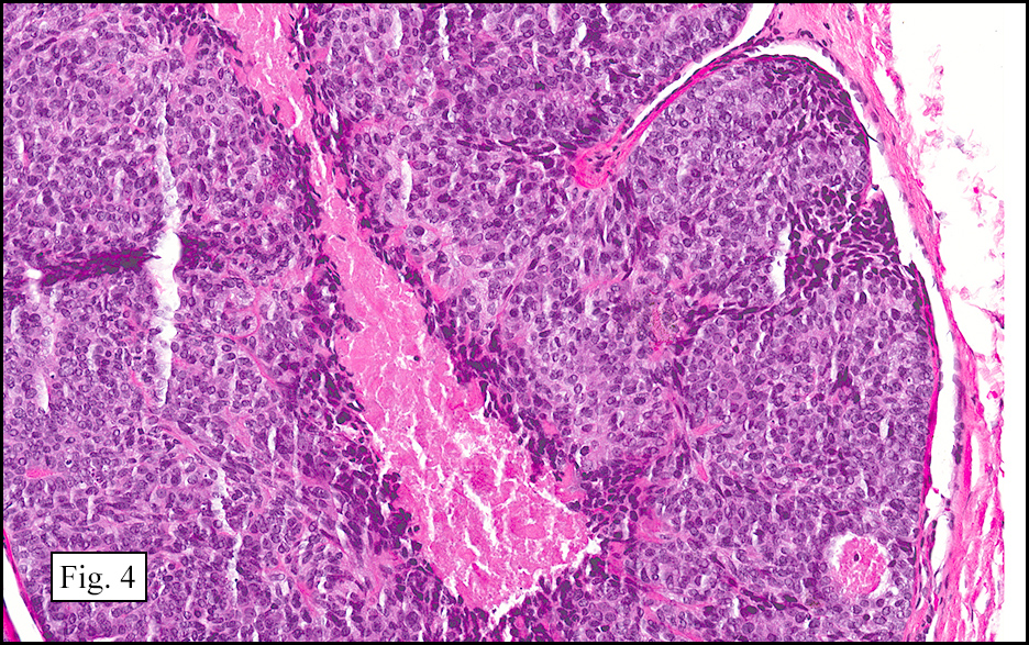
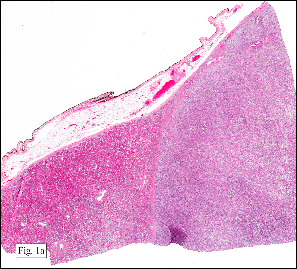
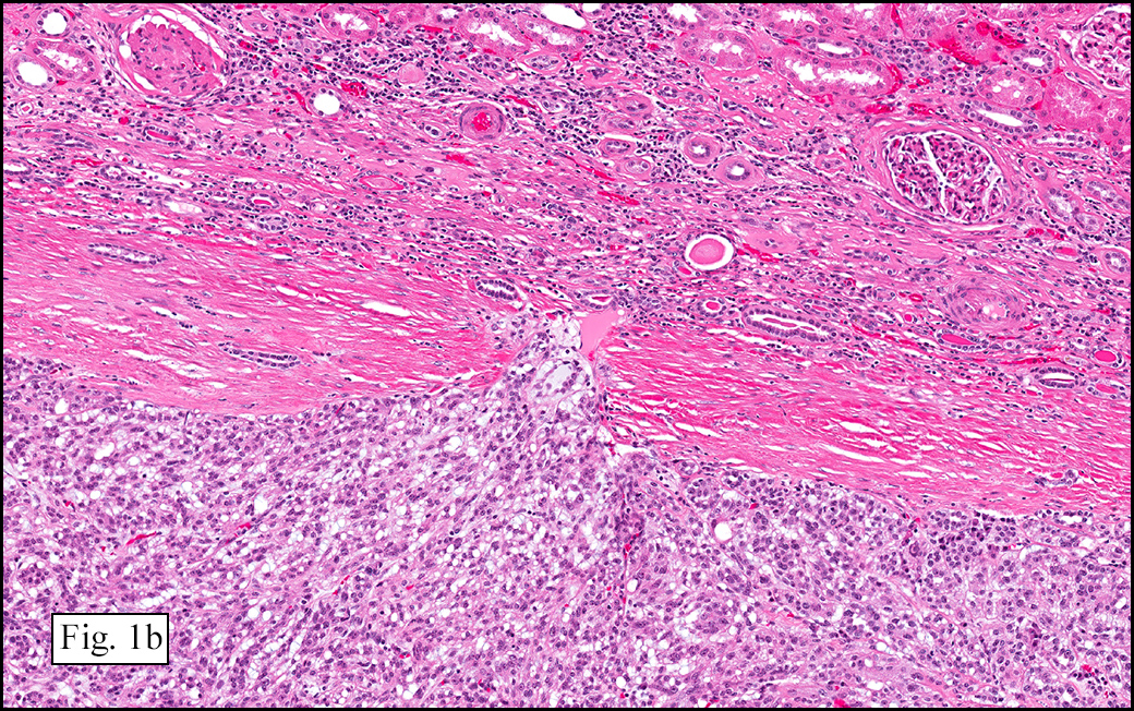
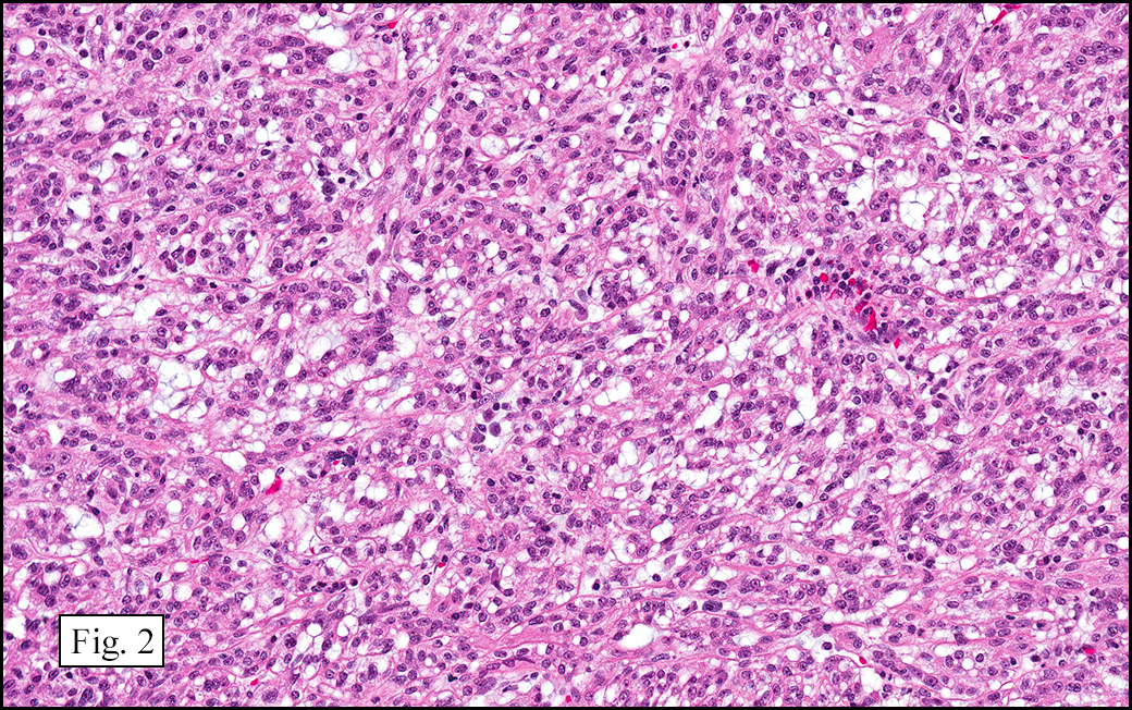
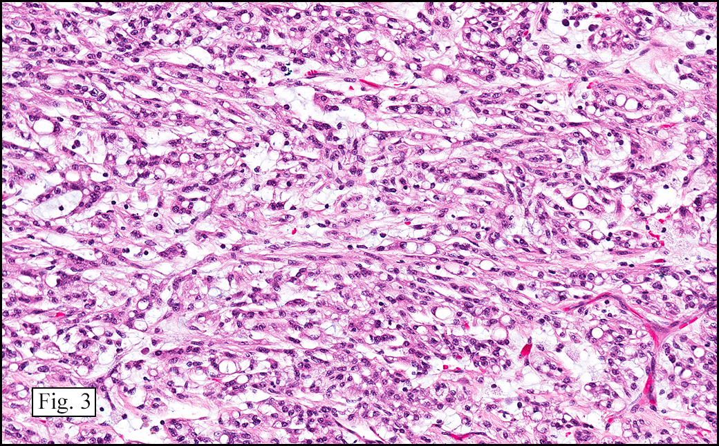
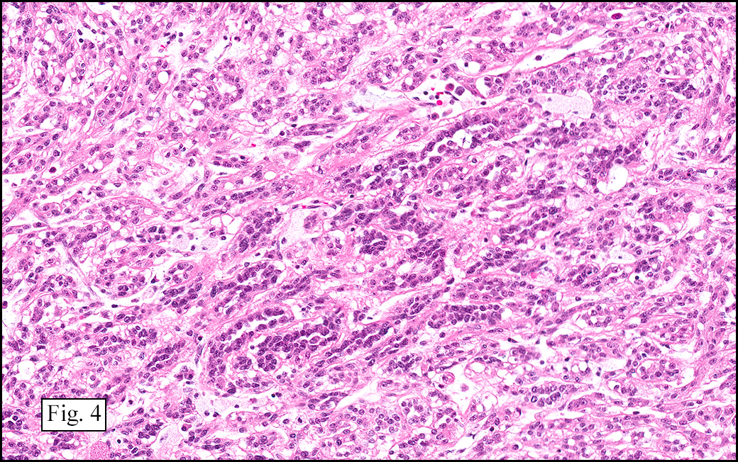
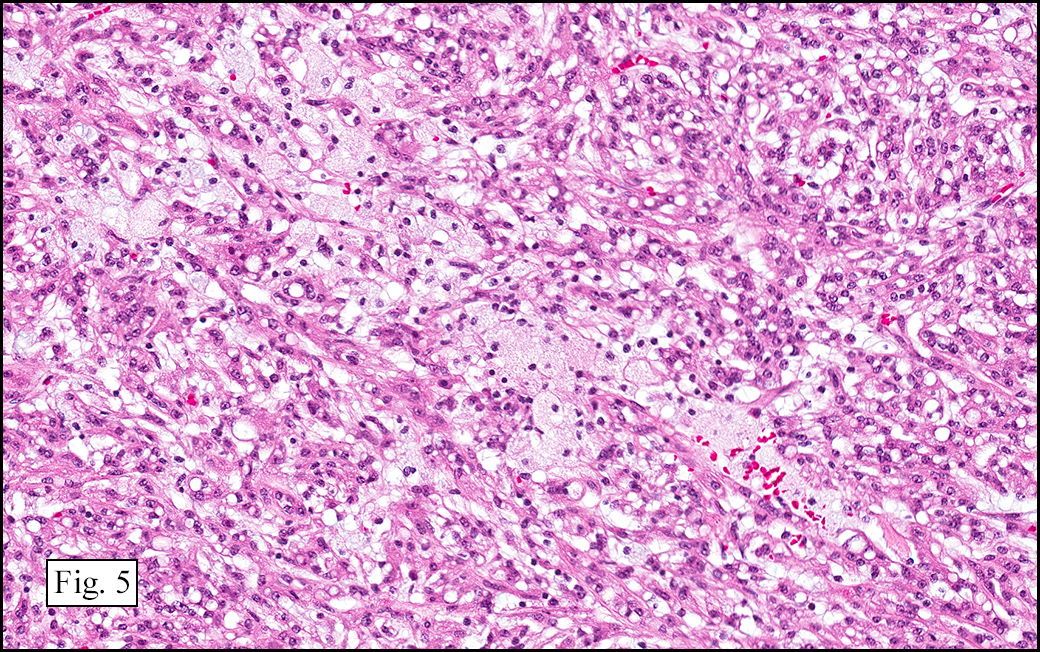
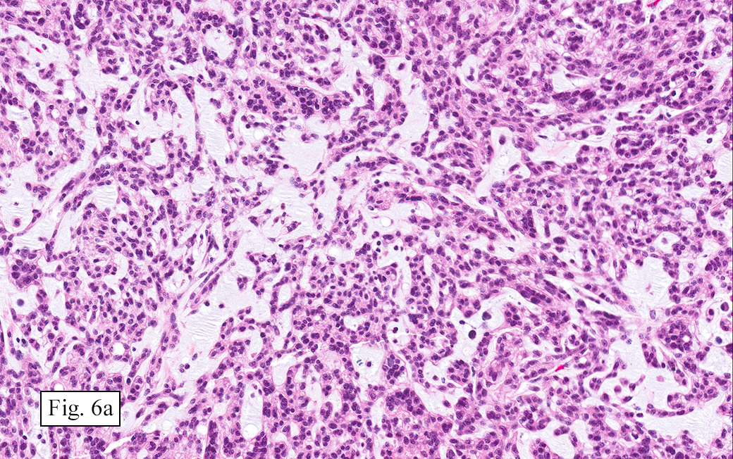
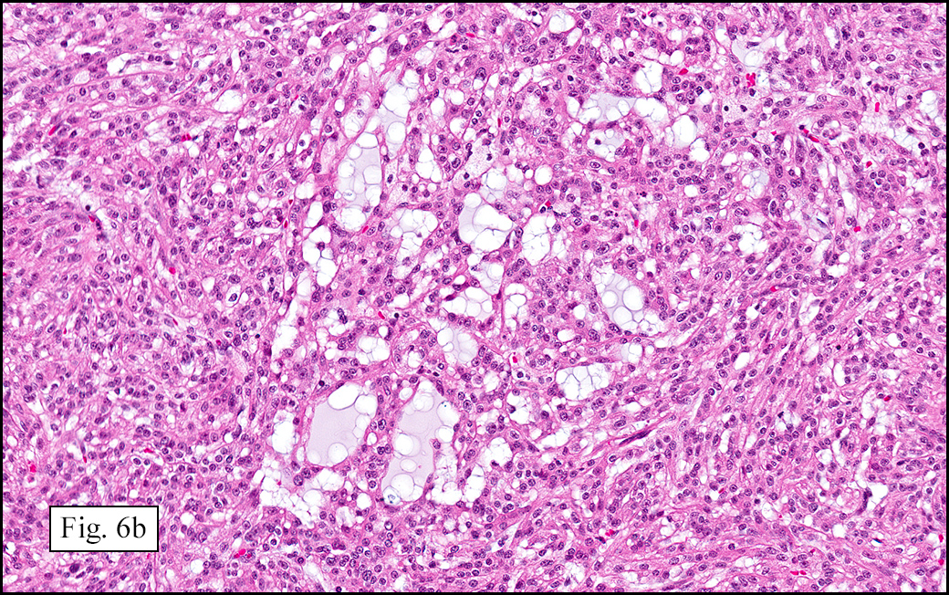
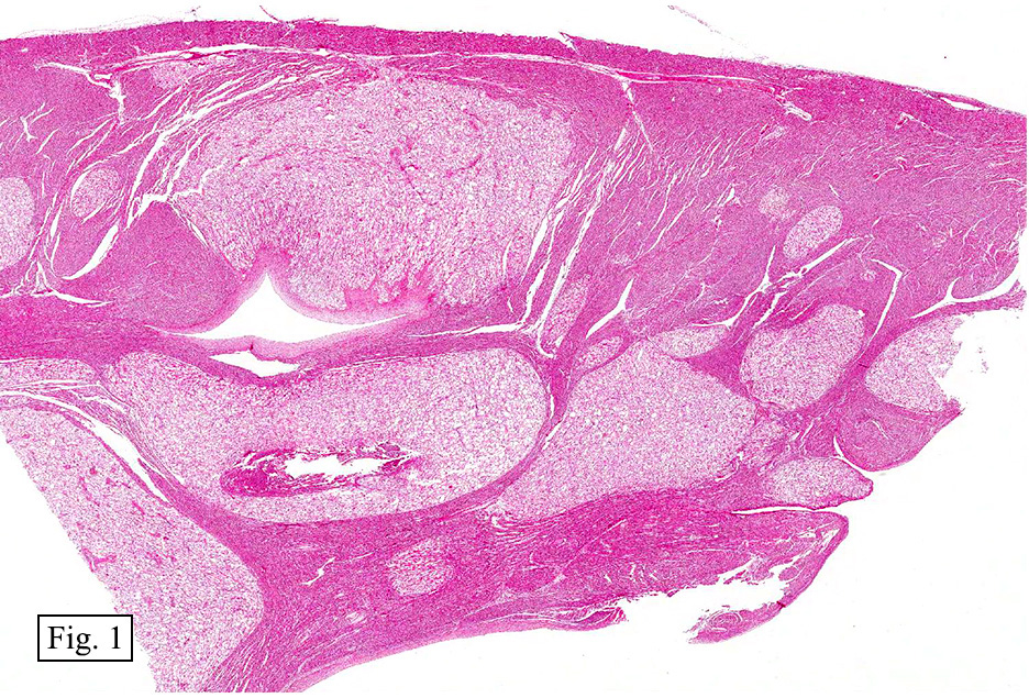
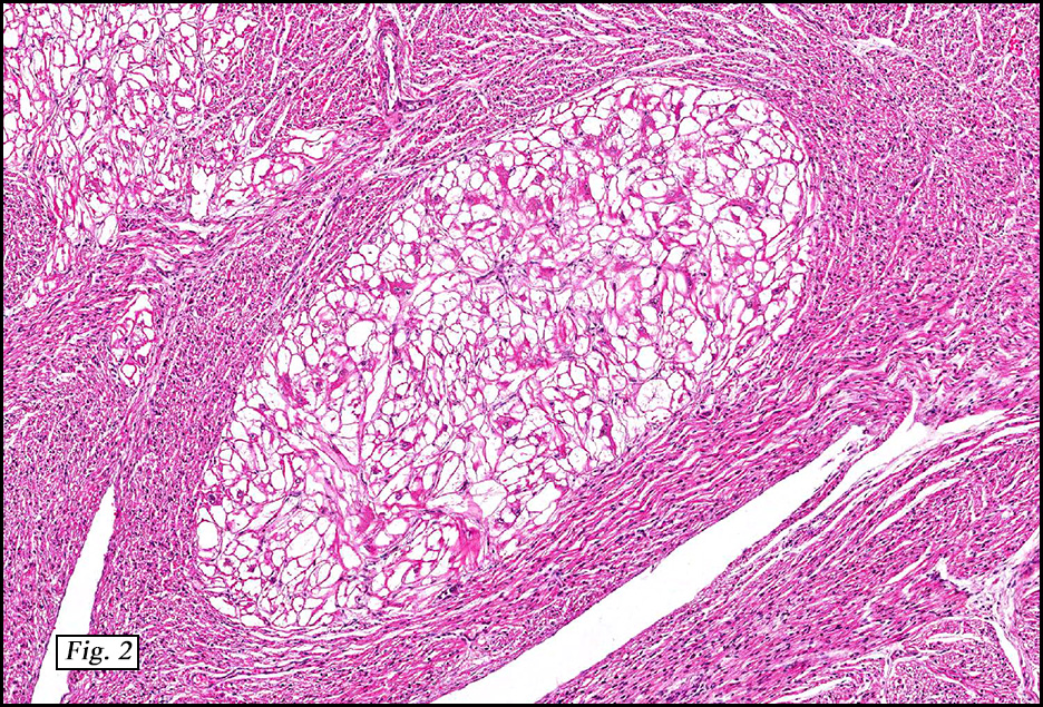
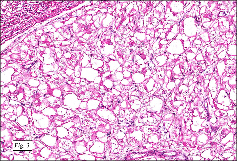
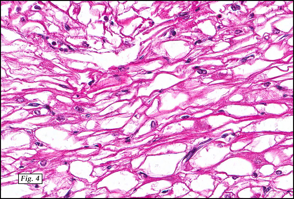
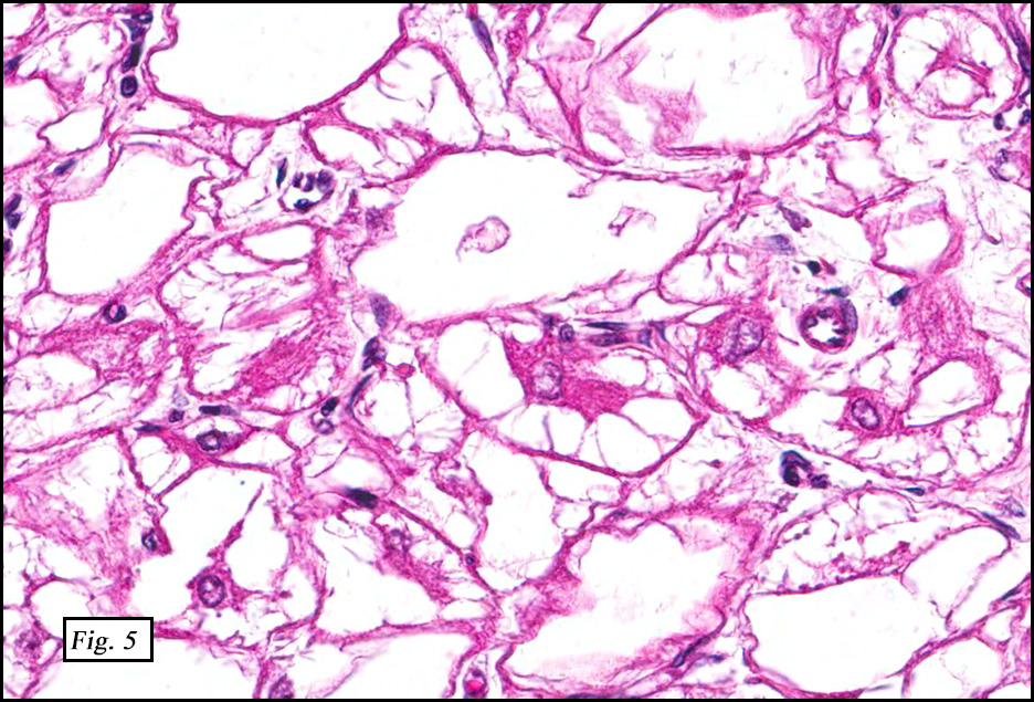
The excised specimen showed a 6 x 5 x 2.5 cm tumor that was composed of tan-brown fibrous tissue. Microscopically, it consisted of sheets of eosinophilic cells with increased vascularity admixed with mature adipose cells (Figs. 1 and 2). Higher power demonstrated round to polygonal, uniform cells with variable granular, eosinophilic, and multivacuolated cytoplasm and central, round nuclei. Neither atypia nor mitotic figures were seen (Fig. 3).

Diagnosis: Hibernoma, Back

Tae Hun Kim, MSIV and Donald R. Chase, M.D.
Department of Pathology & Human Anatomy
Loma Linda University Medical Center,
Loma Linda, California

Discussion: Hibernoma is a rare, benign adipocytic tumor of brown fat, named as such by Gery in 1914 because he felt that the fat resembled that which was found in hibernating animals. Other terms including lipoma of immature adipose tissue, lipoma of embryonic fat, and fetal lipoma have also been used.

Hibernoma arises predominantly in young adults with a peak incidence during the third decade of life but occurrences may span ages 2 to 75 years (mean age of 38). But in general they tend to occur in a younger population than patients with lipomas. Curiously, only 9 of 170 Armed forces Institute of Pathology (AFIP) cases occurred before 18 years of age. The location of these tumors has been thought to be in sites where brown fat persists beyond fetal life, such as neck, back, and retroperitoneum. But the AFIP study demonstrated that almost one third of their cases occurred in the thigh and other sites that lack vestigial brown fat. These observations suggest an altered differentiation pathway towards brown fat rather than a true origin from native brown fat. Like lipomas, hibernomas present as slow-growing, painless, movable tumors that usually arise in the subcutis. Up to 10% can be intramuscular. Because hibernomas usually range from 5-15 cm in diameter, symptoms related to compression of adjacent structures are relatively common.

Ultrasonography demonstrates a well-circumscribed hyperechoic lesion with increased vascularity. On CT, the signal intensity is between that of skeletal muscle and subcutaneous fat. Intravenous contrast administration enhances hibernomas and demonstrates intratumoral septations and vessels. On MRI, signal intensity is less than that of subcutaneous fat on T1-weighted imaging and is increased on T2-weighted imaging. Fat suppression techniques often decreases the signal intensity but is dependent on the fat composition.

Grossly, hibernomas are well-defined, soft tissue masses that are tan to deep red brown due to increased vascularity and high concentration of mitochondrial cytochrome pigments.

Four variants have been described depending upon the amount of regular fat, nature of stroma, and presence of spindle cells:
• Typical (82% of 170 AFIP cases),
• Myxoid (8%),
• Lipoma-like (7%), and
• Spindle cell (2%).

The typical variant demonstrates a distinct lobular pattern consisting of uniform round to oval multivacuolated cells with granular, eosinophilic cytoplasm and central nuclei without atypia and variable amount of white adipose cells.

The lipoma-like variant has a predominance of white adipose cells with scattered hibernoma cells.

The myxoid variant shows hibernoma cells/brown fat in a background of virtually acellular myxoid stroma.

The spindle cell variant is composed of bland spindle cells with multivacuolated hibernoma cells, a hybrid between spindle cell lipoma and hibernoma. This variant often occurs in the neck or scalp. It may have CD34 positivity, while other hibernoma variants are negative.

On electron microscopy, hibernomas are characterized by numerous polymorphous mitochondria.

Immunohistochemically, hibernomas are strongly positive for S-100. They are also positive for Oil Red O and Sudan Black. The karyotypes of hibernomas may be pseudodiploid. Structural rearrangements of 11q13-21 with no consistent chromosome partner are most characteristic. Homozygous deletions of multiple endocrine neoplasia I (MEN1) on 11q13.1 and rearrangements near the GARP gene on 11q13.5 have been implicated in hibernoma pathogenesis.

The differential diagnosis includes:

• Lipomas occur in older adults compared to hibernomas. Lipomas are less vascular and tend to be univacuolated.
• Rhabdomyomas have similar eosinophilic cytoplasm that contains glycogen but lacks lipid vacuoles. Crystals and cross-striations are observed. A desmin stain is usually positive in rhabdomyoma but is negative in hibernoma.
• Granular cell tumors are S-100 positive but lack vacuolated cytoplasm and cytoplasmic lipid vacuoles.
• Well-differentiated liposarcoma (WDLS) lacks brown fat, but may show lipoblasts which can mimic hibernoma cells.
• Myxoid liposarcoma can be confused with the myxoid hibernoma variant as both have multivacuolated cells in a myxoid stroma, but myxoid liposarcoma have mild nuclear atypia, signet ring lipoblasts, and arborizing vessels not found in hibernoma.

Hibernomas regardless of variant typing are treated by complete surgical excision with rare recurrence. Neither metastasis nor malignant transformation have been reported.

Suggested Reading:

Dagher W, et al, Hibernoma presenting as an asymptomatic neck mass, Am J Otolaryngol–Head and Neck Med and Surg (2013), http://dx.doi.org/10.1016/j.amjoto.2013.07.006

Fletcher CDM, Unni KK, Mertens F. World Health Organization Classification of Tumours: Pathology and Genetics of Tumors of Soft Tissue and Bone: Lyon, IARCPress, 2002: 33-34.

Furlong MA, Fanburg-Smith JC, Miettinen M. The morphologic spectrum of hibernoma: a clinicopathologic study of 170 cases. Am J Surg Pathol. 2001;25(6):809 – 14.

Kempson RL, Fletcher CDM, Evans HL, Hendrickson MR, Sibley RK. Atlas of Tumor Pathology: Tumors of the Soft Tissue, 3rd Series Fascicle 30: Washington, D.C., Armed Forces Institute of Pathology, 1998: 197-198.

Liu W, Bui MM, Cheong D. Hibernoma: comparing imaging appearance with more commonly encountered benign or low-grade lipomatous neoplasms. Skeletal Radiol 2013; 42:1073-1078.

Vassos N, et al, Deep-seated huge hibernoma of soft tissue: a rare differential diagnosis of atypical lipomatous tumor/well differentiated liposarcoma. Int J Clin Exp Pathol 2013;6(10):2178-2184.

Weiss S, Goldblum J. Enzinger & Weiss’ Soft Tissue Tumors, 5th ed: Philadelphia, Mosby/Elsevier Inc, 2008; 466-470.

December 2014

<a href=”http://www.cttr.org/forms/Montgomery Registration Form CME.doc”>Some Adventures in Gastrointestinal and Soft Tissue Pathology</a>
December 7, 2014
San Francisco, CA
Elizabeth A. Montgomery, MD
Director of Clinical Gastrointestinal Pathology
Johns Hopkins University

December 7, 2014
8:30 a.m. – 4:45 p.m.

Hyatt Regency San Francisco
5 Embarcadero Center
San Francisco, California, 94111
Phone: (415) 788-1234
Seminar Objectives:
At the conclusion of this seminar, attendees will be able to:
1. Become acquainted with new or newly reclassified entities in gastrointestinal pathology.
2. Emphasize the importance of hollow viscus wall layers from which various neoplasms typically arise.
3. Discuss molecular and immunolabeling pitfalls that arise in diagnosis of both gastrointestinal and soft tissue tumors.
4. Discuss situations in which molecular testing can be useful in the diagnostic workup process.

Materials: (“Attendance Only” includes items 1 and 2)
1. 6 hour lecture (Cover seminar objectives, correlate clinical histories, discuss the role of special studies in diagnosis, therapy, and counseling, and project photographs of the study cases and other illustrative materials)
2. Comprehensive syllabus that includes diagnoses, discussion, and appropriate references from pertinent medical literature
3. Glass slides of representative tumors

March 2014: A 44 year old man with a painful mass in a finger

History: A 44 year old man presented with a five year history of a left index finger nodule. It was mildly painful and had been increasing in size over the past year. The excised specimen was 1.2 x 0.7 x 0.4 cm and had a somewhat gritty firm, yellow tan cut surface.

Microscopically, the tumor was cystic and well circumbscribed (Figs. 1a, 1b). There were papillary regions (Figs. 2a, 2b) with cells showing round to oval vesicular nuclei and cytologic atypia (Fig 3) Mitotic figures and necrosis were present (Fig. 4).

Diagnosis: Aggressive Digital Papillary Adenocarcinoma

Dennis Aaron Reinke M.D. and Donald R. Chase, M.D.
Department of Pathology & Human Anatomy
Loma Linda University Medical center, Loma Linda, California

Discussion: Described by Dr. Helwig in 1977 as “Papillary eccrine adenoma”, the tumor was later renamed “Aggressive Digital Papillary Adenocarcinoma” (ADPA). It is felt to be of sweat gland origin and tends to occur in the digits, palms or soles. ADPA generally presents as a solitary nodule, usually 2 cm or larger and may be painful. The clinical suspicion is giant cell tumor of the tendon sheath, gout, soft tissue infection, pyogenic granuloma, glomus tumor, or ganglion cyst. Only rarely is there a history of trauma or chemical exposure. Most cases tend to occur in older men (males:females = 7:1) with a median age of 52 years, but it has been seen in ages as young as 14.

ADPA are usually cystic masses that involve the dermis and/or subcutis. They are often poorly circumscribed and infiltrative and microscopically show epithelial nodules that have papillary projections and glandular elements. The cells lining the cystic spaces may show atypia, pleomorphism and/or mitoses. The glandular cells can be cuboidal or columnar with round to oval vesicular nuclei. Necrosis may be present. The stroma is variable and may show areas of thin, fibrous septae or thick hyalinized collagen. Clear cell change, squamous metaplasia, and areas with spindle cells are sometimes seen. Most ADPAs are positive for keratin, carcinoembryonic antigen (CEA), and S-100 stains.

Fine needle aspiration cytology may be helpful for early diagnosis of ADPA. Syncytial groups with complex folded branching sheets of pleomorphic epithelial cells with clear anatomical borders are characteristic. There usually is nuclear overlap and crowding. Microscopically, fibrovascular cores may be seen having cells with large pale nuclei with finely granular chromatin and nucleoli. The cytoplasm is dense and cell borders are distinct.

ADPAs are known to locally recur, and metastasis is not uncommon. Studies on the rates of metastasis vary, but appear to range from 4-41.2%. The most common metastatic site is lung; however regional lymph nodes may also be involved.

Treatment of aggressive digital papillary adenocarcinoma should include wide excision or amputation of the digit at the appropriate level. One study suggested that patients receiving this treatment had ~5% likelihood of local recurrence; while those not treated with wide excision or amputation have ~50% chance of local recurrence.

Microscopically, the differential diagnosis includes:

• Aggressive papillary adenoma and aggressive papillary adenocarcinoma have previously been considered as distinct entities, based on criteria such as glandular differentiation, mitotic rate, necrosis, more pronounced cellular atypia, presence of zonal pleomorphism, and presence of bony or vascular invasion. However recent studies have suggested that since rates of metastasis and local recurrence are similar, they should be considered the same disease and treated the same.

• Papillary eccrine adenoma most often occurs in the distal extremities of African Americans. Tubular structures similar to eccrine ducts may often be dilated and contain intraluminal papillomata. Unlike ADPA they do not metastasize.

• Malignant eccrine acrospiromas are characterized by infiltrative architecture, perineural and lymphovascular invasion, atypical mitoses and pleomorphism. It may show solid, papillary, or glandular patterns and appear as a poorly differentiated adenocarcinoma. Atypical cells of ADPA may sometimes be seen next to the benign components of eccrine acrospiromas.

• Metastatic origin: Microscopically, aggressive papillary adenocarcinoma is usually not confused with other skin neoplasms, as the sites of involvement and papillary architecture of the lesions point to the diagnosis. It may however, appear very similar to other metastatic adenocarcinomas. A careful history and use of imaging studies should be pursued to rule out breast, lung, thyroid, ovarian, and other primary malignancy sites. Positive p63 expression tends to occur in primary lesions, while it is often negative in metastases.

In summary, aggressive papillary adenocarcinoma of the digit is a rare tumor that primarily involves tissues of the digits, palms or soles of men over age 50. They show a characteristic papillary cystic pattern with irregular and branching tubuloalveolar and ductal structures lined by epithelial cells that may show atypia, pleomorphism and mitoses. Appropriate identification is important to warrant aggressive surgical intervention to prevent local recurrence and metastasis.

Suggested Reading:

1. Altman CE, Hamill RL, Elston DM. Metastatic aggressive digital papillary adenocarcinoma. Cutis. 2003;72(2):145-7.
2. Holden B, Colome-Grimmer M, Savage C, Stierman K, Pou A. Malignant eccrine acrospiroma with metastasis to the parotid. ENT: Ear, Nose & Throat Journal [serial online]. May 2002;81(5):352.
3. Horii T, Sekiya H, Takatoku K, Hoshino Y. Aggressive digital papillary adenocarcinoma 5 years after surgical resection of adenoma on index finger. European Journal Of Orthopaedic Surgery & Traumatology [serial online]. July 2010;20(5):397-399.
4. Hsu H, Ho C, Chen C, Yang C, Hong H, Chuang Y. Aggressive digital papillary adenocarcinoma: a review. Clinical & Experimental Dermatology [serial online]. March 2010;35(2):113-119.
5. Krishnamurthy J, Patil, B. Aggressive digital papillary adenocarcinoma diagnosed by fine needle aspiration cytology. J Cytol. 2010 July;27(3):109-111.
6. Keramidas E, Miller G, Revelos K, Kitsanta P, Page R. Aggressive digital papillary adenoma-adenocarcinoma. Scandinavian Journal Of Plastic & Reconstructive Surgery & Hand Surgery [serial online]. June 2006;40(3):189-192.
7. Malafa M, Mckesey P, Stone S, Dudley-Walker S, Cockerell C. Sentinel Node Biopsy for Staging of Aggressive Digital Papillary Adenocarcinoma. Dermatologic Surgery [serial online]. June 2000;26(6):580-583.
8. McKee PH, Calonje E, Granter SR. Pathology of the Skin, With Clinical Correlations. [Edinburgh] : Elsevier Mosby, c2005.; 2008:1631-1632.
9. Rosai J, Ackerman LV. Rosai and Ackerman’s Surgical Pathology. Mosby; 2011:138-142.
10. Suchak R, Wang WL, Prieto VG, et al. Cutaneous digital papillary adenocarcinoma: a clinicopathologic study of 31 cases of a rare neoplasm with new observations. Am J Surg Pathol. 2012;36(12):1883-91.
11. Courtiss EH, Ruin DB, Helwig EB. Papillary eccrine adenoma. Plast Reconstr Surg. 60(6), 1977.
12. Duke WH, Sherrod TT, Lupton GP. Aggressive digital papillary adenocarcinoma (aggressive digital papillary adenoma and adenocarcinoma revisited). Am J Surg Pathol 2000 24(6):775-84.
13. Kao GF, Graham JH, Helwig EB. Aggressive digital papillary adenoma and adenocarcinoma. Arch Dermatol. 1984;120:1612.
14. Kao GF, Helwig EB, Graham JH. Aggressive digital papillary adenoma and adenocarcinoma. A clinicopathological study of 57 patients with histochemical, immunological and ultrastructural observations. J Cutan Pathol. 1987;14:129-146.

February 2014: A 75 year old woman with flank pain and a renal mass

History: A 71-year-old healthy woman presented with non-specific right-sided abdominal pain and was subsequently found to have a right renal mass. Upon surgical removal, a firm 5.1 cm tan-yellow mass was found. It extended to the renal hilum.

The tumor was well-circumscribed (Fig. 1a) but focally infiltrated its thin fibrous capsule (Fig. 1b). It consisted of sheets of spindled cells with vacuolated cytoplasm comprising much of the tumor (Figs. 2, 3). Bland-appearing cuboidal cells lined scattered tubular structures (Fig. 4). Histiocytes were commonly found (Fig. 5). Extracellular mucin was also present (Figs. 6a, 6b) confirmed by positive Alcian Blue pH 2.5 and mucicarmine stains. In addition, the tumor showed positive staining for pancytokeratin, CK7, AMACR and RCC (Fig. 7) with weaker focal positivity for CK34bE12. The tumor did not stain with CK20, CD10, CD31, or HMB-45.

Diagnosis: Mucinous Tubular and Spindle Cell Carcinoma (MTSCC) of Kidney

Jenny L. Smith, D.O. and Donald R. Chase, M.D.
Department of Pathology & Human Anatomy
Loma Linda University Medical center, Loma Linda, California

Discussion: In 1997, Dr. MacLennan [MacLennan et al] first described this low-grade, pleomorphic neoplasm of the kidney terming it “mucinous tubulocystic renal carcinoma”. He thought that it was a subset of collecting duct carcinoma, but fifteen years later, in 2002, the World Health Organization consensus conference on the classification of renal neoplasms recognized it as a specific entity and adopted the name “mucinous tubular and spindle cell carcinoma” (MTSCC). Although collecting duct epithelium is favored as site of origin, debate exists if it is actually of loop of Henle origin.

MTSCC is a rare renal neoplasm primarily of adult women. There is a 4:1 female predominance, with an average presentation of 53 years (range: 13 to 82) [Yang et al, Lima et al]. Typically asymptomatic, the tumor is commonly discovered incidentally while imaging for other reasons. But when the tumor is large it may present with flank pain or hematuria and consequential anemia.

Grossly, the tumor is well-circumscribed, gray-white, tan, or yellow and lacks significant hemorrhage or necrosis. Due to variable mucin content, cut surfaces may appear shiny or “mucoid”. It is usually 2.0 to 4.0 cm in size, although tumors less than 1.0 cm to greater than 18.0 cm have been described [Lima et al].

Microscopically, MTSCC consists of compact interconnecting tubules lined by bland low-cuboidal cells with eosinophilic, sometimes vacuolated cytoplasm. Samson, et al, noted vacuolization in 2 cases out of 17 analyzed. There are varying degrees of spindled cells arranged in cords, separated by pale basophilic myxoid stroma. “Mucin poor” variants are sometimes observed, where the spindled component becomes paramount for correct diagnosis [Rosai]. Of note, the spindled cells show low-grade round to oval nuclei, distinct from high-grade nuclei seen in sarcomatoid entities. Occasionally, focal neuroendocrine differentiation may be seen. Although not normally prominent; papillations, chronic inflammation, necrosis, and foam cells may be present. Phenotypically, the tumor cells characteristically express low molecular weight cytokeratins (CK7, CK19), EMA, AMACR, and vimentin. Generally, they do not express RCC, CD10, HMWCK, or CD117 [Ursani et al].

The differential diagnosis is fairly limited:

• Papillary renal cell carcinoma (PRCC) shows areas of compacted tubules and glomeruloid formations but usually lacks spindled cells and mucin. Thought to arise from proximal tubule origin, PRCC expresses CD10, unlike MTSCC.

• Sarcomatoid variant of renal cell carcinoma is a malignant neoplasm in which spindled cells characteristically display high grade cytology.

• Collecting duct carcinoma demonstrates small cysts and tubular structures but the lining cells are generally pleomorphic with high nuclear grades and a frequent hobnail morphology. Instead of having a mucinous background, the stroma of collecting duct carcinoma shows inflammation and desmoplasia.

Mucinous tubular and spindle cell carcinoma is a rare tumor only recently described as a distinct entity. Despite its pleomorphism and spindled morphology, it carries a better prognosis than other renal cell malignancies. Since it behaves less aggressively than other renal cell carcinomas, it is important to separate it from more common RCCs so that they may receive appropriately different treatment.

Suggested Reading:

MacLennan GT, Farrow GM, Bostwick DG. Low-grade collecting duct carcinoma of the kidney: report of 13 cases of low-grade mucinous tubulocystic renal carcinoma of possible collecting duct origin. Urology 1997; 50:679.

Lima et al.: The imaging and pathological features of a mucinous tubular and spindle cell carcinoma of the kidney: a case report. World J Surg Oncol 2013 11:34.

Gregory T. MacLennan GT, Bostwick DG. Tubulocystic Carcinoma, Mucinous Tubular and Spindle Cell Carcinoma, and Other Recently Described Rare Renal Tumors. Clin Lab Med 2005; 25(2): 393-416.

Rosai, J, Ackerman LV. Rosai and Ackerman’s Surgical Pathology. 10th ed. Mosby. 2004: Ch. 17. 1189.

Fine SW, Argani P, DeMarzo AM, Delahunt B, Sebo TJ, Reuter VE, Epstein JL. Expanding the Histologic Spectrum of Mucinous Tubular and Spindle Cell Carcinoma of the Kidney. Am J Surg Pathol 2006;30:1554-1560.

Yang G, Breyer BN, Weiss DA, MacLennan GT. J Urol. 2010 Feb;183(2):738-9.

Zhou M, Netto GJ, Epstein JI. Uropathology. Elsevier/Saunders. 2012: 287.

January 2014: A newborn with multiple cardiac tumors

History: Immediately following delivery to a healthy young woman, a baby boy was intubated for respiratory distress. An echocardiogram was performed on the child, revealing left ventricular enlargement with multiple ventricular masses that partially obstructed the outflow tract. Given the severity of his condition and the extent and location of the masses, the boy was deemed to be a poor surgical candidate. Ventilatory support was withdrawn and the boy expired shortly thereafter.

Gross examination of the heart at autopsy revealed numerous firm, white-tan ventricular mural nodules up to 1.8 cm. Although unencapsulated and apparently well-circumscribed, the nodules involved a substantial portion of the myocardium with some protruding into the ventricular cavity and approximating the mitral and aortic valves.

Microscopically, the tumors were comprised of large, polygonal clear cells which were distinct from the surrounding normal myocardiocytes (Figs. 1, 2, 3). The lesional cells were distended by cytoplasmic vacuoles which surrounded the nuclei (Fig. 3 & 4). Delicate wisps of pink cytoplasm emanated from the small, central nuclei extending to the periphery (Fig. 5). Significant atypia, mitotic figures, and necrosis were not encountered.

Diagnosis: Cardiac Rhabdomyoma

Elif L. Akin, M.D. and Donald R. Chase, M. D.
Department of Pathology & Human Anatomy
Loma Linda University Medical center, Loma Linda, California

Discussion: Cardiac rhabdomyoma (CR) is the most common cardiac neoplasm in the pediatric population, comprising about 60% of cardiac tumors in pediatric autopsy series and an overwhelming 90% of cardiac neoplasms diagnosed in utero. Considered to be a benign hamartoma which has no predilection for either gender, this neoplasm most frequently arises in the ventricles.

Grossly, cardiac rhabdomyomas are distinct, well-circumscribed white-tan nodules which contrast with surrounding myocardium. They can occur as solitary or multiple lesions, ranging in size from a few millimeters up to 10 cm. Although they usually arise in the left ventricular free wall, they can involve the muscle surrounding any of the cardiac chambers.

Microscopically, CR are characterized by nodules of round or polygonal clear cells that are much larger than myocardiocytes and swollen with glycogen-rich vacuoles. Nuclei are small and centrally located and have delicate connections to the perimeter of the cell comprised of eosinophilic wisps of cytoplasm, which are thought to represent invaginations of the cytoplasmic membrane. These so-called “spider cells” are characteristic of cardiac rhabdomyomas. Their peripherally vacuolated, clear appearance is a result of glycogen deposition, which is lost during routine processing. Striations may also be seen. While some CR may show degenerative change, significant nuclear pleomorphism, mitotic activity, and necrosis are not typically present.

Many CR occur as asymptomatic lesions that eventually spontaneously regress. As the regression rate is an estimated 50 to 70%, surgical intervention is often unnecessary, making CR the second most common cardiac tumor in pediatric surgical series (behind cardiac fibroma). When symptomatic, the clinical presentation of children with CR varies considerably depending on the size, extent, and location of the tumor(s). It is not altogether surprising that lesions involving the conduction system may cause arrhythmias and that large CR projecting into the ventricular cavity may cause flow abnormalities and mechanical obstruction. Potential clinical manifestations of problematic CR include murmurs, arrhythmias, and heart failure and may necessitate partial surgical resection, depending on symptom severity.

Although CR can occur as a solitary, isolated lesion, it is most frequently seen in the context of tuberous sclerosis complex (TSC). When associated with TSC, lesions tend to be multifocal and extend into the ventricular cavity. Because of the strong link to TSC, patients with a diagnosis of CR must be assessed for other signs of this disease. Cardiac rhabdomyomas are less often associated with congenital cardiac malformations including tetralogy of Fallot, Ebstein anomaly, and hypoplastic left heart syndrome.

There are several entities (including non-neoplastic lesions) which may be considered in the differential diagnosis of cardiac rhabdomyoma.

• Glycogen storage disease (Pompe disease) is also characterized by vacuolated myocytes. However, the cytoplasmic vacuoles tend to be central, with myofibrils at the periphery. In contrast to CR, glycogen storage disease does not form distinct nodules and lacks spider cells.

• Purkinje cell hamartoma (histiocytoid cardiomyopathy) is a rare pediatric cardiac tumor which also presents as multiple intramural nodules. Although this condition also has a predilection for the left ventricle, the nodules tend to be subendocardial. Unlike CR, the cells of histiocytoid cardiomyopathy (as the name would suggest) resemble histiocytes, are quite a bit smaller than spider cells, and show finely vacuolated cytoplasm filled with mitochondria, rather than glycogen.

• As the most commonly excised pediatric cardiac tumor, the shear frequency of cardiac fibroma makes it another important consideration in the differential diagnosis. While CR may be multifocal, cardiac fibromas are typically solitary mural lesions which resemble fibromatoses. Characterized by bland spindled cells interspersed with collagen bundles, fibromas tend to have infiltrative borders. Additionally, cardiac fibromas may demonstrate focal calcification, a feature not ordinarily observed in CR.

• Lipomatous hypertrophy, in contrast to the above conditions, affects older, usually obese adults. It preferentially involves the atrial septum and is distinguished from CR by the presence of three cell populations including brown and white adipocytes as well as hypertrophic myocytes.

In summary, CR is a benign hamartomatous neoplasm of infancy which typically arises in the left ventricular wall. It is characterized by spider cells: large cells with cytoplasmic vacuolization and central nuclei with strands of eosinophilic cytoplasm extending to the cell borders. While these lesions are usually asymptomatic and spontaneously regress, they can cause serious clinical problems (e.g., heart failure) and may require surgical intervention. Appropriate recognition and diagnosis of CR is important to determine whether surgery is required and to initiate further evaluation for possible tuberous sclerosis complex.

Suggested Reading:

Burke A, Tavora F. Practical Cardiovascular Pathology. Wolters Kluwer Health Lippincott Williams & Wilkins. 2011: 375-380.

Stocker JT, Dehner LP, Husain AN. Stocker & Dehner’s Pediatric Pathology: 3rd ed. Wolters Kluwer Health Lippincott Williams & Wilkins. 2010: 566-569.

Virmani R, Burke A, Farb A. Atlas of Cardiovascular Pathology. W. B. Saunders Company. 1996: 82-91.

Weiss SW, Goldblum JR. Enzinger & Weiss’s Soft Tissue Tumors: 5th ed. Mosby Elsevier. 2008: 583-592.

Scroll to top