June 2011: A 74 year old man with Helicobacter pylori presents with worsening gastric pain

History: A 74-year-old male presented with worsening gastric pain, and a prior history of Helicobacter pylori infection.

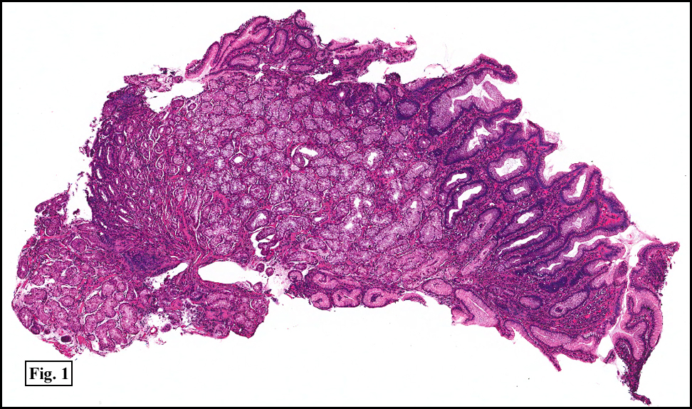
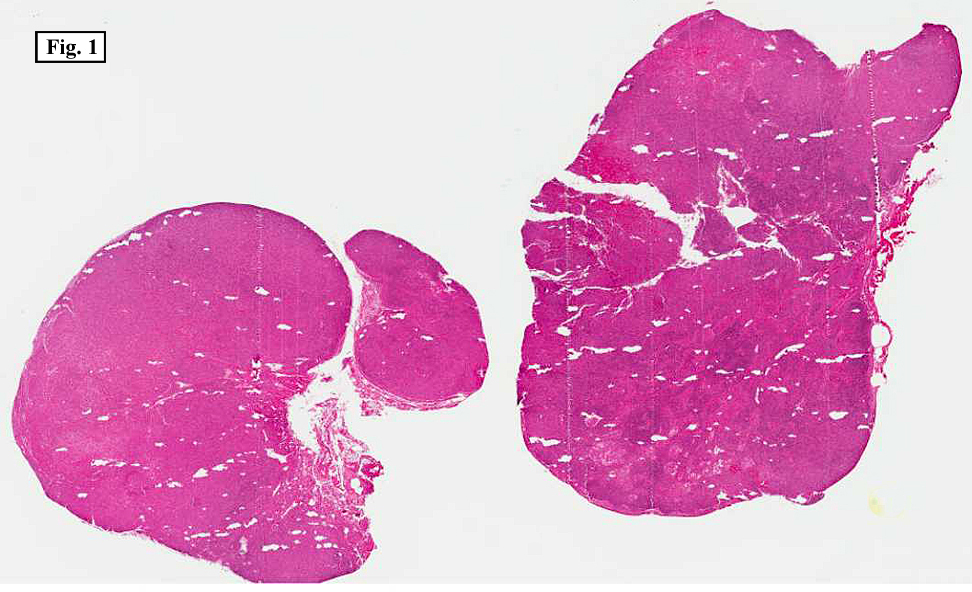
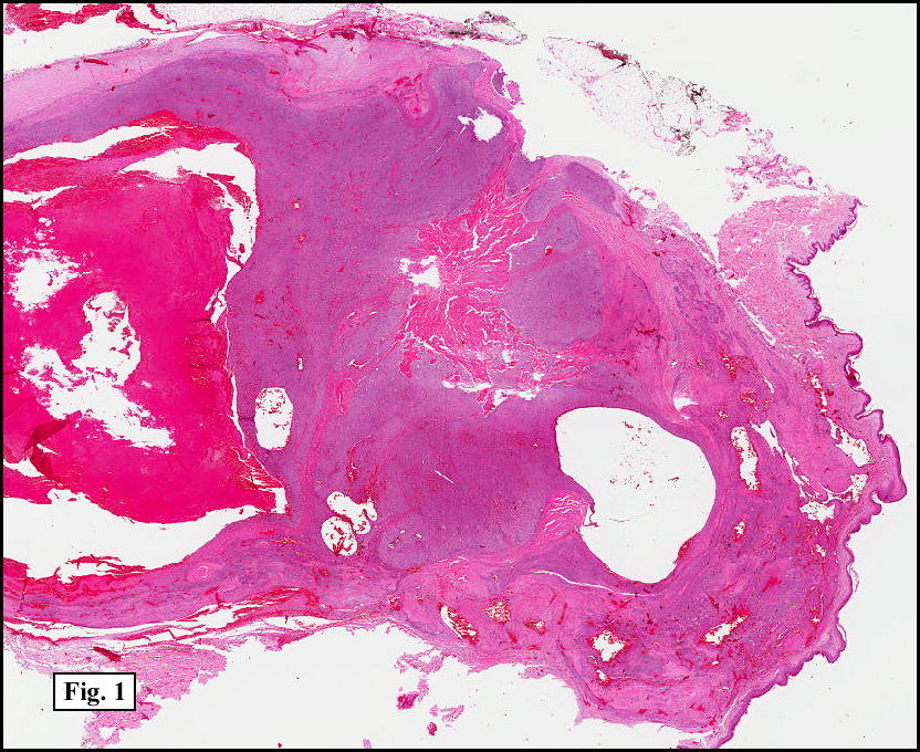
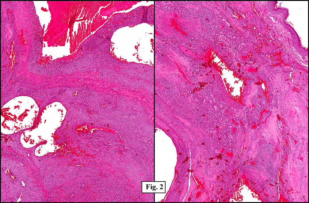
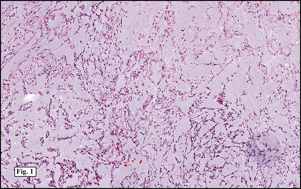
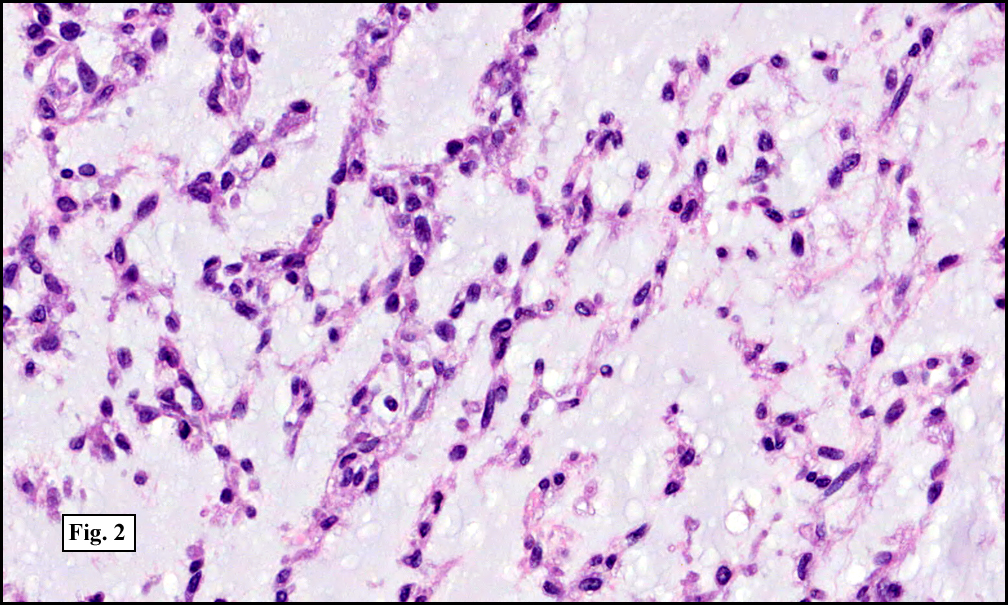
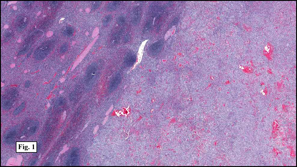
Two years earlier he had an endoscopic biopsy of the gastric body and antrum which revealed moderate acute and chronic inflammation consisting of a superficial mucosal infiltrate of lymphocytes, plasma cells, neutrophils and scattered eosinophils (Fig. 1). A Giemsa stain showed numerous curved bacterial rods present within the gastric crypts and on the surface of the glands (Fig. 2). He was treated with the standard triple therapy (omeprazole, metronidazole and clarithromycin) with resolution of dyspepsia.

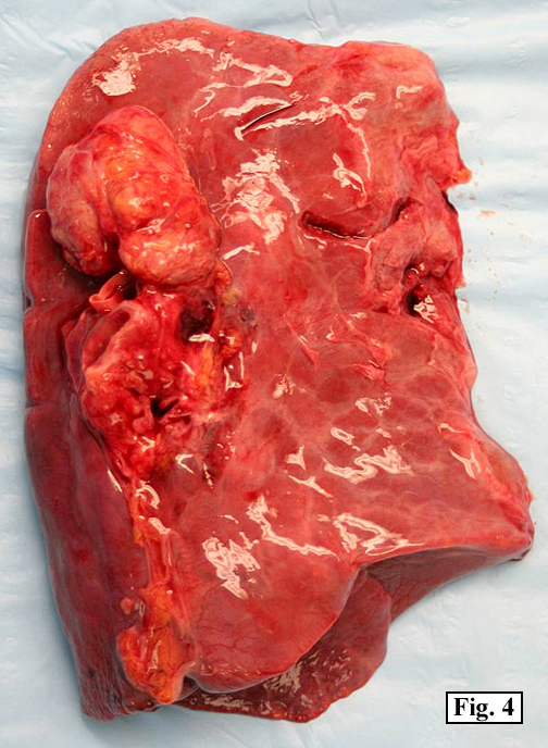
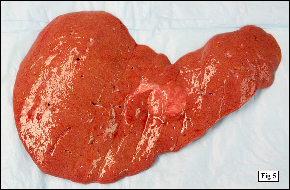
He now presented with a 4 to 5 month history of abdominal pain and anemia. Endoscopic studies revealed a large fundic mass. A biopsy was performed.

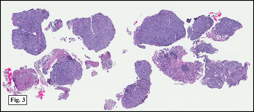
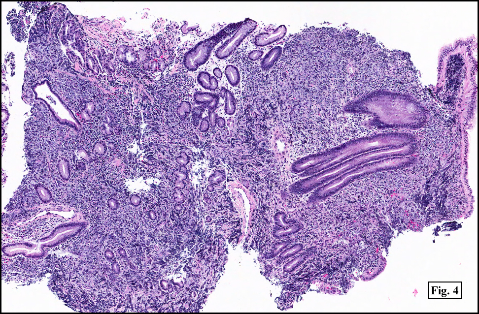
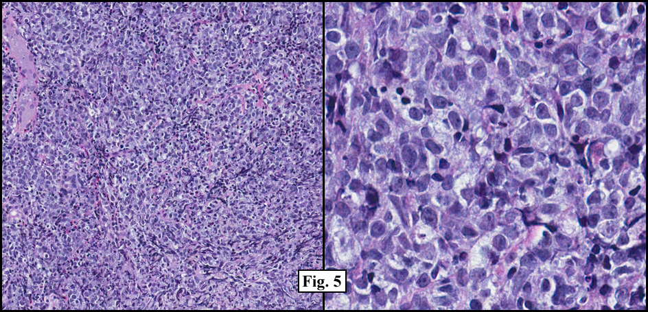
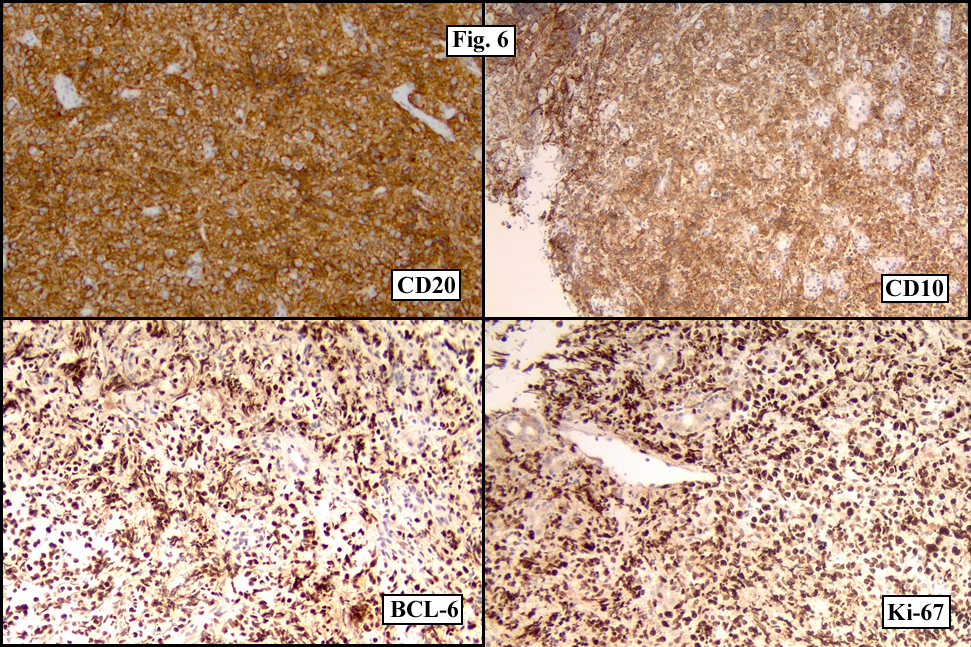
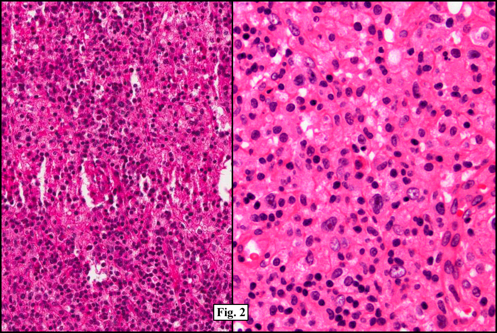
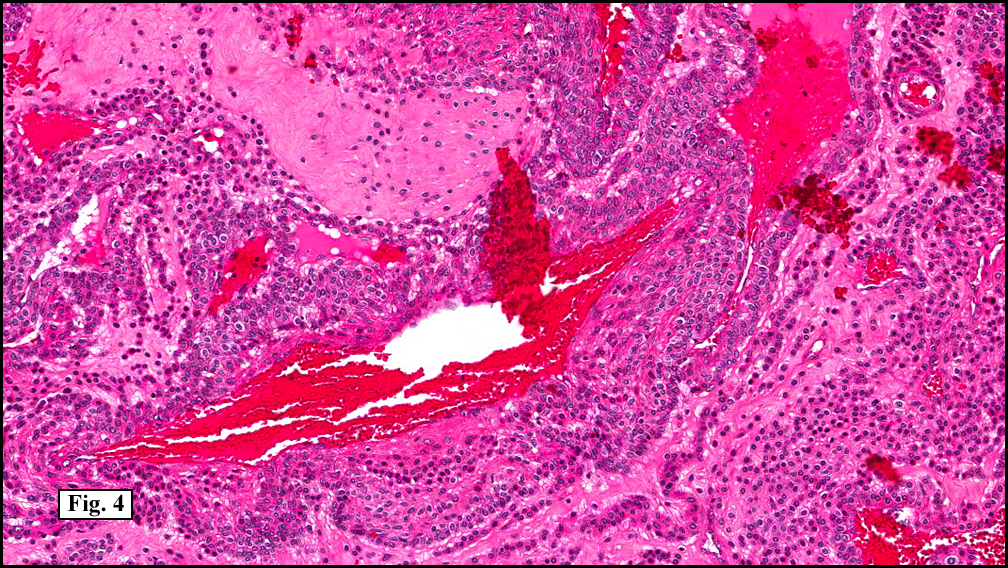
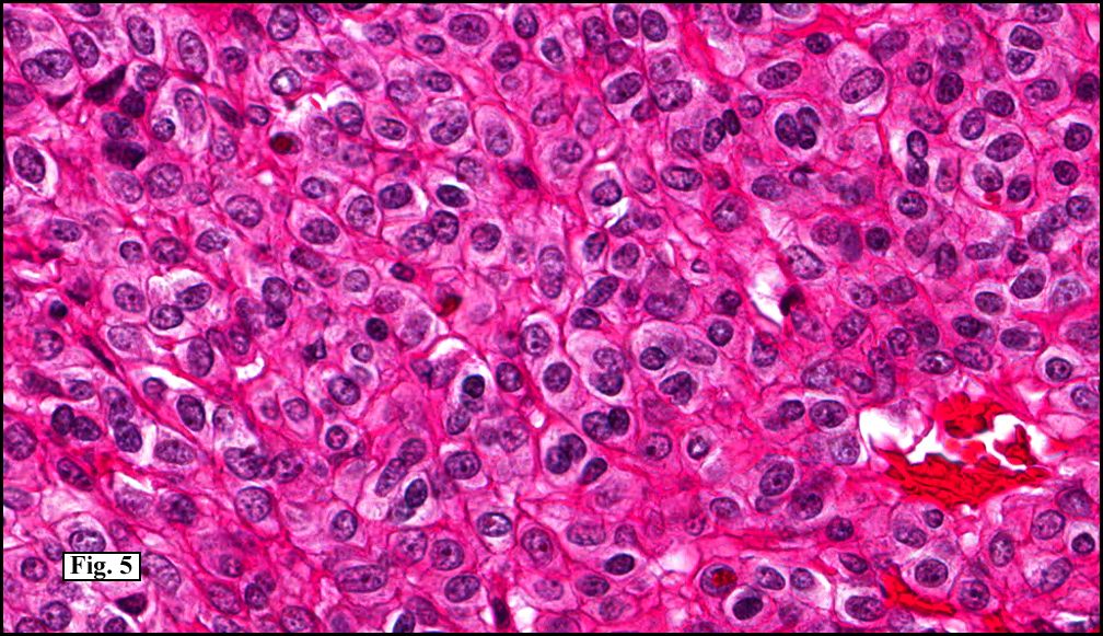
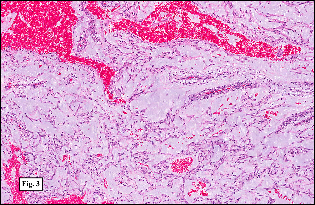
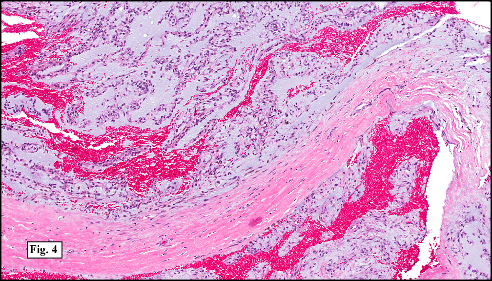
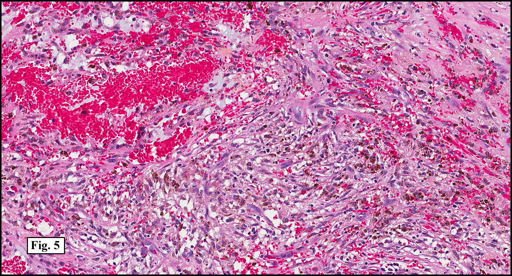
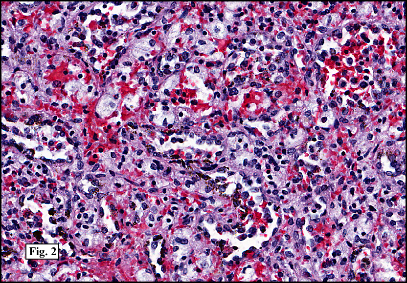
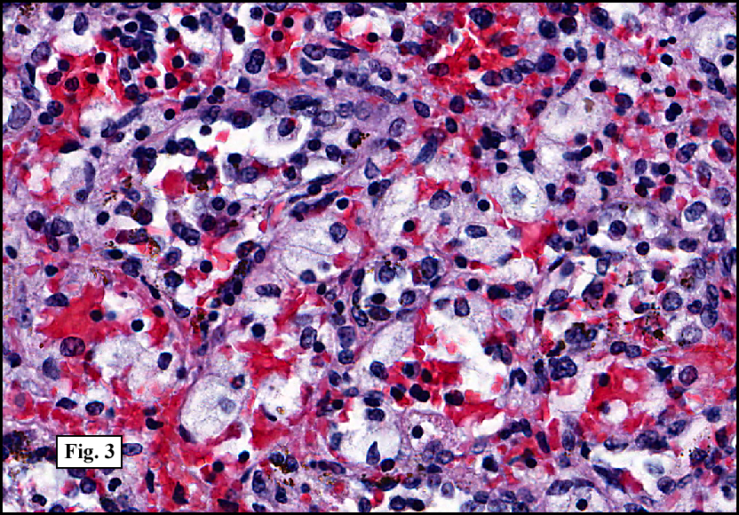
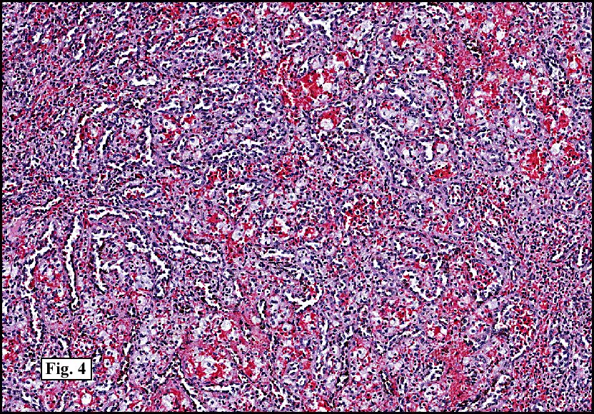
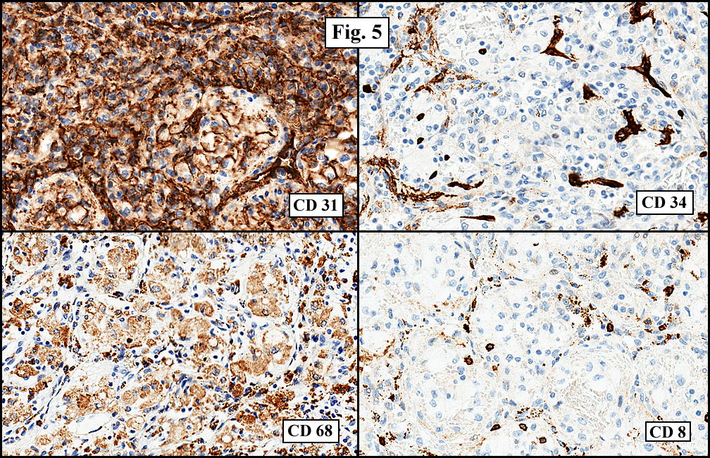
Sections of the gastric mass showed replacement of the lamina propria and distortion of normal gastric glands (Fig. 3) by an infiltrate of monomorphic, medium to large lymphocytes with round nuclei, finely clumped chromatin and multiple basophilic medium-sized, occasionally paracentrally situated nucleoli (Figs. 4, 5). Occasional mitotic figures were seen. Focal superficial erosion with acute inflammation was also present. Giemsa stain was performed and showed no evidence of Helicobacter pylori microorganism. Immunohistochemically, the tumor cells showed expression of CD45, CD20, CD79a, CD10, BCL-6 with a high Ki-67 proliferation index (>95%), but was negative for CD138 and CDX2 (Fig. 6).

CT studies revealed diffuse lymphadenopathy throughout his abdomen and pelvis. A large left groin mass was also present on physical examination. Needle core biopsies were performed and revealed a morphology similar to the gastric lesion with diffuse replacement of normal nodal architecture by a monomorphic population of medium to large lymphoid cells. A flow cytometric study revealed the lymphocytes to show expression of CD20, CD19, CD10 with lambda restriction. A portion of the biopsy material was submitted for FISH analysis showing a subpopulation of cells with a c-myc gene rearrangement.

A follow-up CT of the abdomen and pelvis performed two years following treatment with R-CHOP showed the stomach mass and all the previously enlarged lymph nodes to have disappeared.

Diagnosis: “Burkitt Lymphoma”

Hannah H. Wong, MD, Jeffrey D. Cao, MD, Donald R. Chase, MD
Department of Pathology, Loma Linda University and Medical Center,
Loma Linda, California
California Tumor Tissue Registry, Loma Linda, California

Discussion: Burkitt lymphoma (BL) is a B-cell non-Hodgkin lymphoma which was first described in 1958 by Denis Burkitt in the lymphoid tissue around the tooth of an African child. It comprises up to half of all childhood non-Hodgkins lymphoma; however, is an uncommon form of non-Hodgkin lymphoma in adults, with a reported yearly incidence of 1200 patients in the United States. Although it is generally described to be more common in children and young adult males, the 2007 National Cancer Institute Surveillance, Epidemiology and End Results (SEER) database showed 59% of all adult Burkitt lymphoma cases in the United States were older than 40 years of age. Clinically, Burkitt lymphoma is divided onto three variants based on their clinical presentation, morphology and biology.

• Endemic BL: Most common in children from equatorial Africa, it generally presents in the jaw with eventual spread to extranodal sites, such as the bone marrow and leptomeniges.
• Sporadic BL: There is no geographical limitation to this variant, which is mostly found in children and young adults. The median age in adult patients is 30 years, and there is a slight predilection for males. Sporadic BL most often present as abdominal masses, especially in the ileo-cecal region. However, it may be found in the ovaries, kidneys and breasts. Involvement of lymph nodes has been reported to be more common in adults compared to children.
• Immunodeficiency-associated BL: This variant of BL is primarily limited to patients with human immunodeficiency virus (HIV) infection.

Adult patients with Burkitt lymphoma in the United States most often present with constitutional B symptoms (fever, fatigue, weight loss), bulky abdominal masses and rarely lymph node involvement. The tumor is so aggressive that bone marrow involvement is present at the time of diagnosis 70% of the time, and leptomeningeal involvement presents in up to 40% of patients. A timely diagnosis is vital for this aggressive lesion, and initial therapy is aimed to begin within 48 hours of diagnosis.

Microscopically, there is generalized effacement of normal architecture by a diffuse monotonous population of cohesive, intermediate-sized lymphoid cells with high nuclear to cytoplasmic ratios and smooth round to oval nuclear contours. The nuclei show finely clumped, dispersed chromatin and generally have multiple nucleoli. Frequent mitotic figures and apoptotic bodies are generally present, with frequent benign macrophages filled with ingested apoptotic tumor cells creating the characteristic “starry-sky” appearance. A florid granulomatous reaction may be present in some cases of BL, making recognition of the underlying BL difficult. However, these lesions generally have a good prognosis. The malignant lymphoid cells may also have eccentric basophilic cytoplasm with a single central nucleolus.

Immunohistochemically, the tumor cells generally express B-cell antigens such as CD19, and CD20 as well as CD10, BCL6, CD38, CD77 and CD43. They generally do not express TdT, but may be weakly positive for BCL2. There is generally up to 100% positivity of Ki-67 in most of these lesions. The immunophenotype of BL may be demonstrated by tissue immunohistochemistry or flow cytometry. Gene expression analysis is important in BL, as a large number cases have the diagnostic MYC translocation at band 8q24 of the IG heavy chain region 14q23, t(8;14). Other common translocation patterns in BL include t(2;8) and t(8:22). Despite the aggressive nature of BL, it is a highly curable condition. Patients with BL generally respond well to high doses of systemic chemotherapy, and intrathecal therapy.

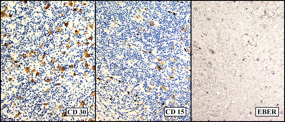
The pathogensis of BL has been associated with the Epstein-Barr virus. Virtually all tumor cells in endemic BL show the presence of EBV within their genome, while EBV positivity is present in approximately 30% of sporadic BL, and 25-40% of immunodeficiency-associated BL. However, there is no known association of BL with Helicobacter pylori (H. pylori). In a review of the current literature there were a total of 4 cases of BL associated with H. pylori, with three of those cases being in children and one in a 53-year-old male. H. pylori is a gram negative rod first identified by Warren and Marshall in 1983. H. pylori has been associated with chronic active gastritis, peptic ulcers and subsequent development of carcinoma, and gastric lymphoid neoplasms, specifically mucosa-associated lymphoid tissue (MALT) lymphomas of the stomach. In fact, H. pylori is listed as a Grade I carcinogen by the World Health Organization.

Gastric MALT lymphoma is a marginal zone B-cell low grade lymphoma generally affecting older adults. It is the most common type of extranodal marginal zone lymphoma and comprises up to 85% of extranodal MALT lymphomas. The association between gastric MALT lymphoma and H. pylori has been well established. The stomach in its normal physiologic state does not possess any organized lymphoid tissue. When H. pylori infects the stomach, it produces a host of antigens which stimulate H. pylori-specific T cells to induce proliferation of B lymphocytes. The proliferation of the B cells then leads to the development of lymphoid proliferations of the MALT type. The role of H. pylori in gastric MALT lymphoma has been further established as the lymphoma rapidly regresses with eradication of H. pylori. It has been proposed that the association between gastric BL and H. pylori may be similar to that of H. pylori and gastric MALT lymphoma.

Despite the possibility of a similar causal relationship between H. pylori and Burkitts lymphoma, BL and gastric MALT lymphoma are quite different entities. Patients with gastric MALT lymphoma often present with abdominal pain, and stage I or II disease unlike BL which is generally stage IV at the time of diagnosis. Bone marrow involvement is rare unlike BL. Microscopically, the lymphoma cells are small to medium in size with slightly irregular nuclei, moderately dispersed chromatin and inconspicuous nucleoli and generally abundant pale cytoplasm; in contrast to BL where the lymphoma cells have scant cytoplasm and multiple prominent nucleoli. The lymphoma cells of gastric MALT lymphoma express CD20 and CD79a but generally do not express CD5, CD10, CD23 and have variable expression for CD43 and CD5. Gastric MALT lymphomas are associated with various translocations such as t(11;18). Treatment for this entity generally starts with treatment of H. pylori by antibiotics, and this has been found to be successful for many cases; however, chemotherapy may be required in some aggressive cases.

Suggested Reading:
Swerdlow SH, Campo E, Harris NL, Jaffe ES, Pileri SA, Stein H, Thiele N, Vardiman JW, Eds. WHO Classification of Tumours of Haematopoietic and Lymphoid Tissues. Lyon, France: IARC; 2008.
Grewal SS, Hunt JP, O’Connor SC, Gianturco LE, Richardson MW, Lehmann LE. Helicobacter pylori associated gastric burkitt lymphoma. Pediatr Blood Cancer. 2007;10:888-890.

Shannon C, Vickers C, Field A, Ward R. Burkitt’s lymphoma associated with Helicobacter pylori. Journal of Gastroenterology and Hepatology. 2000;15:99-103.

Perkins AS and Friedberg JW. Burkitt lymphoma in adults. Hematology. 2008:341-348

Andoh A, Takaya H, Bamba M, Sakumoto H, Inoue T, Tujikawa T, Koyama S, Fujiyama Y, Bamba T. Primary gastric burkitt’s lymphoma presenting with c-myc gene rearrangement. J Gastroenterol. 1998;33:710-715.

Moschovi M, Menegas D, Stefanaki K, Constantinidou CVV, Tzortzatou-Stathopoulou F. Primary gastric burkitt lymphoma in childhood: associated with helicobacter pylori? Med Pediatr Oncol. 2003;41:444-447.

May 2011: A 17 year old boy with history of a heart transplant and recurrent lymphadenopathy

History: A 17 year-old male presented with significant hepatosplenomegaly and a mass in the right lobe of the liver. He had been born with a hypoplastic left heart and diagnosed with velocardiofacial syndrome. At 13 weeks of age he received a heart transplant and was placed on immunosuppressive therapy including; rapamune (sirolimus) and prograf (tacrolimus). Eleven years later he presented with fever, abdominal distension and respiratory distress. A CT scan of his chest was performed and revealed multiple enlarged lymph nodes in the right neck. A biopsy was performed.

Microscopically, there was complete effacement of normal lymph node architecture (Fig. 1) by a heterogeneous population of large, pleomorphic cells with abundant basophilic cytoplasm, one to multiple nuclei, and prominent eosinophilic nucleolus consistent with Hodgkin Reed-Sternberg (HRS) cells, in a background of small lymphocytes, rare histiocytes, plasma cells and eosinophils (Fig. 2). Immunohistochemically, the tumor cells were positive for CD 30, CD 15, EBER (Fig. 3) and weakly positive for PAX-5, and negative for CD 3, CD 20, CD 43 and CD 45. Flow cytometric studies showed no aberrant antigen expression.

The patient’s immunosuppressive therapy was reduced and his lymphadenopathy resolved but he subsequently presented with signs of acute renal failure. A chest and abdominal CT scan revealed a mass in the right lobe of liver. Over the course of his hospitalization he developed sepsis, respiratory failure and eventually expired. On autopsy, prominent mediastinal (hilar and paratracheal) lymph nodes with a fleshy-tan cut surface were identified (Fig. 4). Also, a distinct 3.5 x 3.0 cm fleshy tan nodule was present within the mid-right lobe of the liver (Fig. 5).

Microscopically, sections of the lymph nodes and the liver mass were identical to the prior lymph node biopsy. There was complete effacement of normal architecture by lymphohistiocytic infiltrate consisting of large HRS cells in a background of small lymphocytes, occasional histiocytes, plasma cells and eosinophils. A limited panel of immunostains was performed which revealed the tumor cells to be positive for CD30, CD15 and negative for CD20.

Diagnosis: “Recurrent Classical Hodgkin lymphoma type Post-transplant lymphoproliferative disorder”

Hannah H. Wong, MD, Jun Wang, MD, Donald R. Chase, MD.
Department of Pathology, Loma Linda University Medical Center,
Loma Linda, California
California Tumor Tissue Registry, Loma Linda, California

Discussion: Classical Hodgkin lymphoma type Post-transplant lymphoproliferative disorder (cHL-PTLD) is a rare subtype of PTLD, first described in 1974 in a kidney transplant patient. Post-transplant lymphoproliferative disorders (PTLD) are a group of lymphoid or plasmacytic lesions which develops in roughly 1-5 % of transplant patients secondary to immunosuppression following solid organ, bone marrow and/or stem cell transplantation.

PTLDs are classified by the 2008 WHO classification of Tumors of Haematopoietic and Lymphoid tissues into four main categories:
(1) Early lesions (plasmacytic hyperplasia, infectious mononucleosis-like lesion)
(2) Polymorphic PTLD
(3) Monomorphic PTLD (including B-cell and T-cell neoplasms)
(4) Classical Hodgkin lymphoma-type PTLD

cHL-PTLD has a clinical behavior and morphological presentation identical to classical Hodgkin lymphoma (cHL) and was originally classified in the same category as Hodgkin lymphoma-like PTLD. The 2008 WHO; however, redefined this fourth category of PTLD as classical Hodgkin lymphoma type PTLD and the “Hodgkin lymphoma-like PTLD” is no longer included in this category but rather classified as Polymorphic or Monomorphic B-cell PTLD

Epstein-Barr virus (EBV), a human herpes virus, has been implicated in the development of the majority of PTLD cases, from 60% to up to 100% positivity in early lesions. In fact, only 23-42% of PTLD cases have been found to be EBV negative. EBV infects human lymphocytes, induces cellular proliferation and inhibits apoptosis resulting in the development of malignancies. This virus can remain dormant within cells without viral replication for an in-determined period of time. B-lymphocytes have an internal defense mechanism which prevents viral replication, but allows the expression of a restricted set of viral genes called “latency” genes. These latency genes code for six nuclear and three membrane associated proteins which interfere with cellular control, proliferation and differentiation of B-lymphocytes.

Four latency programs have been described based on the expression of different viral gene combinations in correspondence to the different stages of B-cell activation and differentiation. Each latency program has been associated with a different group of malignancies, except for Latency 0. Latency I is associated with Burkitt lymphoma, latency II with cHL, peripheral T-cell lymphoma, nasal T/NK cell lymphoma, nasopharyngeal carcinoma, and lymphoepithelioma (stomach, thymus). Latency III is associated with infectious mononucleosis, AIDS-related immunoblastic B-cell lymphoma, and PTLD. Table I gives a summary of the four EBV latency programs, viral gene expression and associated diseases.

Table I. EBV latency programs, expressed viral genes and associated diseases

Latency program Expressed viral genes Disease
Latency 0 None None
Latency I LMP-2A/EBNA-1 Burkitt lymphoma
Latency II EBNA-1, LMP-1, LMP-2A, -2B cHL, peripheral T-cell lymphoma, nasal T/NK cell lymphoma
Latency III EBNA-1, -2, -3, -4, -5, -6, LMP-1, -2A, -2B Infectious mononucleosis, AIDS-related immunoblastic B-cell lymphoma, PTLD

Patients with cHL-PTLD present with signs and symptoms identical to classical Hodgkin Lymphoma (cHL) such as fever, weight loss, night sweats and lymphadenopathy. It involves nodal or extranodal sites with a predilection for male and pediatric/adolescent patients. cHL-PTLD has been found to develop much later, even years later in comparison to the other forms of PTLD and has been found more commonly in patients with renal transplants.

Morphologically, cHL-PTLD is virtually identical to cHL. The involved tissue shows a mixed population of small to medium-sized lymphoid cells admixed with scattered large mononucleated, binucleated, and/or multinucleated Hodgkin Reed-Sternberg (HRS) cells, as well as occasional histiocytes, plasma cells, and rare eosinophils in the background. Immunohistochemically, the HRS-cells show the classic expression of CD30 and CD15 antigens, but do not express CD45, CD20 and CD79a. PAX-5 may be positive in some HRS cells, but does not change the diagnosis. EBV-encoded RNA (EBER) in situ hybridization is positive only in the RS-cells. The HRS-cells will also reveal expression of Type II EBV latency.

The main and most important differential diagnosis of cHL-PTLD is Hodgkin lymphoma-like PTLD (HL-like PTLD). The distinction between these two entities is of importance since the biology and treatment of these two entities are quite different. HL-like PTLD has been found to be more similar in its clinical behavior to monomorphic and polymorphic B-cell forms of PTLD than to cHL. Clinically, cHL-PTLD and HL-like PTLD may present with similar symptoms of lymphadenopathy, fever, night sweats and splenomegaly, generally following years of immunosuppression. The morphological features of HL-like PTLD are difficult to differentiate from cHL-PTLD as it also shows large atypical HRS-like cells with a background of small lymphoid cells, occasional histiocytes, plasma cells, and eosinophils. Immunohistochemically, the staining patterns between the two are more distinctive. The HRS-like cells in HL-like PTLD are positive for CD30 and CD20 as well as CD45, but generally negative for CD15. This is in contrast to cHL-PTLD where the HRS-like cells are CD30 and CD15 positive but CD20 and CD45 negative. The pattern of EBER positivity also differs in these two entities, with EBER positivity in the large atypical HRS-like cells, as well as, surrounding by-stander small lymphocytes in HL-like PTLD but only positive in the HRS-cells in cHL-PTLD. HL-like PTLD has also been shown to express a Type III EBV latency pattern, similar to the polymorphic and monomorphic B-cell types of PTLD. HL-like PTLD has been found to respond well to treatment regimens for monomorphic B-cell PTLD such as reduction of immunosuppresion, along with anti-CD20-antibody (rituximab and combinations of chemoimmunotherapy for non-Hodgkin lymphoma (NHL).

Despite the re-classification and better understanding of cHL-PTLD as a distinct type of PTLD with features identical to cHL, there is currently no established treatment protocol. In the reported cases of cHL-PTLD, reduction/withdrawal of immunosuppression along with chemotherapy and radiotherapy have been utilized. Patients treated with standard cHL chemotherapy (such as doxorubicin, bleomycin, vinblastine and dacarbazine [ABVD]) and radiotherapy have been found to do better than those treated as monomorphic PTLD. It has been suggested that cHL-PTLD may respond best to standard cHL treatment protocols.

Table II. Comparison of cHL-PTLD and HL-PTLD

cHL-PTLD HL-like PTLD
Clinical Features Fever, lethargy, lymphadenopathy Fever, lethargy, lymphadenopathy
Histology HRS cells in a background of small lymphocytes, occasional histiocytes, plasma cells and eosinophils HRS-like cells in a background of small lymphocytes, occasional histiocytes, plasma cells and eosinophils
Immunohistochemistry CD30+, CD15+, CD45-, CD20-/+, CD79a- HRS-like cells: CD30+, CD15-, CD45+, CD20+/-, CD79a+
EBV status EBER+ in HRS-cells only EBER+ variably positive in HRS-like cells AND by-stander small lymphocytes
EBV latency Type II latency pattern Type III latency pattern
Treatment ABVD* or other chemotherapies used for Hodgkins lymphoma Reduced immunosuppression, anti-CD20-antibody (rituximab), non-Hodgkin lymphoma chemotherapies (e.g CHOP**

* ABVD – doxorubicin, bleomycin, vinblastine, dacarbazine
**CHOP – cyclophosphamide, adriamycin, vincristine, prednisone

Suggested reading:

Swerdlow SH, Campo E, Harris NL, Jaffe ES, Pileri SA, Stein H, Thiele N, Vardiman JW, Eds. WHO Classification of Tumours of Haematopoietic and Lymphoid Tissues. Lyon, France: IARC; 2008.

Pitman SD, Huang Q, Zuppan CW, Rowsell EH, Cao JD, Berdeja JG, Weiss LM, Wang J. Hodgkin lymphoma-like posttransplant lymphoproliferative disorder (HL-like PTLD) simulates monomorphic B-cell PTLD both clinically and pathologically. Am J Surg Path.2006;30:470-476

April 2011: A 42 year old man with a subcutaneous mass in the right triceps

History: A 42-year-old man presented with a two year history of an enlarging subcutaneous mass in the right triceps. The excised mass was 3 x 1.5 x 1.2 cm.

Microscopically, the tumor was composed mostly of blood vessels with dilated spaces (Fig. 1, 2). Cuffs of uniform rounded cells were present with punched-out nuclei and eosinophilic cytoplasm. Many of the cells were angiocentric (Fig. 3, 4). Oncocytic changes in glomus cells with abundant granular, eosinophilic cytoplasm were also seen (Fig. 5).

Diagnosis: “Glomuvenous Malformation (Glomangioma)”

Cynthia X. Zhao, MD, MPH, Hannah Wong, MD, and Donald R. Chase, MD
Department of Pathology and Human Anatomy, Loma Linda University and
Medical Center, Loma Linda, California
California Tumor Tissue Registry, Loma Linda, California

Discussion: Glomuvenous malformation (GVM), formerly known as glomangioma, is a rare, benign vascular neoplasm first described by Masson in 1924. It is an autosomal dominant condition associated to chromosome 1p21-22. It is considered a variant of classic glomus tumor and accounts for about 20% of glomus tumors. It often arises during childhood, but may occur at any age. No sex predilection is reported.

Clinically, GVM presents as red-blue nodules or plaque-like with a distinct raised hyperkeratonic cobblestone appearance. It may be single or multiple. The process occurs most commonly on the extremities involving skin and subcutaneous tissue and can present as painful or painless nodules.

Histologically, GVMs are characterized by the presence of several layers of smooth-muscle-like glomus cells surrounding distended vascular lumens. Glomus cells are modified smooth muscle cells and involved in heat regulation. They are monomorphic round cells with pale eosinophilic cytoplasm and round to oval nuclei. Immunohistochemically, glomus cells stain positively for vimentin and alpha smooth muscle actin, but negatively for desmin, von Willebrand factor and S-100.

The treatment of choice for isolated cutaneous glomuvenous malformation is surgical excision. Other treatment options include sclerotherapy and laser therapy with ablative or pulsed dye.

Differential diagnoses include:
Classic glomus tumors present in the subungual region, but may also occur at other areas. Histologically, blood vessels are lined by normal endothelial cells and surrounded by a solid proliferation of glomus cells, which are round or oval with round nuclei and acidophilic cytoplasm. The lack of vessel space enlargement differentiates it from glomuvenous malformation.
Aneurysmal fibrous histiocytoma most often occurs on the extremities, and can grow rapidly by spontaneous intralesional hemorrhage. Histological features are large blood-filled spaces surrounded by discohesive fragments of tumor, but devoid of an endothelial lining. Prominent hemosiderin deposition, numerous siderophages and giant cells, and a moderate mitotic rate are noted.
Epithelioid hemangioma is a lesion of vascular origin that was first described in 1969. It is most frequently found in the skin and subcutaneous cellular tissue of the head, particularly around the ears. It microscopy shows an abundant vascular proliferation centered around a larger parent vessel. The classic feature of this lesion is the epithelioid endothelial cells. The endothelial cells have rounded or lobular nuclei and abundant acidophilic cytoplasm, containing occasional vacuoles that represent the formation of a primitive vascular lumen. Abundant infiltration of inflammatory cells consisting mainly of eosinophils and more rarely lymphocytes is frequently noted. Immunohistochemically, CD31, CD34 and the antigen associated with Factor VIII typically demonstrate positive immunostaining in tumors with endothelial differentiation.

Suggested Reading:

1.Weiss SW, Goldblum JR. Enzinger & Weiss’ Soft Tissue Tumors. 5th Ed. Mosby, Inc.
2.Chen AY, Eide M, Shwayder T. Glomuvenous malformation in a boy with transposition
of the great vessels: a case report and review of literature. Pediatr Dermatol. 2009 Jan-Feb;26(1):70-4.
3. Henning JS, Kovich OI, Schaffer JV. Glomuvenous malformations. Dermatol Online J. 2007 Jan 27;13(1):17.
4. Fletcher CD, Unni KK, Mertens F. Pathology and Genetics of Tumours of Soft Tissue and Bone. IARCPress. Lyon, 2002.
5. Boon LM, Mulliken JB, Enjolras O, Vikkula M. Glomuvenous malformation (glomangioma) and venous malformation: distinct clinicopathologic and genetic entities. Arch Dermatol. 2004;140(8):971-6.
6. Mallory SB, Enjolras O, Boon LM, et al. Congenital plaque-type glomuvenous malformations presenting in childhood. Arch Dermatol. 2006;142(7):892-6.
7. Glick SA, Markstein EA, Herreid P. Congenital glomangioma: case report and review of the world literature. Pediatr Dermatol. 1995;12(3):242-4.

March 2011: A 60 year old woman with a mass in her right thigh

History: A 60 year-old woman presented with a progressively enlarging, deeply-seated mass in her right thigh. A 4.5 x 3.5 x 3.0 cm, well-circumscribed, nodular, soft tissue mass was excised.

Microscopically, the tumor was composed of small eosinophilic cells with round to oval nuclei arranged in strands and cords separated by an abundant myxoid matrix (Fig. 1, 2). There were varying degrees of cellularity, ranging from areas with high cellularity and a vaguely storiform pattern to paucicellular areas with stellate, tissue culture-like cells embedded in a thick myxoid background (Fig. 3). Focal bands of mature collagen were seen (Fig. 4, at times, associated with copious hemorrhage and hemosiderin deposition (Fig. 5.). Mitotic figures were difficult to identify.

Immunohistochemical staining was performed to exclude other possible mimicking lesions. The tumor did not stain for S100 protein, cytokeratins, or desmin; ruling out a tumor of melanocytic/neural, epithelial, or myogenic derivation, respectively.

Diagnosis: “Extraskeletal Myxoid Chondrosarcoma”

Brian Willis PSF and Donald Chase MD
Department of Pathology, Loma Linda University and Medical Center,
Loma Linda, California
California Tumor Tissue Registry, Loma Linda, California

Discussion: Extraskeletal myxoid chondrosarcoma (EMC) is a rare morphologically unique malignant soft tissue sarcoma characterized by cords and strands of chondroblast-like cells deposited in an abundant myxoid matrix. Although the nomenclature suggests cartilaginous differentiation, this tumor is currently classified as a tumor of uncertain differentiation due to the lack of convincing evidence that the cells represent actual chondroblasts.

EMC usually arises in the deep tissues of the proximal extremities and limb girdles of adults, primarily during the fifth and six decades, with only a few rare cases encountered in children and adolescents. Men are affected nearly twice as often as women. It has a predilection to develop in the thigh or popliteal fossa, similar to myxoid liposarcoma; however, rare examples of other unusual locations have been reported. Clinical symptoms are non-specific, with most patients presenting with a slowly growing deep-seated mass that may cause pain and tenderness. Radiographic findings are not distinctive; however, it should be noted that because a morphologically identical tumor may occur in bone and correlation with MRI or CT findings are necessary to establish soft tissue origin.

Grossly, conventional EMC is a circumscribed multinodular mass surrounded by a dense fibrous pseudocapsule. Sectioning reveals a gelatinous tan-brown surface divided into lobules by thick fibrous septa, frequently with large amounts of hemorrhage. Most tumors are 4-7 cm in greatest diameter at the time of resection, however, tumor sizes ranging from 1.1 to 25.0 cm have been reported.

Histologically, the tumor consists of nodules of uniform elongated cells in anastamosing cords and strands embedded in an abundant myxoid matrix separated by dense fibrous septa. These cells have small hyperchromatic nuclei with a rim of deeply eosinophilic cytoplasm which may resemble chondroblasts; however, unlike osseous chondrosarcomas there is no differentiation towards cartilage producing cells, and distinct chondrocytes in lacunae are never encountered. Other less common patterns that have been reported include a pseudoacinar pattern and a “cellular variant” with sheets of epithelioid cells with vesicular nuclei reminiscent of Ewings sarcoma. Mitotic figures are rare in most cases, but may be more abundant in undifferentiated cellular forms of the tumor.

Immunohistochemical findings are essentially non-contributory, with positivity for vimentin being the only consistent marker. Varying results have been obtained using S-100 protein, with reports ranging from about 18-40% positivity. In addition, scattered, focal reactivity to keratins and EMA, as well as synaptophysin and neuron-specific enolase have been reported in some cases.

The ultrastructural features are consistent mesenchymal origin, and in some cases feature intracisternal microtubules, a distinct finding that is also present in cases of myxoid chondrosarcoma of the bone. The cytoplasm and rough endoplasmic reticulum contain inclusions of granular material identical to that found in the extra cellular matrix.

EMC is characterized by several distinct cytogenetic findings. The most common and unique clonal abnormality is a balanced translocation t(9;22)(q22;q12) which results in the fusion of the EWS gene on 22q12 with the NR4A3 gene on 9q22. A second less common, but equally specific, translocation t(9;17)(q22;q12) fuses NR4A2 with RBP56. While the molecular consequence of these translocations has yet to be fully elucidated, it appears that the RBP56 and EWS fusion proteins have almost identical oncogenic potential. In a recent study, Filion and colleagues demonstrated that the EWS/NR4A3 fusion protein upregulates the nuclear PPARG gene.

The differential diagnosis of extraskeletal myxoid chondrosarcoma includes:
• Chondroid syringoma
• Parachordoma

Chondroid syringoma is a benign tumor of the skin and subcutaneous tissue that typically occurs in the head and neck. This tumor is of epithelial derivation but features large areas of myxoid stroma with chondrocyte-like cells. It is distinguished from EMC by the presence of duct like structures lined with a two cell layer that stain strongly positive for cytokeratins.

Parachordoma is an unusual entity of uncertain type that may represent a part of a morphologic spectrum that includes myoepithelioma of soft tissue and mixed tumor. This tumor may form lobules with nests of spindled cord-like cells in a myxoid matrix, mimicking the some of the histological findings of EMC; however, it is distinguished by its consistent co-expression of S-100 protein and cytokeratins. Further cytogenetic studies to demonstrate the characteristic t(9,22) translocations of EMC may be of value in particularly difficult cases.

Extraskeletal myxoid chondrosarcoma is a slow-growing tumor that is prone to late local recurrence and may eventually metastasize. While a possibly aggressive clinical course may be suggested by the findings of high grade or cellular variants, the results of large studies fail to correlate cellularity with prognosis. It is therefore, most prudent to treat all suspected cases of extraskeletal myxoid chondrosarcoma with radical local excision with or without adjuvant high-dose radiation to prevent the possibility of future local recurrence.

Suggested Reading:

1. Enzinger FM, Shiraki M. Extraskeletal myxoid chondrosarcoma. An analysis of 34 cases. Hum Pathol 1972; 3: 421-435

2. Meis-Kindblom JM, Bergh P, Gunterberg B, Kindblom LG. Extraskeletal myxoid chondrosarcoma: a reappraisal of its morphologic spectrum and prognostic factors based on 117 cases. Am J Surg Phathol 1999; 23: 636-650

3. Subramanian S, West R B, Marinelli RJ, et al. The gene expression profile of extraskeletal myxoid chondrosarcoma. J Pathol 2005; 206: 433-444

4. Fletcher CD M, Powell G, Mckee PH. Extraskeletal myxoid chondrosarcoma: a histochemical and immunohistochemical study. Histopathology 1986; 10: 489-499

5. Drilon AD, Popat S, Bhuchar G, et al. Extraskeletal myxoid chondrosarcoma: a retrospective review from 2 referral centers emphasizing long-term outcomes with surgery and chemotherapy. Cancer 2008; 113(12): 3364-3371

6. Filion C, Motoi T, Olshen AB, et al. The EWSR1/NR4A3 fusion protein of extraskeletal myxoid chondrosarcoma activates the PPARG nuclear receptor gene. J Pathol. 2009; 217(1): 83-93

February 2011: A 58 year old man with thrombocytopenia associated with a large splenic mass

History: A 58-year-old African-American man with chronic thrombocytopenia for five years was found by abdominal imaging to have a 12.8 x 9.1 x 9.6 cm solid splenic mass encompassing the vast majority of the spleen. A 603 gram diffusely dark red spleen was removed which showed at least six well demarcated nodules. They compressed surrounding tissues. The capsule was intact.

Microscopically, a vaguely nodular to diffuse intra-sinusoidal proliferation of plump, bland-appearing histiocytoid cells with vesicular nuclear chromatin and clear cytoplasm was noted (Fig. 1, 2, 3). Pseudopapillary vascular architecture was present in some areas (Fig. 4). Also present were scattered hemosiderin-laden macrophages, occasional residual lymphocytes and extravasated red blood cells. Immunohistochemical stains (Fig. 5) showed that the intra-sinusoidal histiocytoid cells expressed CD68, CD31, and CD4, but were negative for CD8, CD34, and CD79a. Flow cytometry showed no evidence of a lymphoid neoplasm. Normalization of platelet count was achieved after splenectomy.

Diagnosis: “Littoral Cell Angioma of the Spleen”
Marnelli A. Bautista-Quach MD, Michael Kyle MD, Jeffrey D. Cao MD,
and Donald R. Chase MD
Department of Pathology and Human Anatomy, Loma Linda University and Medical Center
California Tumor Tissue Registry, Loma Linda, CA 92354

Discussion: Splenic masses are commonly due to hematolymphoid neoplasms or metastatic malignancies. However, it is important to recognize the infrequent primary vascular lesions which comprise the majority of non-hematolymphoid splenic neoplasms. Littoral cell angioma (LCA) is a rare splenic vascular tumor with less than 70 reported cases. It was first described by Falk and colleagues in 1991. Computed tomography (CT) with or without contrast generally demonstrates enlarged a spleen with multiple hypoattenuating lesions. However, homogeneous enhancement and thus, isoattenuation is observed when using delayed contrast-enhanced imaging. LCA appears to arise from the sinusoidal lining cells of the red pulp.

Grossly, the spleen consists of multiple focal, relatively uniform, well delineated non-encapsulated dark red to dark brown nodules. Most show distinct margins but cases with poorly defined borders have also been observed. Our case showed a focally nodular spleen with diffusely dark red parenchyma and sinusoidal compression. No distinct demarcation between the uninvolved spleen and mass was identified suggesting almost complete effacement by the tumor.

Histologically, LCA is characterized by a proliferation of anastomosing vascular conduits lined by tall or histiocytoid, bland-appearing cells having either central or eccentrically located nuclei and distinctly clear cytoplasm. Mitotic figures are usually rarely encountered. Pseudopapillary configurations and/or cavernous dilatation of the vessels may also be present. The neoplastic Littoral cells typically co-express CD68 and CD31 indicating both macrophage and endothelial cell features but are negative for CD8 and CD34, whereas the normal endothelial cells lining the splenic venous sinuses are CD34 and CD8 positive. In summary, immunophenotypically, LCA frequently exhibits positivity for CD68, CD31, CD21, CD163, factor VIII, and negativity for CD8 and CD34. Only one reported case failed to expression CD21.

LCA usually has an indolent course; nonetheless, an aggressive type (Littoral cell angiosarcoma) has been encountered, described as having atypical cells, increased mitotic figures and metastatic foci. As such, LCA has been classified as having variable or uncertain biologic behavior. Furthermore, presence of concomitant visceral malignancies, most commonly colorectal adenocarcinoma, or other hematolymphoid neoplasms (33%) occur. An association with congenital and/or immunological disorders is documented in about 17% of the cases.

Splenectomy remains the gold standard in treatment although other modalities, such as partial splenectomy, glucocorticoid therapy, and angioembolization have been described.

Differential Diagnoses:
• Benign
o Hamartoma is mainly a propagation of red pulp with prominent fibrous cords and disordered, endothelium-lined vascular channels. Organized lymphoid follicles are not seen.

o Hemangioma or lymphangioma reflect proliferation of vascular or lymphatic channels lined by a flat, single layer of endothelial cells. Hemangiomas contain blood, while lymphangiomas are filled with proteinaceous substance.

• Uncertain biologic behavior
o Adult Hemangioendothelioma (HAE) is an angiocentric vascular tumor consisting of strands or nests of rounded to slightly spindle-shaped endothelial cells. However, large vascular channels are rarely encountered. Small intracellular lumens mimicking vacuoles or mucin are typically formed in the tumor cells. These cytoplasmic lumens frequently contain red blood cells. Most cases show bland neoplastic cells. Yet, approximately 25% of the cases demonstrate atypia, increased mitotic activity, and focal necrosis. When present, these features are associated with a more aggressive behavior.

o Hemangiopericytoma (HPC)/Solitary fibrous tumor (SFT) is a Proliferation of round, fusiform to spindle-shaped cells (pericytes) supported by and compressing distinct branching, dilated, “staghorn” or “antler-like” sinusoidal vessels which are lined by endothelial cells. A thick layer of collagen usually surrounds the larger vessels. The neoplastic cells, which are located external to the vascular channel basement membrane, are highlighted by a reticulum stain.

• Malignant
o Angiosarcoma of the spleen is a rare entity; however, it is the most frequent malignant non-hematolymphoid neoplasm encountered in the spleen. Increased numbers of large atypical cells with irregular nuclear contour and hyperchromatic nuclei line the disorganized vascular channels. Increased mitotic figures and apoptotic bodies are also present.

• Other considerations:
o Inflammatory Pseudotumor (IPT) or Inflammatory Myofibroblastic Tumor (IMT) was initially described in 1954 from several sites such as the respiratory tract, gastrointestinal tract and liver and recently has been described in virtually all possible sites. However, splenic IPT/IMT is extremely rare. Reportedly, it is well-circumscribed with focal areas of necrosis. The lesion consists of polymorphous lymphocytes, plasma cells and histiocytes and occasionally neutrophils and eosinophils, with propagation of fairly uniform spindle cells that demonstrate anti-smooth muscle actin (SMA) positivity, implying myofibroblastic origin. Some areas of fibroblastic proliferation may show a storiform appearance. Vascular proliferation is also seen.

o Sclerosing Angiomatoid Nodular Transformation of the Spleen (SANT) is a rare, benign entity was first described by Martel and colleagues in 2004. Nodular proliferation of intertwining slit-like vessels lined by bland, plump and occasionally spindled endothelial cells is seen. The vascular nodules are surrounded by prominent collagen bands with interspersed lymphocytes and plasma cells. The absence of the latter features distinguishes this entity from LCA.

o Myoid Angioendothelioma (MAE) was originally described in 1999 by Kraus and Dehner as a benign tumor consisting of small tubular vessels arranged in a sieve-like pattern. The vessels are lined by endothelial cells with small nuclei and scant cytoplasm which express CD34 but are negative for CD8. Bland, intervascular, medium-sized, polygonal to spindle-shaped stromal cells with fibrillary eosinophilic cytoplasm and condensed chromatin are present, in contrast to the histiocytoid cells seen in LCA. Moreover, the stromal cells in MAE characteristically show reactivity with SMA and actin suggesting myoid or myofibroblastic differentiation.

Suggested Reading:

• Abbott RM, Levy AD, Aguilera NS, Thompson WM. From the archives of the AFIP: Primary vascular neoplasms of the spleen: radiologic-pathologic correlation. Radiographics 2004; 24(4):1137-63.
• Arber DA, Stricker JG, Chen YY, Weiss LM. Splenic vascular tumours: a histologic, immunophenotypic and virological study. Am J Surg Pathol 1997; 21:827-35.
• Chourmouzi D, Psoma E, Drevelegas A. Littoral cell angioma, a rare cause of long standing anemia: a case report. Cases J 2009; 4 pages, doi: 10.1186/1757-1626-2-9115.
• Colović R, Suvajdzić N, Grubor N, Colović N, Terzić T. Atypical immunophenotype in a littoral cell angioma. Vojnosanit Pregl 2009; 66(1):63-5.
• Falk S, Stutte HJ, Frizerra G Littoral cell angioma. A novel splenic vascular lesion demonstrating histiocytic differentiation. Am J Surg Pathol 1991; 15:1023-33.
• Hsu CW, Lin CH, Yang TL, Chang HT. Splenic inflammatory pseudotumor mimicking angiosarcoma. World J Gastroenterol 2008; 14(41): 6421-24.
• Karim RZ, Ma-Wyatt J, Cox M, Scolyer RA. Myoid angioendothelioma of the spleen. Int J Surg Pathol 2004; 12(1):51-56.
• Kraus MD, Dehner LP. Benign vascular neoplasms of the spleen with myoid and angiendotheliomatous features. Histopathology 1999; 35:328-36.

Scroll to top