December 2013: A 25 year old woman with an abdominal wall mass

History: A 25-year-old woman presented with a mass on her abdominal wall for almost one year. It was excised, and consisted of homogeneous gray-tan soft tissue fragments.

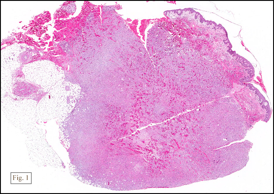
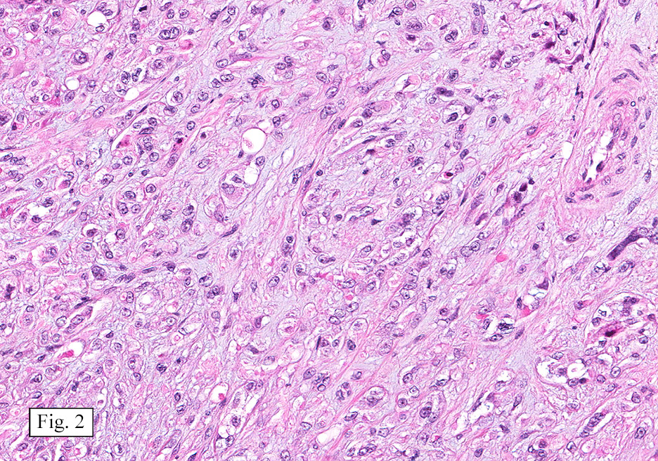
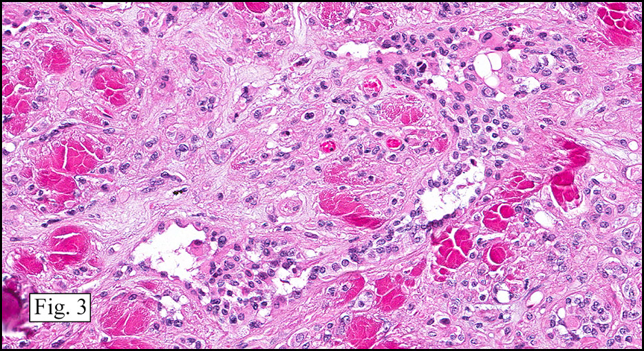
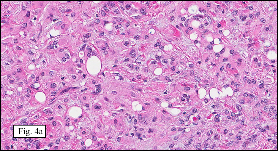
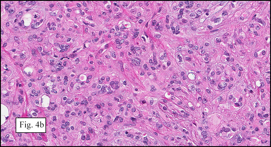
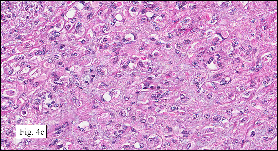
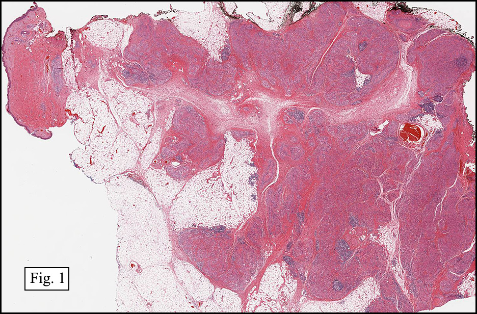
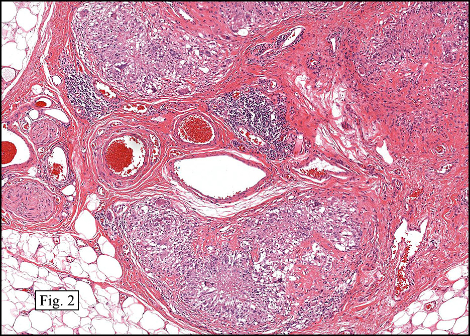
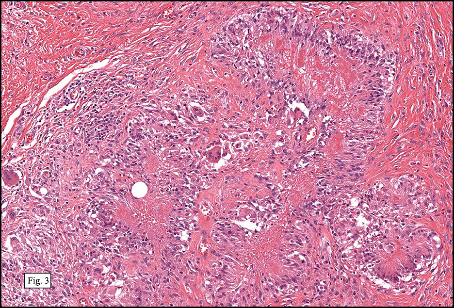
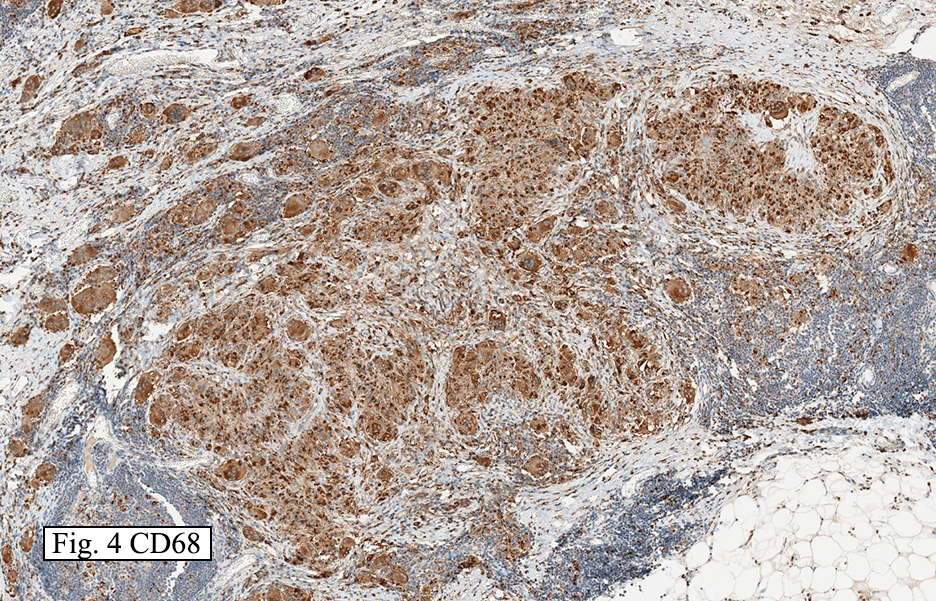
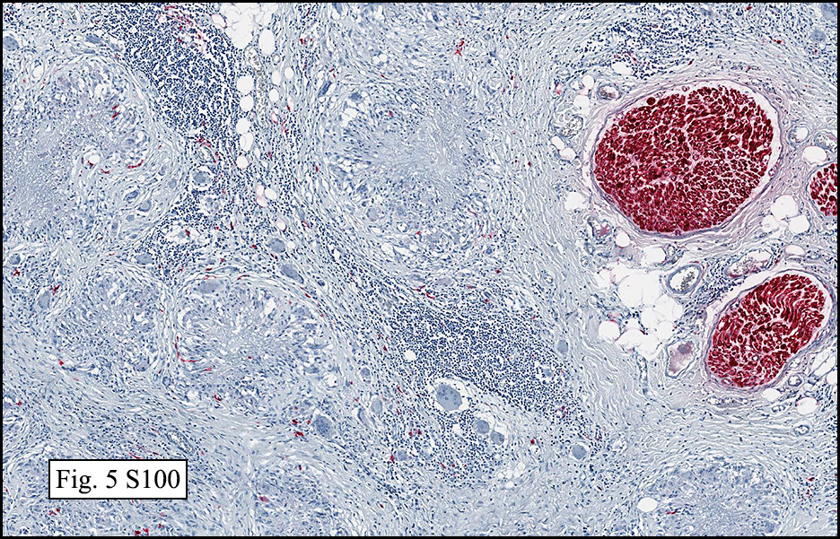
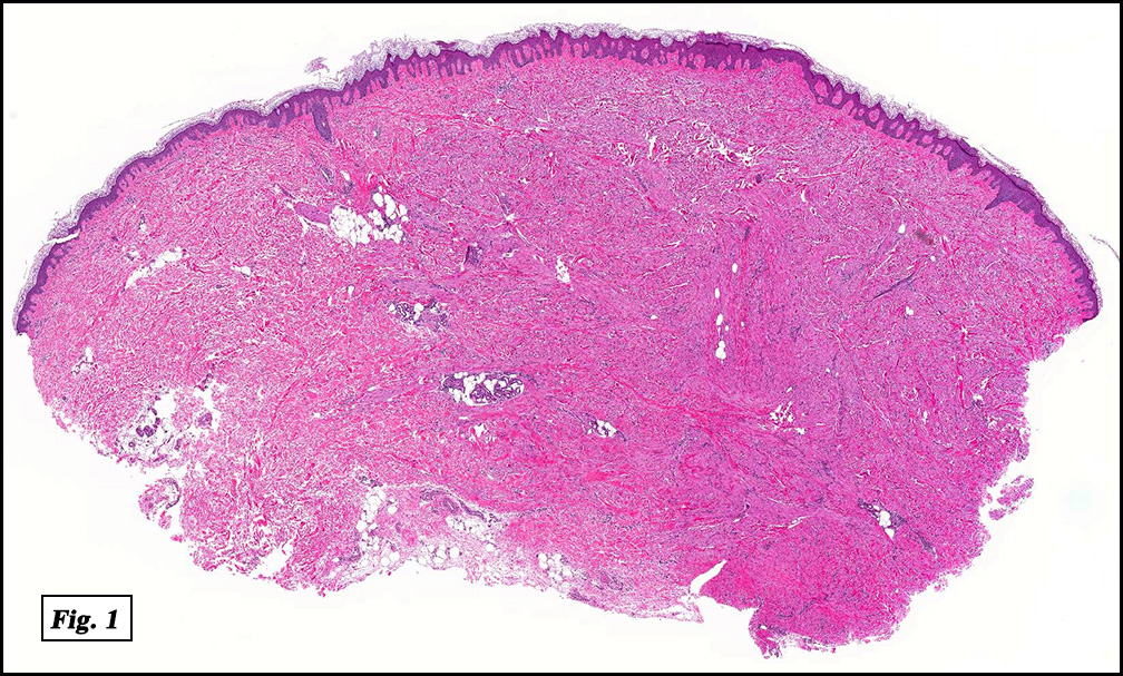
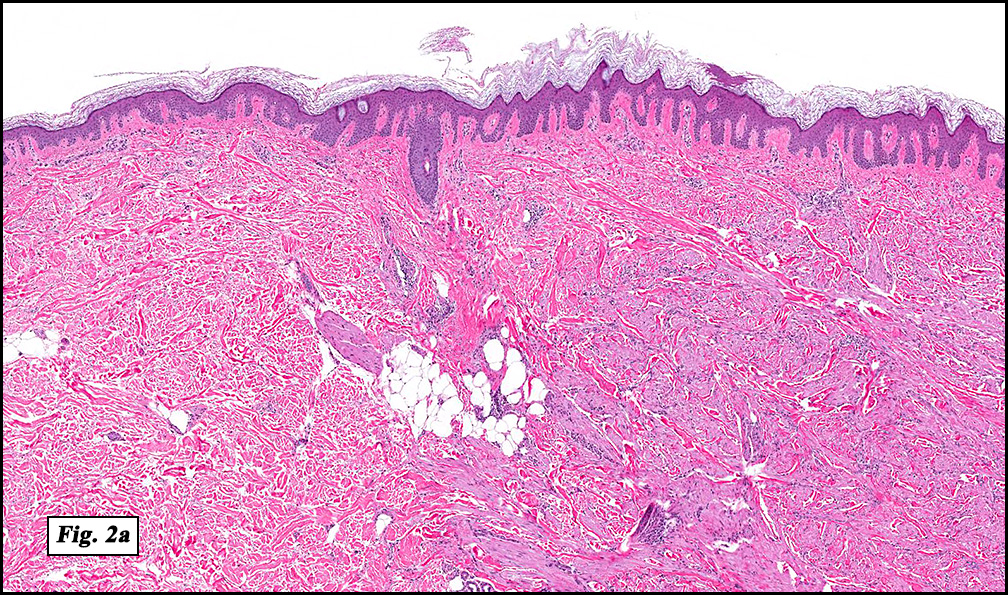
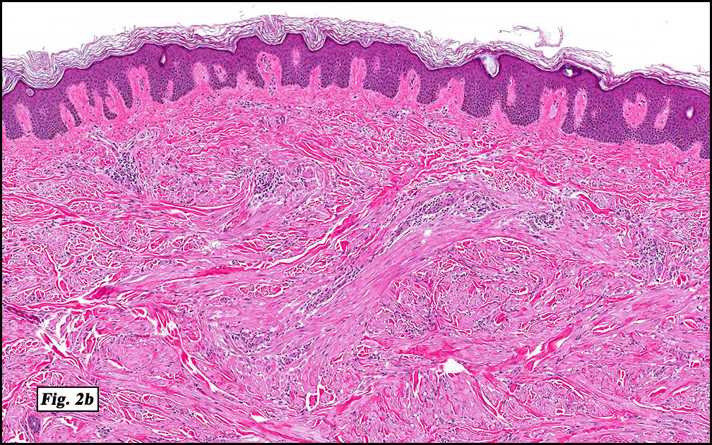
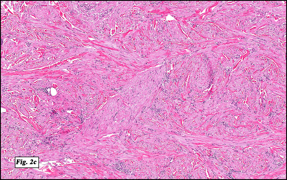
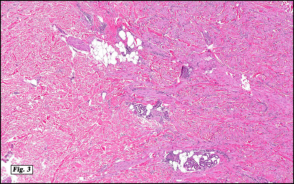
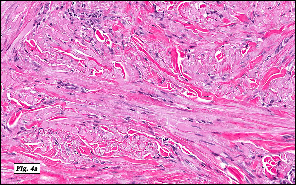
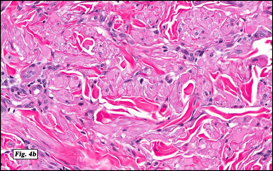
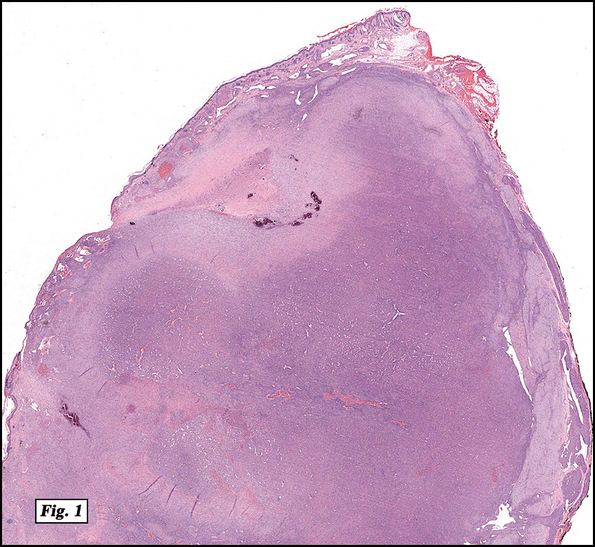
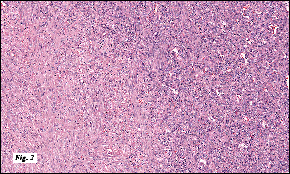
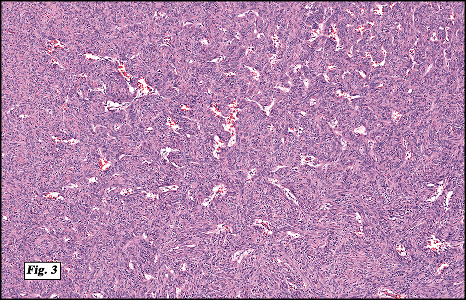
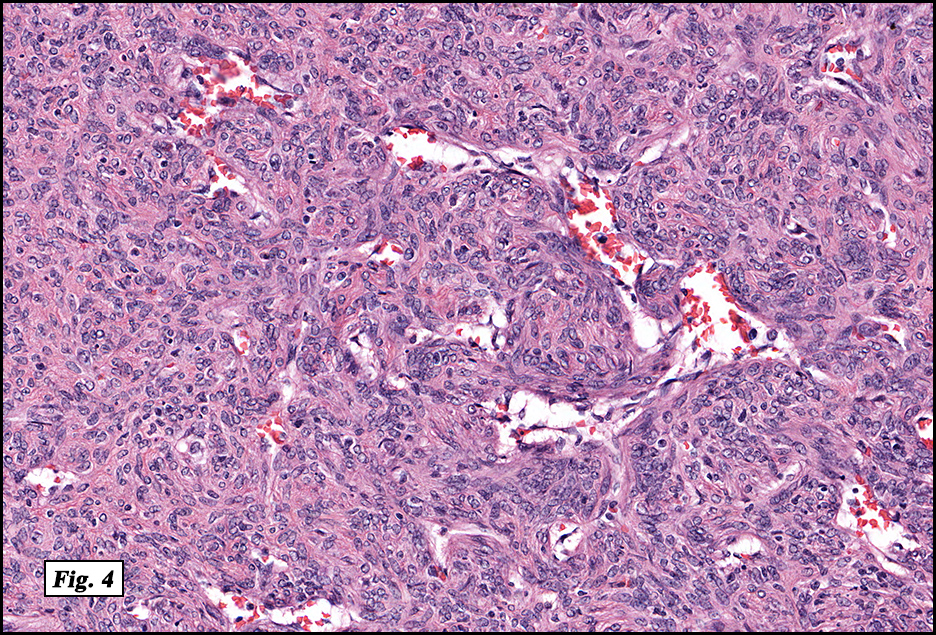
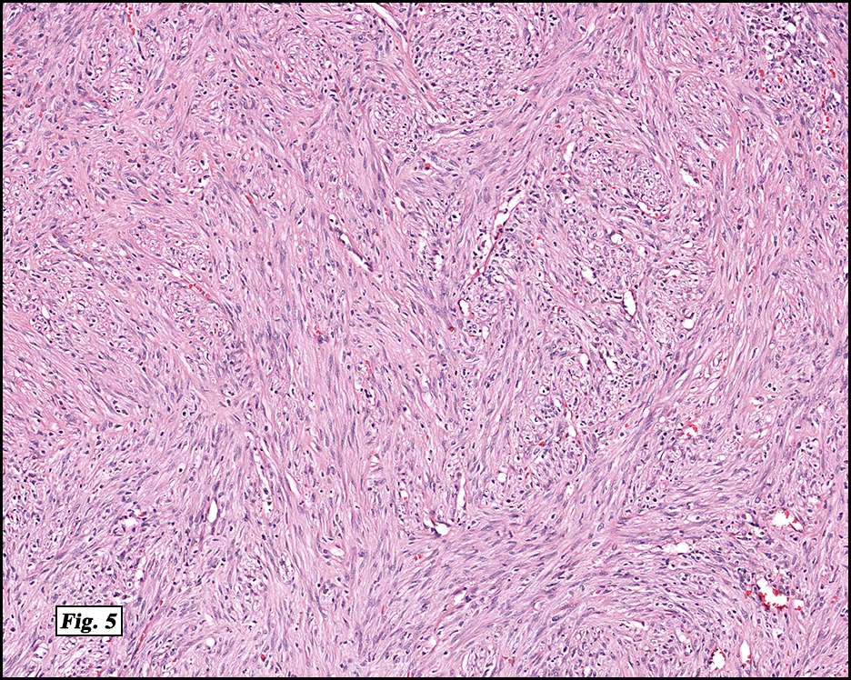
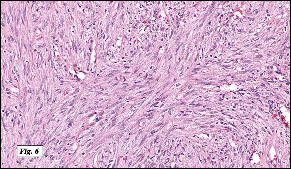
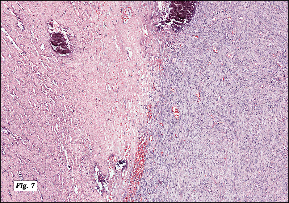
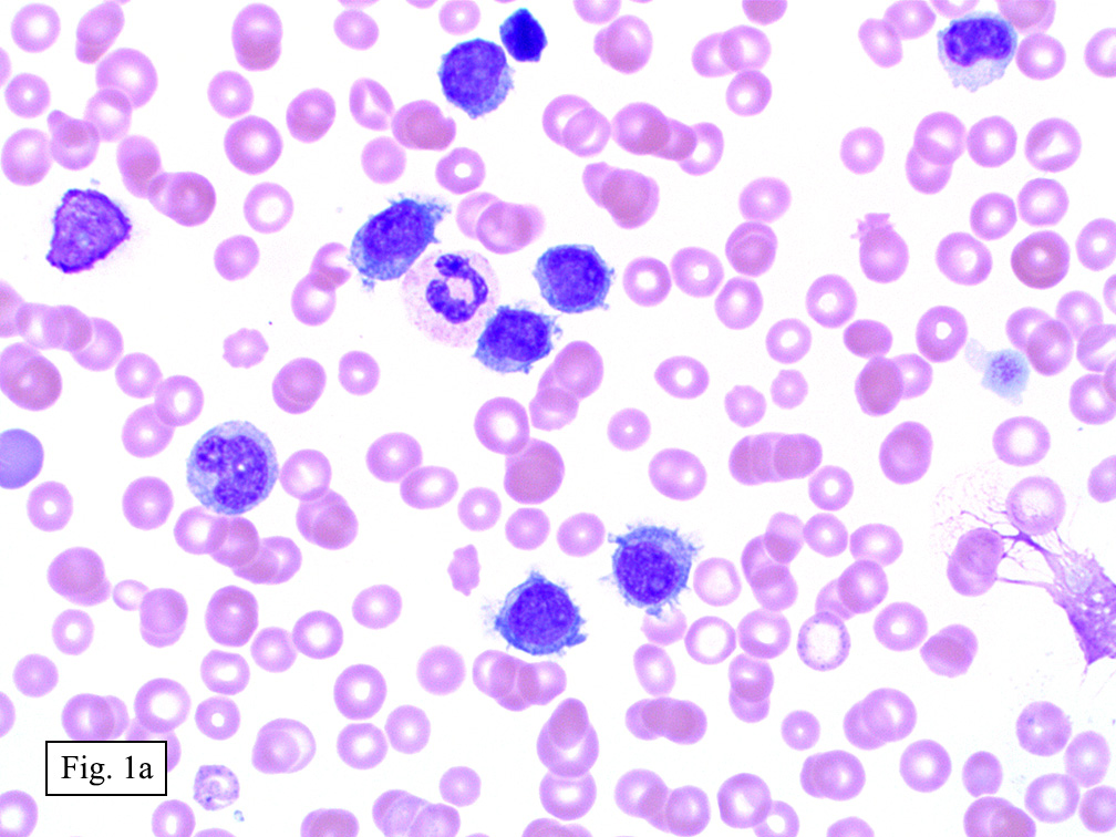
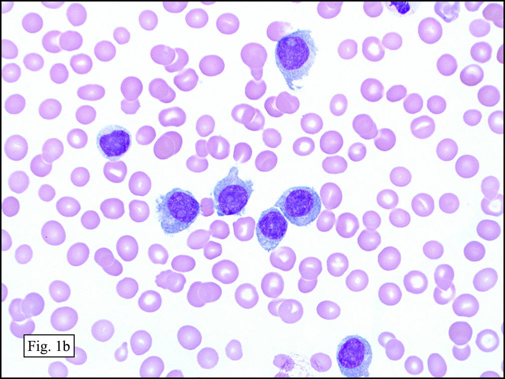
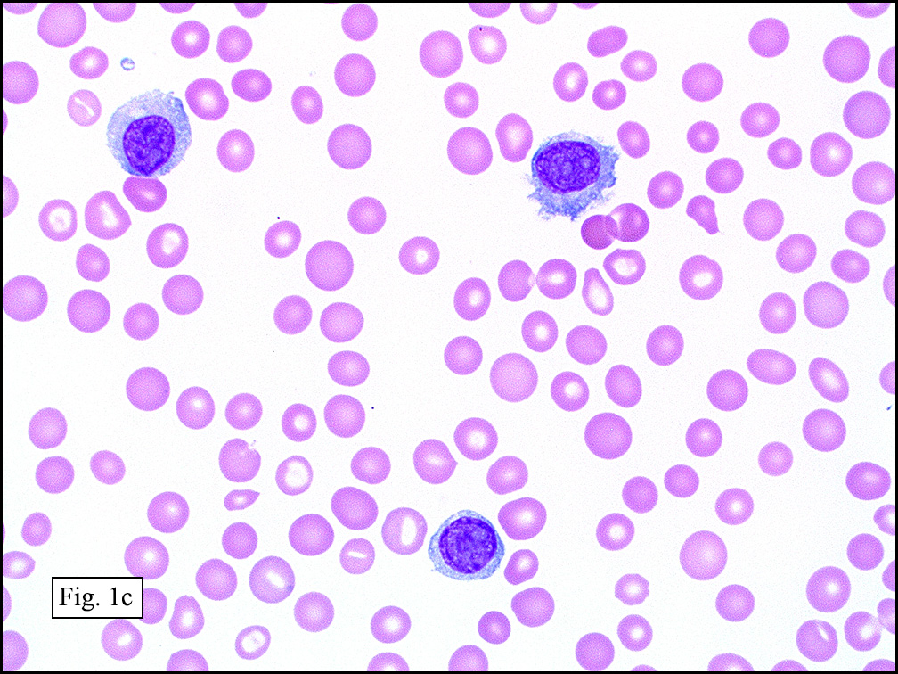
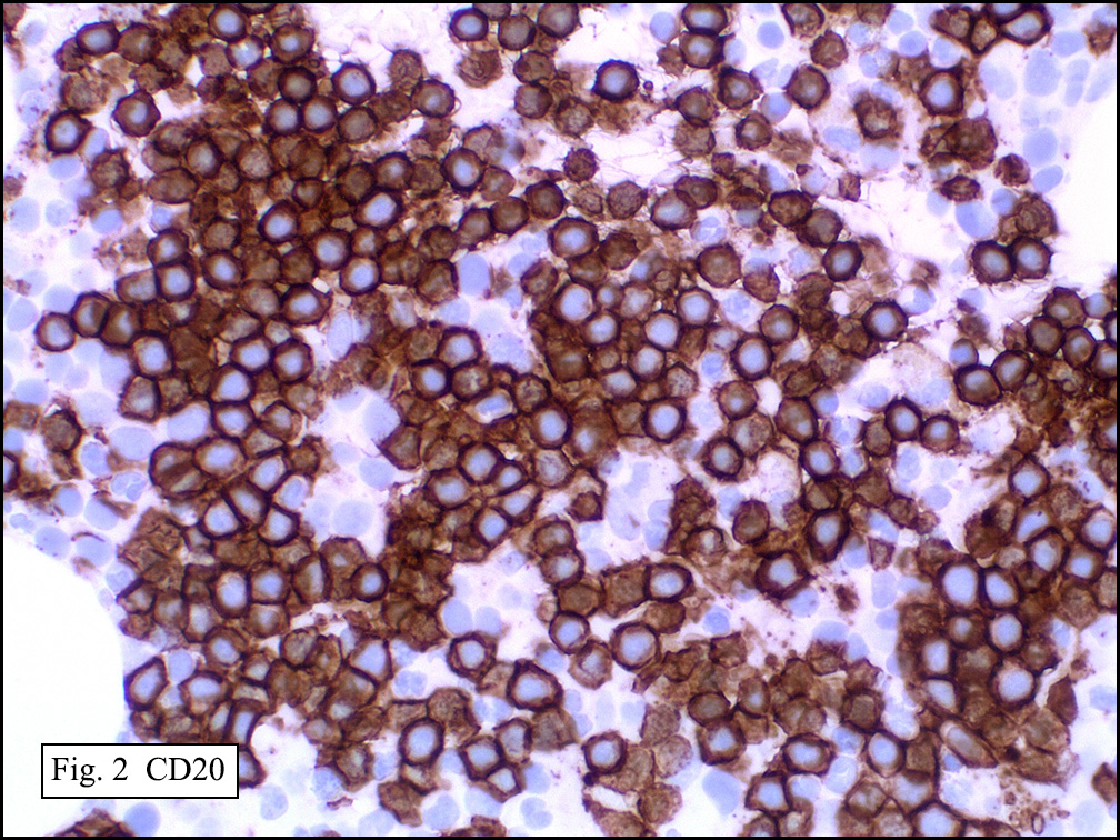
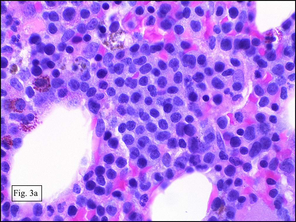
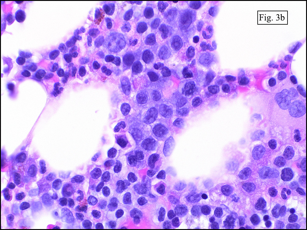
Microscopically, the mass was poorly circumscribed and cellular, located in the dermis and infiltrating skeletal muscle and skin adnexa (Fig. 1). It showed strands and nests of plump cells with eosinophilic cytoplasm in a myxoid background (Fig. 2). In some areas, blood vessel-like structures were present (Fig. 3). Under high amplification these cells had variable-sized vesicular nuclei, prominent nucleoli, cytoplasmic vacuoles and eosinophilic cytoplasm. In most cells the cytoplasmic vacuoles were so prominent that they looked like they had blistered the cells (Fig. 4a, 4b, 4c). Even though nuclear pleomorphism was notable, no atypia or mitotic activity was identified. Immunohistochemical stains showed the tumor cells to be focally positive for factor VIII, but negative for keratin and vimentin.

Diagnosis: Epithelioid hemangioendothelioma

Bingbing Song, M.D. and Donald R. Chase, M. D.
Department of Pathology & Human Anatomy
Loma Linda University Medical Center,
Loma Linda, California

Discussion: Epithelioid hemangioendothelioma (EH) is an angiocentric vascular tumor with metastatic potential and gets the WHO classification as an intermediate grade tumor. It is usually composed of epithelioid endothelial cells with prominent intracytoplasmic vacuoles, arranged in short cords and nests and set in a distinctive myxohyaline stroma.

EH is a rare tumor which may occur in any age group, in either gender and involve virtually any site. It usually presents as a solitary, slightly painful nodule. Deeply situated soft tissue sites may show focal ossification. When EH arises in organs, such as liver and lung, there is a female predilection, possibly due to oral contraceptives.

As original described, EH may arise within or in close approximation to medium-large sized blood vessels. It may mimic an organizing thrombus. Grossly, these tumors have a variegated, white-red color and superficially resemble an organizing thrombus. Some tumors arise from soft tissue instead of blood vessel, but in both types the tumors are composed of short strands or solid nests of round to slightly spindled polygonal endothelial cells. Rarely are large, distinct vascular channels seen, except in the more peripheral regions of the tumor. The tumor cells form small intracytoplasmic lumens, which are seen as clear spaces, or “vacuoles” which can distort or blister the cells. The stroma is usually myxoid or hyaline. Metaplastic bone is occasionally present within large deep lesions and some cases contain prominent osteoclastic giant cells.

Even though most of the tumors appear bland and have no mitotic activity, some tumors contain areas with significant atypia, mitotic activity (>1/10 high power fields), focal spindling of the cells, and/or necrosis. Such features may be related to more aggressive course and poor prognosis. High mitotic figures and tumor size >3.0 cm are associated with a worse prognosis.

The tumor cells are usually positive for CD31, CD34, factor VIII and FLI-1. Focal cytokeratin expression is present in about 25-30%. By ultrastructure, the neoplastic cells are situated on a basal lamina, and possess surface-oriented pinocytotic vesicles and occasional Weibel-Palade bodies.

Genetically, a reciprocal translocation of chromosomes 1 and 3 t(1;3)(p36.3;q25), has been reported.

For the tumors in the soft tissue, the local recurrence rate is 10-15%, the metastatic rate is 20-30%, and mortality is 10-20%. The tumor has more aggressive behavior when it involves visceral organs.

Differential diagnosis includes a variety of vascular tumors and other tumors with epithelioid features in the soft tissue:
• Melanomas and metastatic carcinomas to the soft tissue usually display far more nuclear atypia and mitotic activity than epithelioid hemangioendothelioma.
• Epithelioid hemangioma is composed of multicellular vascular channels which are usually absent in epithelioid hemangioendothelioma and prominent eosinophil infiltration. Mitotic figures are absent.
• Epithelioid angiosarcoma is composed of anastomosing channels lined by highly atypical, mitotically active epithelioid endothelial cells. Necrosis is common.
• Epithelioid sarcoma consists of nodules of rounded eosinophilic cells that surround cores of necrotic debris and collagen. Epithelioid sarcoma usually arises in distal extremity, particularly hand, in the young individuals. The tumor cells are positive for cytokeratin, CD34 (50%), EMA and may be positive for CD31. They are negative for factor VIII and FLI-1.
In summary, epithelioid hemangioendothelioma is an intermediate grade, usually angiocentric vascular tumor with metastatic potential. It can be found in both soft tissue and organs. It is composed of epithelioid endothelial cells with prominent intracytoplasmic vacuoles, arranged in short cords and nests in a distinctive myxohyaline stroma and usually marks for vascular markers.

Suggested Reading:
Fletcher CDM, Unni KK, Mertens F, et al. World Health Organization Classification of Tumor, Pathology and Genetics Tumors of Soft Tissue and Bones. 2002.

Weiss SW, Goldblum JR, et al. Enzinger and Weiss’ Soft Tissue Tumors. 5th edition. 2008.

Kempson RL, Fletcher CDM, Evan HL, et al. Atlas of Tumor Pathology, Tumors of Soft Tissue. 3rd series, 1998.

Mentzel T, Beham A, Fletcher CDM, et al. Epithelioid hemangioendothelioma of skin and soft tissues: clinicopathologic and immunohistochemical study of 30 cases. Am J Surg Pathol.1997 Apr 21(4):363-74.

Deyrup AT, Tighiouart M, Weiss SW, et al. Epithelioid hemangioendothelioma of soft tissue: a proposal for risk stratification based on 49 cases. Am J Surg Pathol. 2008 Jun 32(6):924-7.

Radin DR, Craig JR, Halls JM, et al. Hepatic epithelioid hemangioendothelioma. Radiology 1988 Oct;169(1):145-8.

November 2013: A 54 year old man with a recurrent subcutaneous mass

History: Two years earlier, a now 54 y/o old man was diagnosed as having a schwanomma in his left arm. It was excised, but now he presented with a recurrent subcutaneous mass in the same location. The excised mass was 40 grams, 6.5 x 4.5 x 4.0 cm. The tumor was solid, firm and white-yellow.

Microscopically, the mass was generally circumscribed but had fibrous extensions into the fat. The confluent fibrous bundles formed septae, giving a plexiform appearance (Fig. 1). Inside the septae were numerous multinucleated giant cells, plump mononuclear histiocyte-like cells with abundant eosinophilic cytoplasm, cigar-shaped nuclei and prominent small nucleoli (Fig. 2). The histiocyte-like cells lined up in a palisading pattern in some areas (Fig. 3). Lymphocytes and plasma cells were also seen either singly or in small clusters. Dilated blood vessels were common (Fig. 2). No mitoses, necrosis nor significant pleomorphism were seen. The overlying skin was normal.

Immunohistochemical stains showed the giant cells and histiocyte-like cells to be strongly positive for CD68 (Fig. 4), but negative for S-100 (Fig. 5) and cytokeratin.

Diagnosis: Plexiform Fibrohistiocytic Tumor (Plexiform Fibrous Histiocytoma)

Bingbing Song, M.D. and Donald R. Chase, M. D.
Department of Pathology & Human Anatomy
Loma Linda University Medical Center,
Loma Linda, California

Discussion: Plexiform fibrohistiocytic tumor (PFT) is composed of network of fibrocytes alternating with nodules of giant cells, histiocytes, and chronic inflammatory cells. Though the tumor mostly usually occurs in female patients younger than 30 years, one third have been reported in children less than 10 years old. A small number have also been reported in older patients. The WHO views these tumors as being of “intermediate grade”.

PFTs are usually slowly growing, solitary, painless nodules that have a propensity to occur in the upper arm. They are usually located in the subcutis and are 1-3 cm in diameter. Their cut surface is usually gray-white and firm.

A striking microscopic feature is bundles of fibrous tissue forming a syncytium, with scattered regions of giant cells and mononuclear histiocyte-like cells. These elements are present in varying amounts resulting in three subtypes:

• Fibrohistiocytic subtype: composed mainly of nodules of mononuclear histiocyte-like cells and multinucleated giant cells.
• Fibroblastic subtype: composed mainly of elongated clusters and short fascicles of fibroblast-like cells
• Mixed subtype: composed of both patterns in equal proportion.

The tumor generally forms long extensions which radiate into the surrounding soft tissue. The fibrous tissue is composed of spindled fibrocytes with small nuclei. Nodules are composed of either exclusive histiocyte-like cells or a mixture of histiocyte-like cells and osteoclast-like multinucleated giant cells. The histiocyte-like cells can be either plump/round or spindled with plenty eosinophilic or pale granular cytoplasm. Hemorrhage and hemosiderin are common in the nodules and help to identify this lesion. Chronic inflammation of plasma cells and lymphocytes is often present around the nodules. Dilated blood vessels can be prominent in the tumor, especially in the fibrous tissue. Cellular atypia and pleomorphism are minimal. Mitotic activity is often every low. Vascular invasion is observed in 10-20% of cases. The giant cells and histiocytes-like cells are positive for CD68, and negative for S-100, keratin, factor VIII and CD45. Fibroblasts decorate for vimentin and smooth muscle actin.

PFTs often recur locally, but rarely metastasize. When metastatic to the lung, small fibrohistiocytic nodules usually are found in subpleural locations and/or peribronchiolar regions.

The differential diagnosis includes a variety of fibroblastic/fibromyoblastic tumors and nerve tumors of the soft tissue.

• Plexiform Schwannoma (PS) also has a plexiform pattern and nuclear palisading. But the cells in the nodules are more spindled with no giant cells present. Unlike PFH, PS is strongly positive for S-100.
• Neurothekeoma (NT) also displays a plexiform pattern and is located subcutaneously. But the nodules mostly contain spindle cells instead of plump histiocyte-like cells. Usually the cellularity in the nodules is lower and the stroma is more myxoid with no giant cells seen. These cells express S-100, but not CD68. Some neurothekeomas are more cellular and strongly positive for CD68 but not S-100. This variant of NT has been suggested to have a common histogenesis as PFH.
• Benign dermal and subcutaneous fibrous histiocytomas may contain foam cells and are better circumscribed than PFH.
• Fibrous hamartoma of infancy generally occurs in the similar age group. However, the tumor contains primitive cells within a unique myxoid stroma (“organoid bodies”), but does not have giant cells.

• The absent pleomorphism and rare mitotic activity help to differentiate PFT from
Atypical fibroxanthoma,
Giant cell sarcoma of soft parts, and
Pleomorphic MFH (Pleomorphic Sarcoma).

In summary, plexiform fibrous histiocytoma is an intermediate grade, subcutaneous tissue tumor with a fibrous network containing nodules of giant cells, histiocyte-like cells and intermixed chronic inflammation. The tumor cells strongly express CD68, but not S-100, cytokeratin or factor VIII. It often recurs but rarely metastasizes.

Suggested Reading:

Fletcher CDM, Unni KK, Mertens F, et al. World Health Organization Classification of Tumors, Pathology and Genetics Tumors of Soft Tissue and Bone. 2002.

Kempson RL, Fletcher CDM, Evan HL, et al. Atlas of Tumor Pathology, Tumors of the Soft Tissue. 3rd series, 1998.

Moosavi C, Jha P, Fanburg-Smith JC. An update on plexiform fibrohistiocytic tumor and addition of 66 new cases from the Armed Forces Institute of Pathology, in honor of Franz M. Enzinger, MD. Ann Diagn Pathol. 2007; 11(5): 313-9.

Taher A, Pushpanathan C. Plexiform fibrohistiocytic tumor: a brief review. Arch Pathol Lab Med. 2007; 131(7): 1135-8.

Jaffer S, Ambrosini-Spaltro A, Rosai J, et al. Neurothekeoma and plexiform fibrohistiocytic tumor: mere histologic resemblance or histogenetic relationship? Am J Surg Pathol. 2009;33(6):905-13.

October 2013: A 59 year old woman with a painful leg nodule

History: A 59-year-old healthy woman presented with a solitary painful pink nodule on her right anterior leg. The 1.0 x 0.7 cm nodule had persisted, unchanged, for the last 4 years. An excisional biopsy was performed for symptomatic relief.

Microscopically, the tumor was a dermal-based proliferation with a distinct grenz zone, overlying acanthosis, and no obvious connection to the epidermis (Figs. 1, 2a, 2b). It was comprised of fascicles of spindle cells with ample eosinophilic cytoplasm haphazardly intersecting and surrounding nearby adnexal structures, including a hair follicle (Figs. 2a, 2b, 2c). Nonencapsulated and poorly circumscribed, the packets of spindle cells interdigitated with the adjacent normal reticular dermis (Fig. 3). Upon closer inspection, the spindle cells had cigar-shaped, blunt-ended nuclei and were interwoven with bundles of bright red collagen (Figs. 4a, 4b). The tumor cells showed no significant pleomorphism, mitotic activity, nor necrosis. Immunohistochemistry showed positively for desmin and actin.

Diagnosis: Leiomyoma of pilar arrector origin

Elif L. Akin, M.D. and Donald R. Chase, M. D.
Department of Pathology & Human Anatomy
Loma Linda University Medical Center,
Loma Linda, California

Discussion: Cutaneous leiomyomas were first described in the early 1800s as “painful subcutaneous tubercles” or “tuberculosum dolorosum”. Today, they are usually designated as “leiomyoma of pilar arrector origin” (LPAO) and are clinically grouped with several other cutaneous tumors that may present with localized pain.

LPAO is a relatively uncommon lesion that usually occurs on the extensor surfaces or trunk of young adults, with no predilection for gender. Lesions often manifest as multiple, slow-growing, small pink papules. Occasionally, they may follow a dermatomal distribution or coalesce into plaques. Clinically, LPAO is associated with pain (occurring either spontaneously or with applied pressure), sensitivity to cool temperatures, and/or pruritus. These symptoms are thought to be related to either nerve compression or muscle contraction. Of interest, Fisher & Helwig described three patients in 1963 who experienced lesional pain that was apparently elicited by strong emotions.

As it arises from the arrector pili muscle, LPAO is comprised of packets and bundles of smooth muscle cells intersecting in a chaotic, haphazard arrangement. The tumor usually has a clear grenz zone, does not involve the epidermis, but may have overlying epidermal hyperplasia. The smooth muscle cells have ample eosinophilic cytoplasm and cigar-shaped or vesicular nuclei. While there may be focal areas of mild atypia related to degenerative changes, significant nuclear pleomorphism, mitotic activity, and necrosis are not typically present. LPAO is usually poorly circumscribed, merging into the surrounding tissue, and is often found near a hair follicle.

The mainstay therapy for LPAO is surgical excision and pain management. Even though this is regarded as a benign neoplasm with no potential for malignant transformation, complete surgical excision can be challenging in patients presenting with multiple lesions; otherwise, surgery is usually curative. Alternative therapies including laser ablation and cryotherapy have also been employed.

Recently, a small number of patients presenting with multiple cutaneous leiomyomas of pilar arrector origin have been found to have an autosomal dominant syndrome called “multiple cutaneous and uterine leiomyomatosis” (MCUL, aka Reed syndrome), which predisposes women to the early development of uterine leiomyomas. Interestingly, a small subset of patients and families with MCUL also has associated papillary or collecting duct renal cell carcinoma, a condition known as hereditary leiomyomatosis and renal cell cancer (HLRCC). Both MCUL and HLRCC have been linked to a germline loss-of-function mutation of the fumarate hydratase (FH) gene. This gene encodes an enzyme in the Krebs cycle, which normally functions as a tumor suppressor. Alam et al. have concluded that most patients with cutaneous leiomyomas have an underlying FH mutation. This renders the early identification and correct diagnosis of LPAO quite clinically significant, as it may prompt additional work-up for other conditions such as uterine fibroids and/or renal cancer.

The differential diagnosis includes a variety of benign and malignant fibrohistiocytic and smooth muscle tumors:

• Cutaneous fibrous histiocytoma (dermatofibroma) may also demonstrate a haphazard arrangement of cells. Inflammatory cells, giant cells, and xanthoma cells may also be seen, however, they are absent in LPAO.

• Smooth muscle hamartoma is also comprised of smooth muscle bundles; however, it tends to occur as a single, larger lesion presenting in the lower back of children or young adults and may also be associated with Becker’s nevus.

• Genital leiomyoma is a more common type of cutaneous leiomyoma, which occurs on the nipple, scrotum, or vulva. Unlike LPAO, it tends to demonstrate increased cellularity and is usually well circumscribed.

• Angioleiomyoma usually occurs in females, most often in the lower leg. It is a deeper lesion than LPAO, arising in the superficial subcutis. The smooth muscle cells in an angioleiomyoma surround numerous thick-walled vessels. LPAO, in contrast, does not have prominent vasculature.

• Leiomyosarcoma usually demonstrates significant nuclear pleomorphism and mitotic activity, features that are, by definition, not associated with LPAO.

In summary, leiomyoma of pilar arrector origin is an uncommon benign neoplasm that usually occurs on the extensor surfaces of young adults. While it may occur as a solitary lesion, patients usually present with multiple pink sub-centimeter painful papules/nodules. Surgical excision is typically curative. Correct diagnosis of this lesion is important because it may be a presenting feature of multiple cutaneous and uterine leiomyomatosis and/or hereditary leiomyomatosis and/or renal cell cancer, two hereditary conditions which are associated with germline mutations in the fumarate hydratase gene. Early recognition of these cutaneous leiomyomas can help ensure that patients receive appropriate gynecologic and genitourinary evaluation.

Suggested Reading:

Alam NA, Olpin S, Leigh IM. Fumarate hydratase mutations and the predisposition to cutaneous leiomyomas, uterine leiomyomas and renal cancer. Br J Dermatol. 2005; 153: 11-17.

Calonji E, Brenn T, Lazar A, McKee PH. McKee’s Pathology of the Skin with Clinical Correlations: 4th ed. Elsevier Saunders. 2011; 2: 1696-1697.

Chronic unilateral eruption of painful, erythematous papules and nodules. JAMA Dermatol. 2013; ():2. doi:10.1001/jamadermatol.2013.3358a.
Fisher WC, Helwig EB. Leiomyomas of the skin. Arch Dermatol. 1963; 88: 510-520.

Fletcher CD. Diagnostic Histopathology of Tumors: 3rd ed. Churchill Livingstone Elsevier. 2007: 1562-1563.

Stout AP. Solitary cutaneous and subcutaneous leiomyoma. Am J Cancer. 1937; 29: 435-469.

Weiss S, Goldblum J. Enzinger & Weiss’s Soft Tissue Tumors: 5th ed. Mosby/Elsevier Inc: Philadelphia. 2008: 518-520.

September 2013: A four day old infant boy with a forehead lesion

History: A four day old infant boy underwent a biopsy of a firm 2.5 x 1.0 cm purplish macule on his forehead. Microscopically the lesion showed a nodular growth pattern and biphasic zonality with an abrupt interface between the central hypercellular component and peripheral paucicellular component (Figs. 1, 2). Higher power views of the central region showed a hemangiopericytoma-like pattern with abnormal staghorn vessels (Figs. 3, 4). The hypocellular region around the perimeter showed spindled eosinophilic cells with a fascicular growth pattern, resembling smooth muscle cells or myofibroblasts (Figs. 5, 6). A small area of necrosis and calcification was present (Fig. 7).

Diagnosis: Myofibroma (Myopericytoma)

Summer Blount MD, Donald R. Chase MD
Department of Pathology and Human Anatomy,
Loma Linda University Medical Center, Loma Linda, California
California Tumor Tissue Registry, Loma Linda, California

Discussion: Although myofibromatosis was initially described in 1951 by Williams and Schrum as a type of congenital fibrosarcoma, the entity was quickly reclassified by Stout as congenital generalized fibromatosis when it was shown that this tumor lacked malignant potential. Chung and Enzinger later noted that the cellular makeup was primarily of myofibroblasts and renamed the lesion “infantile myofibromatosis”, and adopted the term “myofibroma” when the lesion was solitary. Later, the term “infantile” was dropped in recognition that these tumors may occur later in life.

The 1998 WHO classification of this tumor separated it into two entities as part of a morphologic spectrum. One was termed myopericytoma (MPC) and the other as a myofibroma (MF). Then the 2013 WHO classification combined a group of similar tumors under the rubric “myopericytoma” (MPC). This class of tumors is characterized by concentric vascular proliferation of spindled cells showing differentiation towards perivascular myoid cells. These entities include solitary myofibroma, myofibromatosis, infantile hemangiopericytoma, angioleiomyoma and glomus tumor. It is still unclear if these are distinct entities, or a spectrum of the same tumor.

The presented tumor is generally termed a “myofibroma.” It is the most common fibrous tumor of childhood, accounting for 20% to 25%. These lesions usually present between birth and five years of age, with a slight male preponderance. However, recent studies have shown that myofibromas may be found in any age group including children and adults. When solitary, they are usually seen on the head and neck, commonly the forehead, parotid region or scalp, and usually range from only a few millimeters in diameter up to several centimeters in diameter. They are confined to the dermis and subcutaneous tissue and have a rubbery to firm touch and a white-gray to pink-purple cut surface. Clinically, they present as painless purple macules which may be mistaken for a hemangioma.

Outcome is determined by disease extent at diagnosis. Individuals with fewer lesions and no visceral involvement have an excellent prognosis. These tumors may spontaneously regress in younger children, particularly those less than 2 years of age. Most cases are sporadic; however, both autosomal recessive and dominant patterns of inheritance have been described. Treatment is generally limited to complete or partial excision with a local recurrence rate of less than 10%. In cases where surgery might cause significant morbidity, chemotherapy has been used to reduce the tumor size and alleviate associated symptoms.

Microscopically, myofibromas have a characteristic nodular appearance that is biphasic with areas of hyper and hypocellularity, resulting in dark and light staining. The darkly stained, hypercellular areas are centrally located and consist of whorling round to oval cells with slightly pleomorphic, hyperchromatic nuclei. There is a predominant pattern of HPC-like thin walled vessels surrounded by small spindled cells with vesicular nuclei, eosinophilic cytoplasm and indistinct cell boarders. Commonly, coagulative type necrosis, calcification and hemorrhage are present. Some tumors with extensive central necrosis mimic necrotic granulomas.

The lighter staining paucicellular peripheral areas consist of plump myoid spindled cells with elongated nuclei and moderately abundant eosinophilic cytoplasm. These spindle cells are arranged in short fascicles often within hyaline-myxoid stroma. Of note, the identification of peripheral myoid-appearing cells can be useful when differentiating myofibroma from other entities.

In the majority of cases the histology of myofibroma is characteristic and additional studies do not need to be utilized. Immunohistochemistry can be used to distinguish it from other considerations or entities when necessary. Myofibromas stain positively for smooth muscle actin and vimentin, and to a lesser extent muscle-specific actin. In the central portion, smooth muscle actin highlights the myofibromatous pattern without reactivity in the contiguous HPC-like foci.

Differential diagnosis:

• Infantile fibromatosis is considered in patients with multiple lesions. The individual nodules have similar features as myofibromas, except that they occur not only in the dermis, subcutis, and soft tissue, but also in muscle, bone, and internal organs. Myofibromatosis is subdivided into multifocal and generalized forms in which visceral involvement is diagnostic for generalized myofibromatosis.

If only the central portion of the lesion is biopsied, various sarcomas may enter the differential diagnosis. Extensive vascular proliferation and necrosis would raise concern for entities such as congenital infantile fibrosarcoma, solitary fibrous tumor, dermatofibrosarcoma protuberans, peripheral primitive neuroectodermal tumor, or other sarcomas. In general, immunostains that will be helpful include S-100, CD99 and cytokeratins.

• Congenital infantile fibrosarcoma (CIFS) usually occurs during the first year of life, with some diagnosed antenatally. Clinically, infantile fibrosarcoma presents as a solitary rapidly enlarging mass, sometimes grotesque in proportion to the child’s size. The superficial and deep soft tissues of the distal extremities are the primary site in nearly two-thirds of cases, with the remainder found on the trunk, head and neck. Microscopically CIFS is highly cellular and may have a storiform or herringbone pattern. The cells are predominately spindled, but have a high nuclear to cytoplasmic ratio with hyperchromasia, tumor necrosis, and frequent mitoses. CIFS lacks the biphasic zonal pattern and peripheral myofibroblasts seen in myofibroma. Genetic analysis can be critical for differentiation between CIFS and myofibroma. There is a known characteristic translocation t(12;15) identified in CIFS, with 40-80% being congenital. Treatment is usually complete surgical excision. However, with appropriate treatment there is a relatively good prognosis when compared to other sarcomas. This tumor has a mortality rate of less than 5%.

• Infantile hemangiopericytoma usually occurs within the first year of life as a solitary lesion located in the subcutis or oral cavity. Older children may have tumors within muscle or mediastinum. Microscopically, infantile hemangiopericytoma closely resembles adult HPC/SFT, but differs in that the superficial lesions are multilobulated, often with distinct intravascular and perivascular satellite nodules outside the main tumor mass. Features such as increased mitotic activity and focal necrosis generally do not indicate a poor prognosis in infantile HPC as it does with the adult form. Treatment is similar to that of myofibroma, with possible spontaneous regression or local excision.

• Fibrous histiocytoma shows a pronounced proliferation of polymorphous cells arranged in a storiform pattern often with a histiocytic component, multinucleated cells and iron pigment. These cells may be smooth muscle actin (SMA) positive like myofibroma, but may also express factor XIIIa and CD68.

• Inflammatory myofibroblastic tumor (IMT) is composed of spindled myofibroblasts within a variable background of collagen and inflammatory cells. IMT often shows rearrangements of the 2p23 chromosomal region, involving the ALK gene, and thus often stains for ALK-1, and lacks an HPC-like pattern.

Suggested Reading:
1. Weiss SW, Goldblum JR. Enzinger & Weiss’s Soft Tissue Tumors. 5th ed. Philadelphia: Mosby-Elsevier, 2008:264-269.
2. Stocker JT, Dehner LP. Pediatric Pathology. 3rd ed. Philadelphia: Lippincott Williams-Wilkins 2011:1052-1056, 1072.
3. Solitary, multifocal and generalized myofibromas: clinicopathological and immuno-histochemical features of 114 cases. Oudijk, Lindsey; den Bakker, Michael A; Hop, Wim C J; Cohen, Marta; Charles, Adrian K; Alaggio, Rita; Coffin, Cheryl M; de Krijger, Ronald R. Histopathology. May2012, Vol. 60 Issue 6B, pE1-E11.
4. Myofibroma of the cheek: A case report. Kassenoff, Tali L.; Tabaee, Abtin; Kacker, Ashutosh. ENT: Ear, Nose & Throat J. Jun2004, Vol. 83 Issue 6, p404-407.
5. Myopericytoma: a unifying term for a spectrum of tumours that show overlapping features with myofibroma. A review of 14 cases. Dray, M. S.; McCarthy, S. W.; Palmer, A. A.; Bonar, S. F.; Stalley, P. D.; Marjoniemi, V.; Millar, E.; Scolyer, R. A J Clin Pathol. Jan2006, Vol. 59 Issue 1, p67-73.
6. The Spectrum of Pediatric Fibroblastic and Myofibroblastic Tumors. Hicks, John; Mierau, Gary. Ultrastructural Pathol. Sep-Dec2004, Vol. 28 Issue 5/6, p265-281.
7. Malignant myopericytoma: expanding the spectrum of tumours with myopericytic differentiation. McMenamin, M E; Fletcher, C D M. Histopathology. Nov 2002, Vol. 41 Issue 5, p450-460.
8. Myopericytoma: A Pleural-Based Spindle Cell Neoplasm Off the Beaten Path. Edgecombe, Allison; Peterson, Rebecca A.; Shamji, Farid M.; Commons, Susan; Sekhon, Harman; Gomes, Marcio M. Int J Surg Pathol. 04/01/2011, Vol. 19 Issue 2, p247-251.
9. Solitary fibrous tumour and haemangiopericytoma: evolution of a concept. Gengler, C.; Guillou, L. Histopathology. Jan2006, Vol. 48 Issue 1, p63-74.
10. Fletcher CDM, Unni KK, Mertens F, eds. Myofibroma and Myopericytoma. In: WHO Classification of Tumors of Soft Tissues and Bone. Lyon: IARC Press 2013:89-90, 118-120.

August 2013: A 90 year old man with an elevated white count

History: A 90-year-old man with a complex past medical history presented to the emergency department after a fall. He was found to have an elevated white cell count (36 x 109/L) without monocytopenia which prompted blood smear review. The blood smear demonstrated frequent medium-sized lymphocytes with round, occasionally clefted nuclei with mildly condensed chromatin, prominent nucleoli, and moderate amounts of pale, basophilic cytoplasm showing multiple hair-like projections (Figs. 1a, 1b, 1c). Flow cytometry was performed on the peripheral blood and showed a kappa-restricted population of lymphocytes expressing CD19, CD20 (Fig. 2), FMC7, CD103, CD11c. A CD25 were negative. A bone marrow biopsy was subsequently performed and showed an immunophenotypically identical population comprising about 35% of bone marrow constituents (Figs. 3a, 3b).

Diagnosis: Hairy cell leukemia variant

Laura Denham1, MD; Jeff Cao1, MD; Donald R. Chase1,2, MD

1. Department of Pathology and Human Anatomy, Loma Linda University
Medical Center, Loma Linda, California
2. California Tumor Tissue Registry, Loma Linda, California

Discussion: Hairy cell leukemia variant (HCLv) is a B-cell lymphoproliferative neoplasm first described by Cawley et al. in 1980 and later termed the “prolymphocytic variant” of hairy cell leukemia. It is a rare disorder, comprising approximately 10% of hairy cell leukemias and 0.4% of all chronic lymphoproliferative disorders, corresponding to about 60-75 cases per year in the United States. As described below, the name is somewhat of a misnomer, as this entity is no longer thought to be related to classic hairy cell leukemia. HCLv is currently a provisional diagnosis in the WHO 2008 and is categorized under “splenic lymphoma/leukemia unclassifiable”.

Patients with HCLv tend to present in their 60’s and 70’s with symptoms of abdominal discomfort secondary to splenomegaly. In addition, they may also complain of weight loss and weakness. Hepatomegaly is present in a subset of patients. There is a slight male predominance, but less so than in classic hairy cell leukemia (cHCL).

Laboratory findings include leukocytosis without monocytopenia. This picture is different than cHCL which generally demonstrates monocytopenia without lymphocytosis. Anemia and thrombocytopenia may also be present in some cases. Cytopenias appear to be the result of hypersplenism rather than bone marrow replacement by a malignant infiltrate.

In contrast to the rare and sometimes difficult to find hairy cells of cHCL, circulating hairy cells or HCLv usually comprise greater than 50% of nucleated cells within the peripheral blood. These cells exhibit moderate to abundant amounts of pale blue cytoplasm with hairy projections. The central, round nucleus commonly contains somewhat condensed chromatin and a prominent nucleolus. Clefted or irregular nuclei are occasionally seen.

Immunophenotypically, HCLv marks as mature B-cells, with expression of CD19, CD22 and FMC7. They are negative for CD10. Unlike cHCL, the variant form does not express CD25 or Annexin A1, and they variably express CD11c (~87% positivity), and CD103 (~60% positivity). Tartrate resistant alkaline phosphatase (TRAP) is also weakly or variably expressed. In regards to clonality, they are more commonly lambda light chain restricted than kappa light chain restricted.

Bone marrow examination shows a hypercellular marrow with an interstitial infiltration of neoplastic lymphoid cells. Involvement of these cells is typically of only a mild degree, and may be quite subtle. Occasional clusters or nodules may be present. Sinusoidal involvement has been reported, but is usually focal. In contrast to the cHCL, reticulin deposition is only slightly to moderately increased, resulting in an easily attainable aspiration when compared to the notoriously difficult aspiration of reticulin-laden cHCL. Splenic involvement typically causes red pulp expansion by neoplastic lymphoid cells with resulting white pulp follicle atresia.

The differential diagnosis for HCLv includes classic hairy cell leukemia, splenic diffuse red pulp small B-cell lymphoma and B-cell prolymphocytic leukemia.

• Classic hairy cell leukemia: As previously stated, monocytopenia may be present, however, lymphocytosis is negligible. There is a strong male predominance, and patients often present with signs of infection. The peripheral blood may show only rare, hairy cells. CD25 and Annexin A1 are positive in cHCL, and reticulin fibrosis is usually moderate to marked.

• Splenic diffuse red pulp small B-cell lymphoma (also known as splenic lymphoma with villous lymphocytes): This entity is also a provisional diagnosis in WHO 2008 characterized by a mild lymphocytosis composed of small lymphocytes with round nuclei displaying condensed chromatin and occasional small nucleoli. The cytoplasm is scanty, with villous projections. Plasmacytoid cytology may be seen. Bone marrow infiltrates predominately involve sinusoids, with occasional interstitial/nodular involvement. Splenomegaly is typical and shows diffuse red pulp expansion with absence of follicular replacement. The cells mark with CD20, DBA.44 and are negative for CD25, CD103, CD11c, CD123 and Annexin A1.

• B-cell prolymphocytic leukemia: Similarly to HCLv, patients often present with splenomegaly and leukocytosis. However, the lymphocytes of PLL demonstrate greater than 55% prolymphocytes which are of medium cell size, round nucleus with condensed chromatin and prominent nucleoli. The cytoplasm is scant and typically lacks projections. An interstitial or nodular infiltrate is seen in the bone marrow. In contrast to HCLv, splenic involvement is characterized by white pulp nodule expansion. Cells are positive for typical B-cell markers CD19, CD22, and FMC7.

The most important reason to differentiate HCLv from classic hairy cell leukemia is that the variant form does not respond to interferon α and has historically shown poor response to purine analogs (pentosin and cladribine) and alkylating agents (chlorambucil and cyclophosphamide). Patients have had better response to splenectomy, which decreases the volume of disease and improves the cytopenias caused by hypersplenism.

Like its classic counterpart, HCLv is has a chronic course with many patients succumbing to diseases other than their leukemia. The median survival for HCLv is about 9 years, compared to cHCL which is around 12 years. Rate of progression is low (~6%), and has been associated with p53 deletion. When transformed, patients present with abdominal masses, weight loss and fever. Circulating blasts are present with a phenotype similar to that seen in the peripheral blood at initial presentation.

In summary, hairy cell leukemia variant shows some features overlapping with classic hairy cell leukemia, such as hairy lymphocytes in the peripheral blood, expression of CD103 and CD11c in some cases and splenic involvement. However, there are notable differences between the two. The variant form presents with lymphocytosis without monocytopenia, and does not express CD25 or Annexin A1. In addition, there is lack of significant reticulin fibrosis within the bone marrow. Most importantly from a clinical perspective is the poor response of hairy cell leukemia variant to medications typically used to treat the classic from of hairy cell leukemia.

Suggested Reading:

1. Charin Y, Brandwein J, Pantalony D, Chang H. Hairy cell leukemia variant with features of intrasinusoidal bone marrow involvement. Arch Pathol Lab Med. 2005; 129:395-398.

2. de Totero D, Tazzari PL, Raspardori D, di Celle PF, Carbone A, Gobbi M, Foa R. Phenotypic analysis of hairy cell leukemia: “variant” cases express the interleukin-2 receptor beta chain, but not the alpha chain (CD25). Blood. 1993;82(2):528-535.

3. Kanellis G, Garcia-Alonso L, Camacho FI, Garcia JF, Mollejo M, Montes-Moreno S, Garcia-Vela JA, Piris MA. Hairy cell leukemia variant. J Hematopathol. 2011;4:13-16.

4. Matutes E, Wotherspoon A, Brito-Babapulle V, Catovsky D. The nature history and clinic-pathological features of the variant form of hairy cell leukemia. Leukemia. 2001;15:184-193.

5. Matutes E, Wotherspoon A, Catovsky D. The variant form of hairy-cell leukemia. Best Practice and Research Clinical Haematology. 2003;16(1):41-56.

6. Piris M, Foucar K, Mollejo M, et al. (2008) in WHO Classification of Tumours of Haematopoietic and Lymphoid Tissues, Splenic B-cell lymphoma/leukaemia, unclassifiable, eds Swerdlow SH, Campo E, Harris NL,et al. (IARC Press, Lyon, France), pp 191–193.

7. Sainati L, Matutes E, Mulligan S, de Oliveira MP, Rani S, Lamper IA, Catovsk D. A variant form of hairy cell leukemia resistant to α-interferon: clinical and phenotypic characteristics of 17 patients. Blood. 1990;76(1):157-162.

Scroll to top