December 2015: A 74 year old woman with abdominal pain

History: 74-year-old gravida 1, para 1 female with a history of hysterectomy and bilateral salpingoopherectomy for severe pelvic endometriosis who presented with abdominal pain. A CT scan demonstrated a 6 cm pelvic mass lying between the bladder and rectum. Fecal occult blood test was positive. The patient underwent attempted resection of the pelvic mass, as well as a sigmoid colon resection.

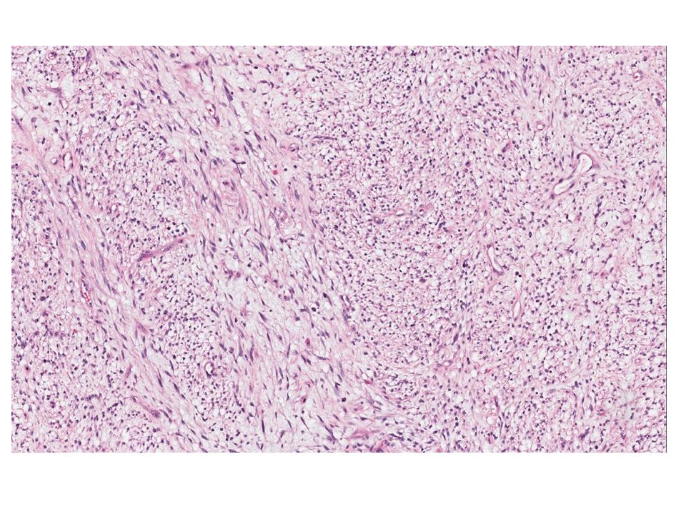
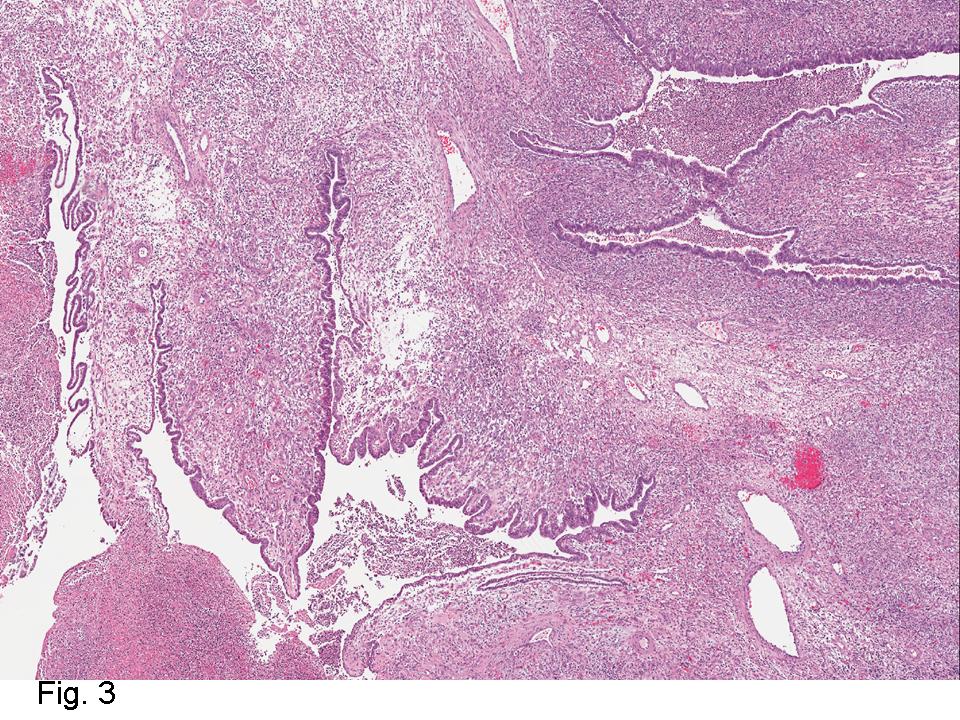
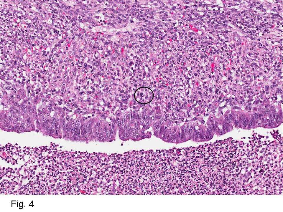
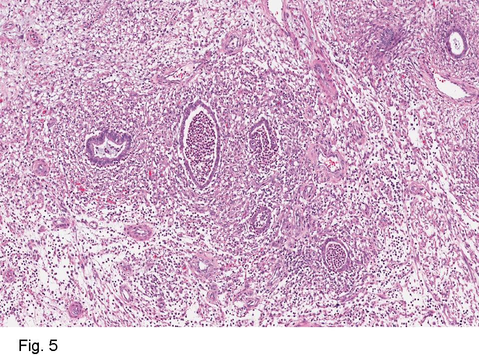
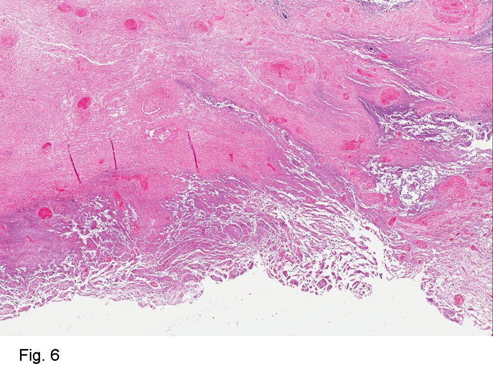
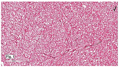
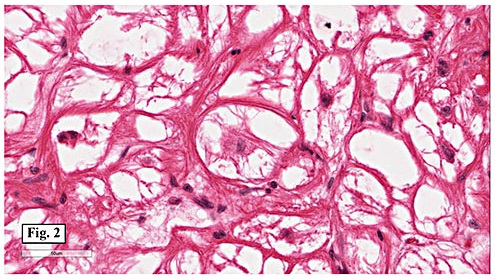
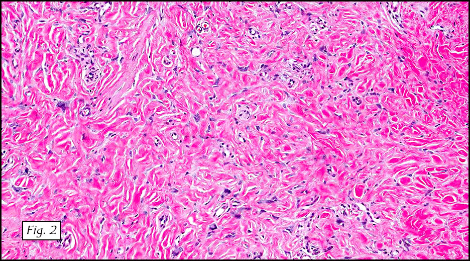
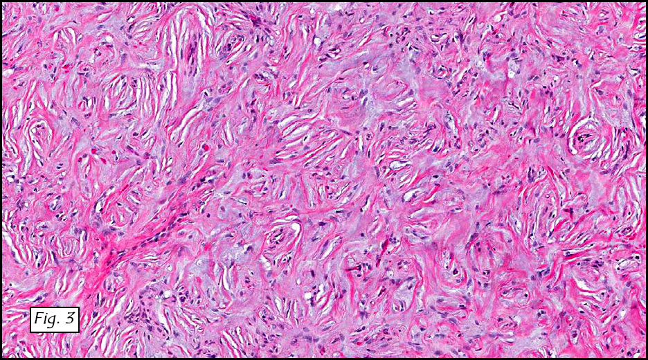
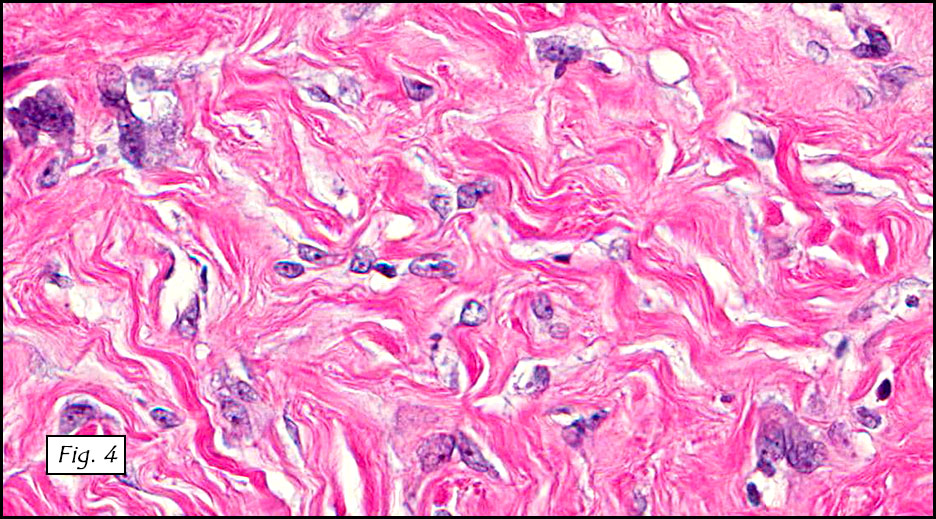
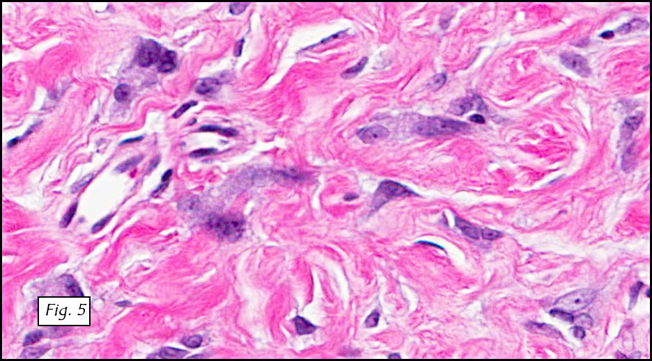
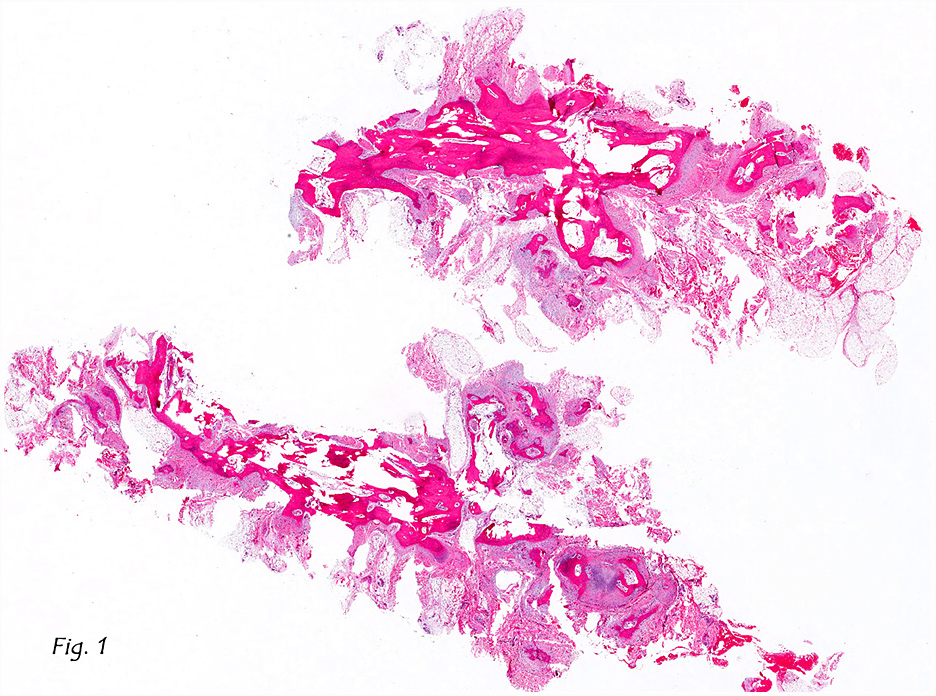
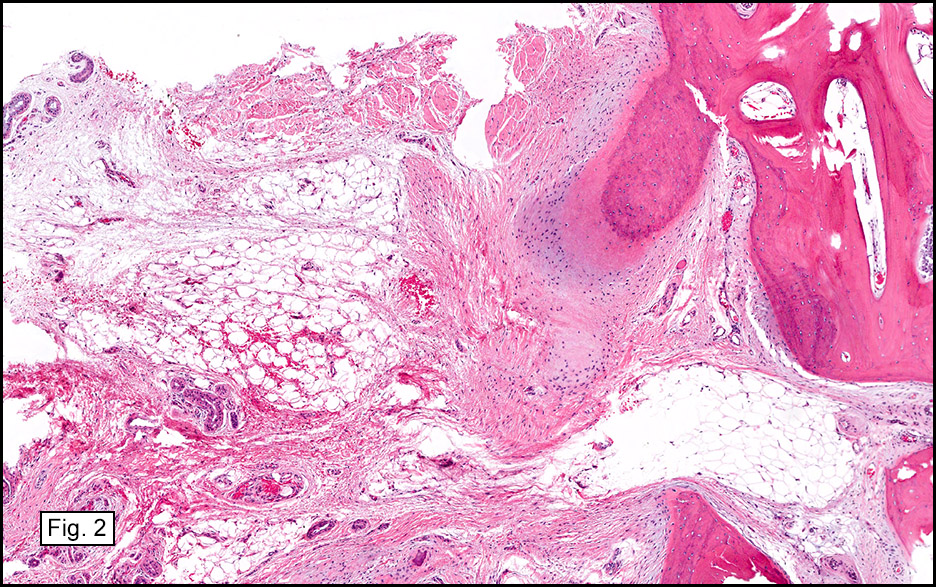
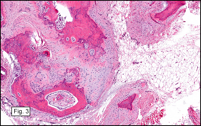
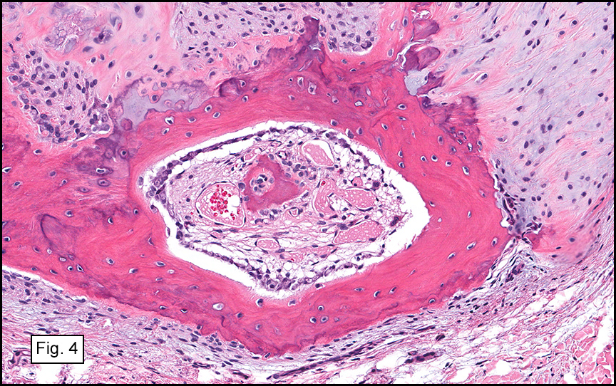
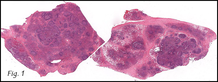
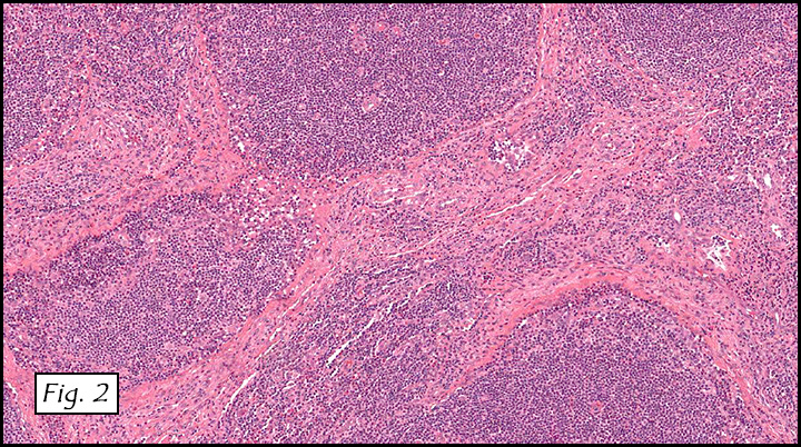
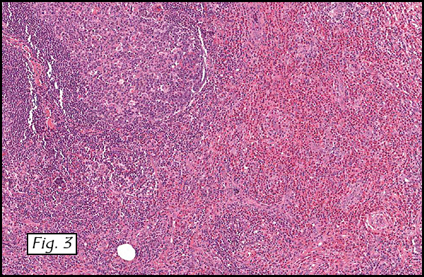
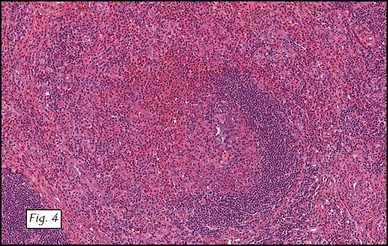
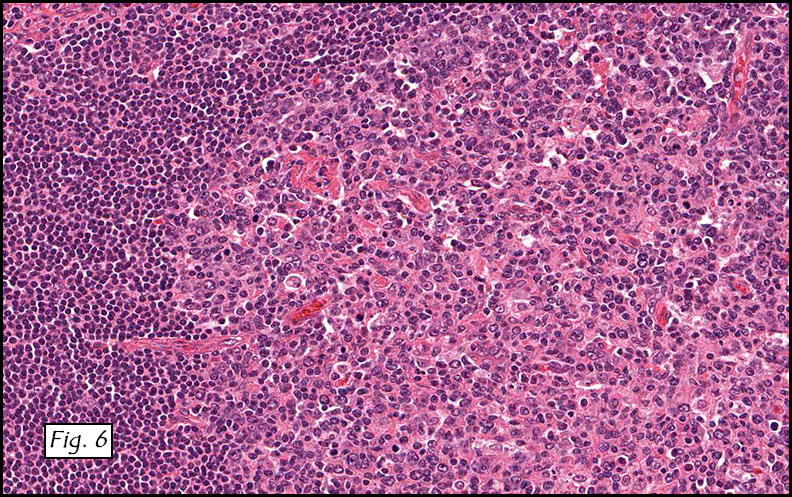
Gross examination of the resection specimens demonstrated a 10 cm mass that is adherent to the sigmoid colon mass. Also received was a 3 cm aggregate of fragments of tumor designated as pelvic tumor. Microscopically, the tumor comprised large expanses of a spindled mesenchymal neoplasm with relatively monotonous nuclear features, moderate atypia, and indistinct cytoplasm (Fig.1). Occasional areas showing true epithelial gland formation were present, with surrounding hypercellular condensations of the atypical mesenchymal elements (Fig.2). There was prominent stromal overgrowth, resulted in polypoid epithelial projections forming “leaf-like’ low-power appearance (Fig.3). Mitotic figures were easily identified (up to 8 per 10 HPF) (Fig.4). Foci of endometriosis in the bowel serosa were also present (Fig.5). The overlying bowel mucosa was ulcerated, but was without dysplasia (Fig.6).

Diagnosis:

Extrauterine Mullerian Adenosarcoma with Sarcomatous overgrowth
Suneetha Chintalapati, M.D., Donald R. Chase, M.D. and Jeremy K Deisch, M.D.
Department of Pathology and Human Anatomy
Loma Linda University Medical Center, Loma Linda, California
California Tumor Tissue Registry, Loma Linda, California

Discussion: Mullerian Adenosarcoma is an uncommon mixed epithelial-mesenchymal tumor of low-grade malignancy that affects the female genital tract. Mullerian adenosarcoma is regarded as a lesion with intermediate pathologic features and prognosis, lying between benign adenofibroma and carcinosarcoma.
Extrauterine Mullerian adenosarcoma is very rare, and is presumed to arise in foci of endometriosis, given its frequent co-existence (as seen in this case). Extrauterine examples are more aggressive than uterine tumors, due to ease of access to the peritoneal cavity, as it is not retarded by the thick myometrial wall. The recurrence rate following surgery is higher. Extrauterine Mullerian adenosarcomas are the second most common malignancy arising in extraovarian endometriosis, following clear cell carcinoma. Exogenous or endogenous estrogen may have some role in the transformation process, whereas the effectiveness of combined estrogen–progesterone hormone replacement therapy is still to be determined.

There are currently two recognized sets of criteria for the pathologic diagnosis of malignant transformation of endometriosis. Sampson’s criteria for malignant transformation of endometriosis requires that there is a focus of endometriosis in close proximity to malignancy, that the tumor shows a histological appearance compatible with endometrial origin, and that that other primaries are appropriately excluded. The criteria set forth by Clement and Scully requires a mitotic count of two or more per ten high-power fields, the presence of stromal cellular periglandular cuffs, intraglandular protrusion of cellular stroma, and at least a moderate degree of nuclear atypia of the mesenchymal elements.

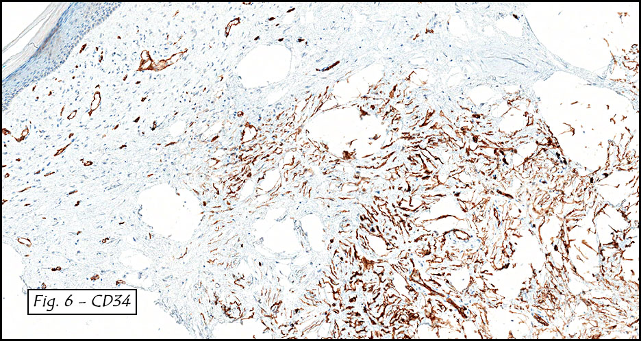
Pathologically, Mullerian adenosarcomas are biphasic tumors, in which there are glands lined by benign or mildly atypical Mullerian-type epithelium, intimately mixed with a proliferating mesenchymal component that is a low grade sarcoma, with cellularity more pronounced at the periphery of the glands, forming periglandular cuffs. The immunoprofile is similar to endometrial stromal sarcoma: CD10, WT1, ER and PR are usually positive.
The immunoreactivity for adenosarcomas is similar to that of adenofibromas, which are associated with favorable outcome, as well as other benign lesions such as endometrial polyps and endometriosis. The significant histologic and immunophenotypic overlap between adenosarcomas and adenofibromas suggest that some of the tumors currently classified as adenofibromas, on the basis of their low mitotic count and lack of significant nuclear atypic, are, in fact, well-differentiated adenosarcomas.

Adenosarcomas with sarcomatous overgrowth differ histologically and prognostically from typical adenosarcomas. Sarcomatous overgrowth requires the presence of stromal predominance be present in at least 25% of the tumor volume. In tumors with sarcomatous overgrowth, the mortality rate is as high as 75%, with a similar malignant potential to high grade uterine sarcomas. These tumors may show a high proliferative index by Ki-67 immunostaining, and may show strong p53 staining and loss of CD10 and progesterone receptor immunoexpression.

Malignant transformation of gastrointestinal endometriosis is rare, but when it occurs, it predominantly affects postmenopausal patients. Recognition of these lesions is important as primary gastrointestinal neoplasms are managed differently than those arising in endometriosis. An adenosarcoma arising from endometriosis should be considered in the differential diagnosis of a pelvic mass, even one appearing in the colon wall, because ectopic endometrial tissue has the potential to exist throughout the peritoneal cavity.

Suggested Reading:

Atlas of Gynecologic Surgical Pathology – Clement, Young, 2014, third edition, 256-259.

Clement PB, Scully RE. Extrauterine mesodermal (mullerian) adenosarcoma: A clinicopathologic analysis of five cases. Am J Clin Pathol 1978;69:276–83.

Mullerian adenosarcoma arising from rectal endometriosis, Chunseok Yang etal, Ann Coloproctol. Oct 2014, 30(5): 232-236.

Mullerian Adenosarcoma of the uterus with sarcomatous overgrowth, reecha singh et al, Clin Med Insights case rep. 2010; 3:27-30.

Neoplastic and Pre-neoplastic changes in gastrointestinal endometriosis; Rhonda k. yantiss metal; Am J Surg Pathol 24(4): 513-524, 2000.

Rosai and Ackerman’s Surgical Pathology, 2011, Tenth edition, Volume 2, 1507-1508.

August 2015: A five year old girl with a cardiac conduction problem

History: A 5-year-old girl presented with cardiac conduction problem. Workup revealed a mass in the right atrium.

Microscopically, the mass consisted of enlarged myocytes with abundant, vacuolated or clear cytoplasm (Fig. 1). At higher magnification, the tumor cells had strands of cytoplasmic myofibrils radiating from the central nucleus to the cell periphery forming so-called “spider cells” (Fig. 2). Cross striations were commonly seen (Fig. 3). Neither necrosis nor mitotic figures were present.

Diagnosis: Rhabdomyoma of the atrium

Li Lei, M.D. and Donald R. Chase, M.D.
Department of Pathology and Human Anatomy
Loma Linda University Medical Center, Loma Linda, California
California Tumor Tissue Registry, Loma Linda, California

Discussion: Primary cardiac tumors are rare. In the young pediatric group, the commonest cardiac tumors are rhabdomyomas (45-63%), followed by fibromas (20%) and myxomas (10-15%); while in teenagers and adults, myxomas account for about two-thirds of primary cardiac tumors.

As its name implies, rhabdomyoma is a benign tumor of striated muscle differentiation. It is considered a hamartoma or malformation other than a true neoplasm, largely because the tumor cells have a very low proliferative index … essentially, they do not proliferate.

Cardiac rhabdomyoma almost exclusively affects infants and young children. About 75% of cases occur before the age of one year. It can occasionally be seen as early as 20 weeks gestational age. Arrhythmia is the most common presentation, though many patients are asymptomatic. Lethal arrhythmia or valvular orifice obstruction can cause sudden cardiac death.

Approximately 51-88% of cardiac rhabdomyomas are associated with tuberous sclerosis, and it may be the earliest sign of tuberous sclerosis in a family. On the other hand, about 43-72% of children with tuberous sclerosis have or will develop cardiac rhabdomyomas. A minority of cases are sporadic. Occasionally, association with folliculin mutation and Birt Hogg Dube syndrome has been reported.

Cardiac rhabdomyomas are often multiple, particularly those associated with tuberous sclerosis. Up to 10% of cases are solitary. It typically presents as intramural lesion in the ventricles, but may protrude into the ventricular cavity as a pedunculated or sessile mass. Interventricular septum and atria can also be involved. Macroscopically, it is well-circumscribed but nonencapsulated, with a homogeneous whitish or grayish cut surface.

Histologically, the cells are big and polygonal. Large cytoplasmic vacuoles are separated by strands of cytoplasm extending radially between nucleus and cell membrane, giving a characteristic “spider cell” appearance. The vacuoles are rich in glycogen which is PAS positive and diastase sensitive. Cross-striations are also routinely seen. Mitotic figures and necrosis are absent.
Ultrastructurally, the cells have abundant glycogen and rare, small mitochondria. Cellular junctions resembling intercalated disks are all around the periphery instead of exclusively at the cell poles as seen in normal cardiomyocytes.

Immunohistochemically, the tumor cells express muscle specific actin (MSA), myoglobin, desmin and vimentin. As with other tumors associated with tuberous sclerosis, cardiac rhabdomyoma is often positive for HMB45 but is usually negative for S100.

The differential diagnosis of cardiac rhabdomyoma includes:

• Histiocytoid cardiomyopathy is extremely rare in patients over the age of 2 years. It presents as subendocardial yellowish nodules or plaques. The large polygonal cells have coarsely granular, foamy to pale eosinophilic cytoplasm resembling histiocytes. They show cholinesterase immunoreactivity but do not express histiocytic antigen(s). The granules are bizarre mitochondria under electron microscope.
• Cardiac hibernoma consists of granular to multivacuolated adipocytes, which are typically S100 positive. The cytoplasmic vacuoles are small and stain for neutral fat. “Spider cells” and cross-striations are lacking.
• Cardiac myxoma often occurs on the left side of the atrial septum. It arises from endocardium as a polypoid mass and does not involve underlying myocardium. Cut surface is gelatinous. Perivascular cuffing by tumor cells within myxoid stroma is commonly seen.
• Cardiac rhabdomyosarcoma is anecdotal in children, with only a few cases reported in the literature. Invasive biologic behavior and malignant histologic features help differentiate. Extensive clinical workup excluding metastasis is necessary before rendering the diagnosis.

Cardiac rhabdomyoma has a tendency to regress spontaneously over time, especially throughout the first year. Approximately 54-100% of cases undergo complete or partial regression. Surgery is therefore reserved for seriously symptomatic patients. If the risk of radical resection outweighs the benefit, a partial resection may be satisfactory given the regressing potential of cardiac rhabdomyoma. In inoperable cases, mammalian target of rapamycin (mTOR) inhibitors such as everolimus and sirolimus have been tried and appear promising.

Suggested Reading:

Goldblum J, Folpe A, Weiss S. Enzinger & Weiss’ Soft Tissue Tumors, 6th ed: Philadelphia, Elsevier Inc, 2014; 591-2.
Suvarna SK. Cardiac Pathology: A Guide to Current Practice. Springer 2013; 201-21.
Basso C, Valente M, Thiene G. Cardiac tumor pathology. Springer 2013; 59-62.
Burke A, Virmani R. Pediatric heart tumors. Cardiovasc Pathol. 2008;17:193-8.
Bondavalli D, White SM, Steer A, Pflaumer A, Winship I. Is cardiac rhabdomyoma a feature of Birt Hogg Dubé syndrome? Am J Med Genet A. 2015;167A:802-4.

June 2015: A man with a 2.3 cm nodule on the right shin

History: A 37-year-old man presented with a well circumscribed, unencapsulated, bluish pink 2.3 x 2.3 cm nodule on his right shin. A 2.0 x 2.0 cm shave biopsy was taken (Fig. 1).

Microscopically, the tumor was dermal-based and involved adnexal structures. It consisted of disorganized collagenous fibers (Fig. 2) admixed with scattered hyperchromatic pleomorphic cells. Myxoid stromal change and storiform patterns were also seen (Fig. 3).

The pleomorphic cells showed enlarged, spindled to stellate-shaped nuclei with small basophilic nucleoli (Fig. 4). These atypical cells showed marked variation in nuclear size and shape. Generally they had indistinct cytoplasmic borders. Many of the more bizarre cells showed enlarged hyperchromatic nuclei (Fig. 5). Multinucleated giant cells with jumbled nuclei were occasionally present. Mast cells, adipocytes, hemosiderin or other pigments were absent. Neither necrosis nor mitotic figures were identified.

A CD34 stain highlighted the atypical cells, stroma and blood vessels (Fig. 6).

Diagnosis: Pleomorphic Fibroma of the Skin

Li Lei, M.D., Ph.D., Camilla Cobb, M.D. and Donald R. Chase, M.D.
Department of Pathology and Human Anatomy
Loma Linda University Medical Center, Loma Linda, California
California Tumor Tissue Registry, Loma Linda, California

Discussion: Pleomorphic fibroma (PF) of the skin, as its name implies, is a cutaneous lesion characterized by pleomorphic cells in a fibrotic stroma. A lesion of similar morphology may be seen in tendons and has been termed “PF of tendon sheath”. PF was first described in 1989 by Dr. Kamino et al. Since then only a handful of cases have been reported. The incidence and prevalence of PF are unclear.

Clinically, PF affects adults in a wide age range (27-75 years) with a median of 50 years. There is a slight predilection for females. No racial difference has been documented. Patients often present with a slowly-growing, solitary, cutaneous nodule usually involving the extremities, followed by the trunk and the head and neck. It is usually asymptomatic but may occasionally be associated with dull pain and/or pruritus. On physical examination, the nodule is small (< 2 cm), polypoid or sessile, and indurated. It is well-circumscribed but non-encapsulated, with a whitish solid firm cut surface. Histologically, PF is a hypocellular dermal lesion composed predominantly of dense, haphazardly arranged collagen. Interspersed between the collagen fibers are spindle-shaped or stellate cells and focal multinucleated giant cells. The most characteristic feature is degenerative nuclear atypia, presented as large, bizarre, hyperchromatic nuclei with smudged chromatin and small nucleoli. Mitotic figures are rare or absent. Necrosis is not seen. There is minimal inflammatory infiltrate. Many times the stroma is densely sclerotic, in fact PF may intimately coexist with sclerotic fibroma with a thin transitional area in between. Because of this, the term “Pleomorphic Sclerotic Fibroma” has been suggested as an alternative term which encompasses a proposed spectrum. It is not uncommon for PF to show focal or diffuse myxoid stromal changes. Although these changes usually lack clinical significance, Dr. Weedon and colleagues reported a case of myxoid PF which transformed into a grade III myxofibrosarcoma. Immunohistochemically, the cells are strongly positive for vimentin, and variably positive for CD34, smooth muscle actin, muscle-specific actin and CD99. They are negative for CD31, desmin, S-100, CD68 and cytokeratin. The differential diagnosis of PF includes: • Atypical fibroxanthoma: This tumor almost always involves sun damaged skin of the elderly and grows rapidly. Histologically, it is hypercellular and composed of spindled to bizarre cells and xanthoma cells. Mitotic figures are brisk and can be atypical. CD10 positivity is seen in 95% of cases. Approximately half of the cases are CD68 positive. • Dermatofibroma with atypical cells is usually more cellular than PF. Atypical cells are intermixed with spindle cells and histiocyte-like cells. Hemosiderin and siderophages are often present. It is typically CD34 negative. Another helpful tool is pan-monocytic/macrophage marker Ki-M1p which is positive in dermatofibroma with atypical cells but negative in PF. • Giant cell fibroblastoma usually affects children. The characteristic feature is pseudovascular spaces that are lined by a discontinuous row of CD34 positive atypical spindle cells and floret-like giant cells. Cytogenetic studies show supernumerary ring chromosome derived from t (17; 22). • Pleomorphic hyalinizing angiectatic tumor can be distinguished by clusters of ectatic thin-walled vessels with fibrin deposition. Pleomorphic stromal cells often contain intracytoplasmic hemosiderin and/or intranuclear pseudoinclusion. A mixed inflammatory infiltrate is usually present in the background. • Atypical lipomatous tumor (ALT): Normal adipose tissue may occasionally get entrapped in PF, causing diagnostic confusion with dermal ALT. At the molecular level, PF lacks MDM2 amplification by fluorescent in situ hybridization as well as MDM2 protein expression by immunohistochemistry, whereas MDM2 amplification is 93.5% sensitive for diagnosing ALT. Interestingly, Dr. Rosai and Dr. Aguilar reported a case with PF in the dermis and ALT in the contiguous subcutis. Whether or not this is purely coincidental is unclear. • Pleomorphic lipoma: The typical locations include posterior neck, shoulder and back. The bizarre cells display a wreath-like arrangement of nuclei around deeply eosinophilic cytoplasm, giving them a floret-like appearance. Mast cells are often present. CD 34 is typically positive. Cytogenetic studies show loss of 16q or 13q. • Ancient schwannoma is usually large and located in deep structures such as the retroperitoneum. Besides nuclear atypia, other degenerative changes including cyst formation, calcification, hemorrhage and hyalinization are also present. Immunostaining for S-100 highlights most of tumor cells. CD34 marks only blood vessels. • Neurofibroma with nuclear atypia shows isolated mild nuclear. It is usually more cellular than PF and composed of polymorphous proliferation. Collagen fibers have a “shredded carrots” appearance. Mast cells, and occasionally pseudomeissnerian bodies, are present. Immunostaining for S-100 picks up a subset of cells. • Spindle cell/sarcomatoid squamous cell carcinoma typically occurs on sun-damaged skin of elderly men as a raised, ulcerated nodule. Keratin pearls, connection to epidermis and epidermal lesion are discriminating morphological features. Strong immunoreactivities for both p63 and high molecular weight cytokeratins confirm keratinocytic derivation of tumor cells. • Desmoplastic melanoma is often deceptively amelanotic with prominent interstitial fibrosis. However, it has a predilection for sun-damaged skin of the elderly, particularly head and neck areas. Morphologically, it can be distinguished by neurotropism, nodular lymphocytic aggregates, lymphovascular invasion and in situ components such as lentigo maligna. Immunohistochemically, though negative for HMB45 and Melan A, it is positive for S-100 and SOX10. • Desmoplastic fibroblastoma, sometimes referred to as collagenous fibroma, usually arises in deep subcutaneous tissue or skeletal muscle. The scattered spindle or stellate-shaped cells lack atypia and CD34 expression. The etiology of PF remains unknown. Patients usually report no history of trauma or other stimuli to the area. The immunoprofile of PF suggests origin from myofibroblasts and/or dermal dendrocytes. Cytogenetic and molecular studies are currently sparse. Nuclear atypia, in the absence of other evidence suggestive of malignancy, is considered a degenerative phenomenon that can be seen in many benign neoplasms including some of the aforementioned as well as non-neoplastic entities such as ischemic fasciitis. It is important to recognize that degenerative atypia does not adversely affect the prognosis. Therefore, over-treatment should be avoided. Simple excision is usually curative for PF, with local recurrences rarely seen in incompletely excised lesions. To the best of our knowledge, metastasis has never been reported. Suggested Reading:
Goldblum J, Folpe A, Weiss S. Enzinger & Weiss’ Soft Tissue Tumors, 6th ed: Philadelphia, Elsevier Inc, 2014; 216-218.
Brenn T. Pleomorphic dermal neoplasms: a review. Adv Anat Pathol. 2014;21:108-30.
Al-Zaid T, Wang WL, Lopez-Terrada D, Lev D, Hornick JL, Hafeez Diwan A, Fletcher CD, Lazar AJ. Pleomorphic fibroma and dermal atypical lipomatous tumor: are they related? J Cutan Pathol. 2013;40:379-84.
Aguilar C, Rosai J. Pleomorphic fibroma of the skin, atypical lipomatous tumor, or both? Int J Surg Pathol. 2011;19:63.
Dore A, Robertson I, Williamson R, Weedon D. Progression of a myxoid pleomorphic fibroma to myxofibrosarcoma. Australas J Dermatol. 2003;44:287-90.
Rudolph P, Schubert C, Zelger BG, Zelger B, Parwaresch R. Differential expression of CD34 and Ki-M1p in pleomorphic fibroma and dermatofibroma with monster cells. Am J Dermatopathol. 1999;21:414-9.

May 2015: An adult male with a subcutaneous soft tissue mass involving a heel

History: A painful, one cm nodule was found the subcutaneous tissue of the right heel of an adult male. Radiographs showed it to be ill-defined and radio-opaque. The nearest bone was the calcaneus, but it was not involved. There was no history of trauma.

The lesion, at low magnification, consisted of an admixture of adipose tissue, osteoid, trabecular mature bone, and fibrous stroma (Figs. 1, 2). Prominent osteoid, bone and abortive cartilaginous elements were present (Fig. 3). Zones of osteoid were surrounded by cellular or myxoid fibrous stroma and were sometimes rimmed by osteoblasts (Fig. 4).

Diagnosis: Panniculitis Ossificans, heel

Bing Wang, M.D., and Donald R. Chase, M.D.
Department of Pathology and Human Anatomy
Loma Linda University Medical Center, Loma Linda, California
California Tumor Tissue Registry, Loma Linda, California

Discussion: Panniculitis Ossificans (PO) is also referred to as a “fibro-osseous lesion” or “heterotopic ossification”. It has been subclassified based upon its location [1]:

Unlike usual panniculitis, which may show inflammatory infiltrates involving the septa [4], PO commonly lacks inflammation. If inflammation is present it is usually minimal and mostly evident in the tissue surrounding the lesion [2].

PO usually affects young, physically active adolescents and adults. Most cases involve the limbs, and some are seemingly incited by injury. The initial symptoms may include pain or tenderness followed by soft tissue swelling. Later, the swollen tissue becomes circumscribed and indurated. Calcification usually starts three to six weeks following injury [2].

Radiographs show a series of changes, initially with an increase in soft tissue density or opacity. Calcification starts around the end of third week, and as the lesion becomes increasingly calcified, it becomes well-circumscribed with either a solitary region of calcification or scattered regions of calcification [2].

Histologically, PO is usually well-circumscribed and is characterized by the presence of a distinct zonal pattern that reflects different components of this entity, including: normal-appearing adipose tissue, fibrous stroma, osteoid and mature bone. The adipose tissue consists of normal lobules of mature fat. Fat necrosis usually is not seen. Foci of osteoid formation are scattered between the lobules of adipose tissue, and are usually surrounded by a cellular or myxoid fibrous stroma. Trabeculae of mature bone usually begins in the middle of osteoid islands. The content of the mature bone formation depends on the stage of the lesion. Sometimes osteoid and mature bones are rimmed by layers of osteoblasts showing little variation in size and shape. The osteoid and bone are separated from the surrounding tissue by a zone of loose or compressed fibrous tissue. The fibrous tissue consists of fibroblasts and myofibroblasts which may display a mild degree of cellular pleomorphism and rather prominent mitotic activity. In addition, there may be prominent vascular proliferation, focal hemorrhage, or fibrin deposition [2].

Panniculitis ossificans is a benign lesion and its etiology is not fully understood. It is usually caused by some kind of injury. It is proposed that injuries may generate an environment which produces certain proteins such as bone morphogenic protein, which in turn, can induce pluripotential mesenchymal cells to turn into osteogenic cells, which can then form osteoid, which in turn develops into mature heterotopic ossification [3].

There is no convincing evidence of malignant transformation of panniculitis ossificans [2]. The early stage of PO has immature and highly cellular zones that may be confused with extraskeletal osteosarcoma. However, extraskeletal osteosarcoma usually occurs in older patients; in contrast panniculitis ossificans often occurs in younger active people. Unlike panniculitis ossificans, extraskeletal osteosarcoma shows marked cellular atypia and infiltration of neighboring tissues in a destructive manner. Panniculitis ossificans is well-circumscribed with distinct zones of different components. In some cases, PO may contain entrapped atrophic muscle, which may cause confusion with another entity in the same category, myositis ossificans. The difference lies in how much muscle or adipose tissue is present in the lesion [2].

Suggested Reading:
1. Ackerman LV. Extra-osseous localized non-neoplastic bone and cartilage formation (so – called myositis Ossificans): clinical and pathological confusion with malignant neoplasms. J Bone Joint Surg Am 1958; 40-A(2): 279-298.
2. Goldblum JR, Folpe AL and Weiss SW. Soft tissue tumors, sixth edition. Pg 926-932.
3. McCarthy EF, Sundaram M: Heterotopic ossification: a review. Skeletal Radiol.34:609-619 2005.
4. Requena L. Normal subcutaneous fat, necrosis of adipocytes and classification of the panniculitides. Semin Cutan Med Surg 2007; 26:66–70.

April 2015: A 32 year old man with a soft tissue mass adjacent to the right humerus

History: A 32-year-old man presented with a mass in the right arm. It involved the humerus and upon excision was 8.2 x 6.5 x 3.5 cm and weighed 52 grams. It consisted mostly of fatty tissue but had a 3.0 x 2.5 cm, vaguely circumscribed nodule centrally located.

H&E staining of the central lesion revealed marked hyperplasia of lymphoid follicles with expanded germinal centers and well-formed mantle zones (Fig. 1). Some of the follicles had intervening fibrosis (Fig. 2). Germinal centers contained lymphocytes admixed with dendritic cells, polykaryocytes and eosinophilic proteinaceous deposits. Eosinophils were widely interspersed between the follicles with focal micro-abscess formation (Fig. 3), or occasionally infiltrated germinal centers with resultant follicular lysis (Fig. 4). The lesion was well-vascularized. Numerous post-capillary venules lined by flat endothelial cells were present between follicles and within germinal centers (Figs. 5, 6). Epithelioid endothelial cells were not seen.

Diagnosis: Kimura Disease, arm

Li Lei, M.D., Ph.D. and Donald R. Chase, M.D.
Department of Pathology and Human Anatomy
Loma Linda University Medical Center, Loma Linda, California
California Tumor Tissue Registry, Loma Linda, California

Discussion: Kimura disease (KD), named after Kimura for recognizing the vascular component of the entity in 1948, was first described in China in 1937 by Kim and Szeto as eosinophilic hyperplastic lymphogranuloma. It is a chronic inflammatory process characterized by benign reactive lymphoid proliferation with significant eosinophilia, secondary vascular proliferation and fibrosis.

KD is endemic in Asia, particularly in East and Southeast Asia. Other racial groups including Caucasian, African American and Hispanic may also be targeted. There is a striking male predominance with reported male-to-female ratio ranging from 3.5:1 to 19:1. Most of the affected patients are in their twenties to forties.
KD typically presents as a deep subcutaneous mass measuring 2 – 6 cm in size, but giant tumors up to 13 cm have been reported. Commonly it presents as cervical lymphadenopathy with a classic example being a peri-auricular mass involving the parotid gland. Less common sites include the extremities (like our case), axilla, groin, epiglottis, chest wall, spermatic cord and peripheral nerve.

Laboratory tests usually show peripheral blood eosinophilia and elevated serum immunoglobulin (IgE). Imaging is usually nonspecific, but sometimes may be seen as multiple ill-defined, enhancing lesions around the parotid gland with associated lymphadenopathy.
Clinically, up to 16% of cases are associated with proteinuria, nephrotic syndrome, membranous glomerulonephritis, bronchial asthma, ulcerative colitis or necrotizing eosinophilic vasculitis. Patients are otherwise asymptomatic.

Histologically, the tumor consists of dense lymphoid aggregates with prominent germinal centers, which contain IgE-bearing dendritic reticulum cells, polykaryocytes (Warthin-Finkeldey type), nuclear debris and a delicate eosinophilic matrix. Interfollicular dense eosinophilic infiltrate is a common feature and may form microabscesses. Plasma cells, small lymphocytes and mast cells are often increased in the paracortex. There is a moderate proliferation of postcapillary venules which are lined by flat endothelial cells. Long-standing lesions usually develop hyaline fibrosis and become less vascular. Affected lymph nodes demonstrate same histologic features with preserved architecture.
The differential diagnosis prominently includes “Epithelioid hemangioma” (EH), also known as “angiolymphoid hyperplasia with eosinophilia”, a benign vascular neoplasm which shares many features of KD. Both entities affect young to middle-aged adults, presenting in the head and neck. Both have an inflammatory component, a vascular component and increased numbers of eosinophils, but there are differences:

Despite the above differences (table) these two entities appear intertwined. Some lesions have characteristic features of both making specific classification almost impossible. Moreover, KD and EH can coexist in the same patient. Patients with one entity can develop the other during follow-up. These phenomena suggest that EH and KD may be a part of a spectrum of reactive vascular lymphoid proliferations.

In addition to EH, other differentials of KD include:

• Angioimmunoblastic T cell lymphoma at its early phase can have marked follicular hyperplasia with germinal centers, eosinophilia and prominent high endothelial venules. However, patients are often older and present with fever and generalized lymphadenopathy. Lymph node architecture is usually effaced. Perivascular clustering of neoplastic clear cells and increased follicular dendritic cell meshworks are helpful clues. Molecular study shows monoclonal T cell receptor gene rearrangements in the majority of the cases or IgH rearrangements in the minority.
• Langerhans cell histiocytosis can present as indolent lymphadenopathy in young adults. Microscopically, lymph node architecture is effaced by mixed infiltrates of Langerhans cells and eosinophils. CD1a and S100 immunoreactivity help to confirm the diagnosis.
• Follicular lymphoma: Primary cutaneous follicular lymphoma generally affects the elderly. Follicles consist of predominantly centrocytes with admixed centroblasts. In cases of secondary cutaneous involvement by follicular lymphoma, a pertinent medical history, back-to-back follicles with no cell polarization and t(14;18) translocation help reach the correct diagnosis.
• Hodgkin lymphoma has a bimodal age distribution with the first peak at 15-35 years of age. Lymph nodes have a “fish-flesh” cut surface. Despite lymphocytes, eosinophils, and collagen fibers in the background, CD30 and CD15 positive Hodgkin Reed-Sternberg cells are pathognomonic.

The etiology of KD remains unknown. Multiple clues such as elevated serum IgE, peripheral eosinophilia and association with other autoimmune diseases favor immune system hypersensitivity as the underlying pathophysiology. The endemic epidemiology of KD may reflect either inherent genetic susceptibility or an antigenic trigger unique to the geographic area.
Patients with KD normally have a favorable outcome. If untreated, the mass may remain stable or spontaneously regress. The standard care is simple surgical excision. Other options include laser therapy, radiotherapy and systemic corticosteroids. Local recurrence rate vary from 14 – 44%. Characteristics which may predict recurrence include: size ≥ 3.5 cm, peripheral eosinophilia ≥35%, Notch-1 upregulation and Ki-67 proliferation index ≥ 3% outside the germinal center. Metastases are very rare and therefore anecdotal.

Suggested Reading:
Goldblum J, Folpe A, Weiss S. Enzinger & Weiss’ Soft Tissue Tumors, 6th ed: Philadelphia, Elsevier Inc, 2014; 649-654.
Rosai J. Rosai and Ackerman’s Surgical Pathology, 10th ed: Philadelphia, Elsevier Inc, 2011; 1805.
Kung IT, Gibson JB, Bannatyne PM. Kimura’s disease: a clinico-pathological study of 21 cases and its distinction from angiolymphoid hyperplasia with eosinophilia. Pathology. 1984;16:39-44.
Deng WY, Ye SB, Luo RZ, Yan SM, Gao YF, Yang YZ, Guo ZM, Chen YF. Notch-1 and Ki-67 receptor as predictors for the recurrence and prognosis of Kimura’s disease. Int J Clin Exp Pathol. 2014;7: 2402-10.
Buder K, Ruppert S, Trautmann A, Bröcker EB, Goebeler M, Kerstan A. Angiolymphoid hyperplasia with eosinophilia and Kimura’s disease – a clinical and histopathological comparison. J Dtsch Dermatol Ges. 2014;12:224-8.
Liu XK, Ren J, Wang XH, Li XS, Zhang HP, Zeng K. Angiolymphoid hyperplasia with eosinophilia and Kimura’s disease coexisting in the same patient: evidence for a spectrum of disease. Australas J Dermatol. 2012;53:e47-50

Scroll to top