January 2011: A 16 year old young woman with a thigh mass

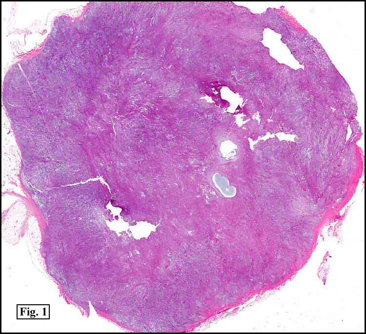
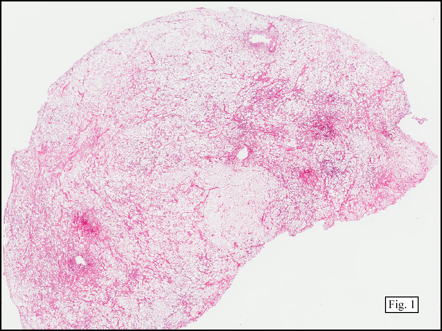
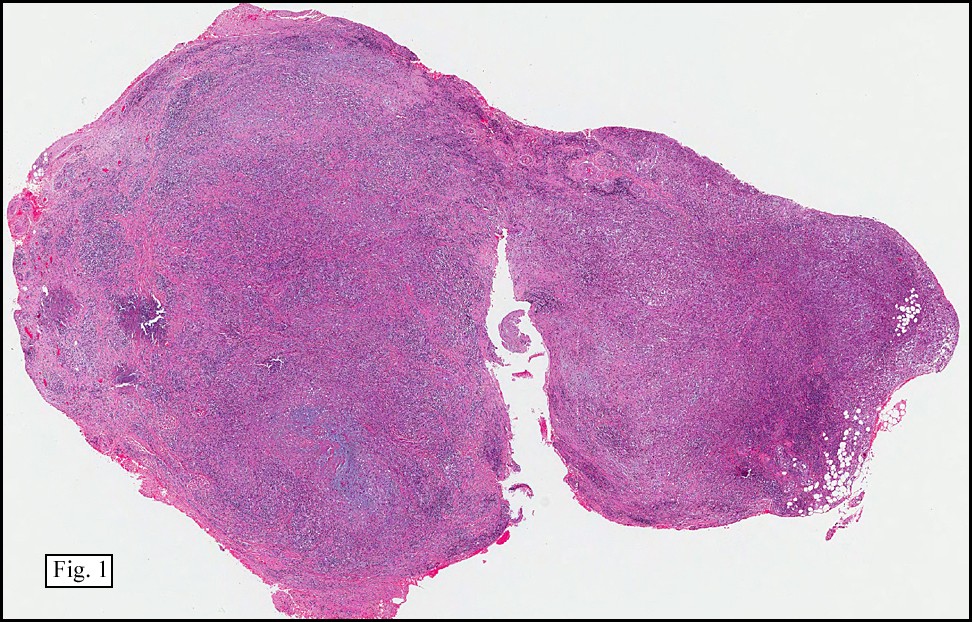
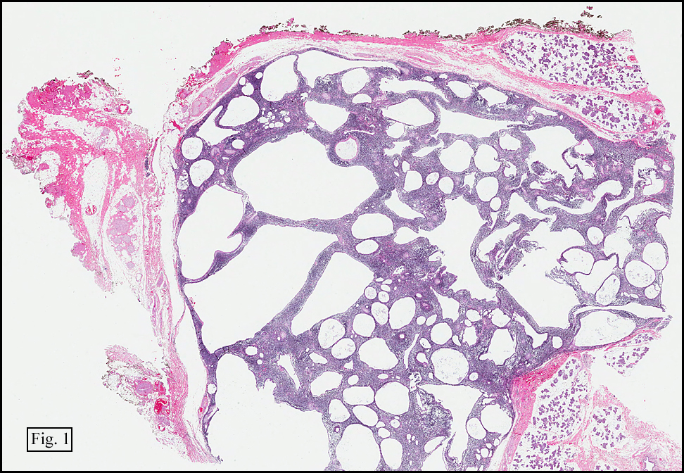
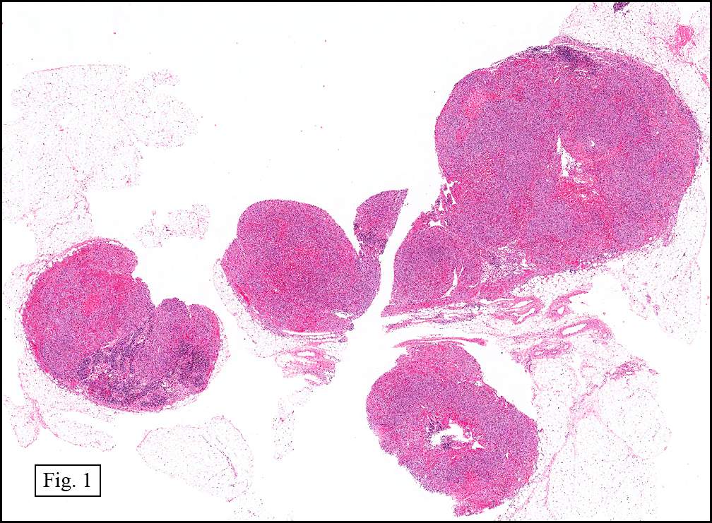
History: A 16-year-old young woman presented with a six month history of a posterior left thigh nodule. The mass was associated with mild pain and swelling. Surgery revealed a 3 cm well-defined and partially encapsulated nodule without apparent involvement of adjacent muscle (Fig. 1).

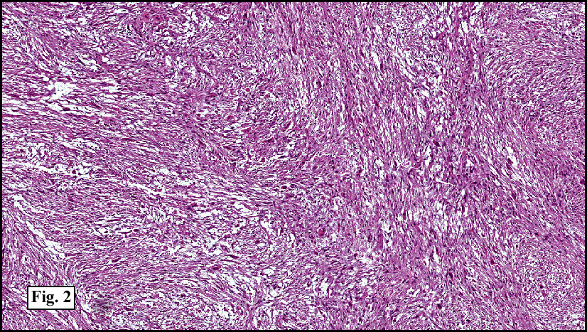
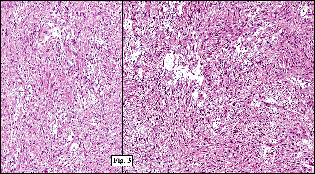
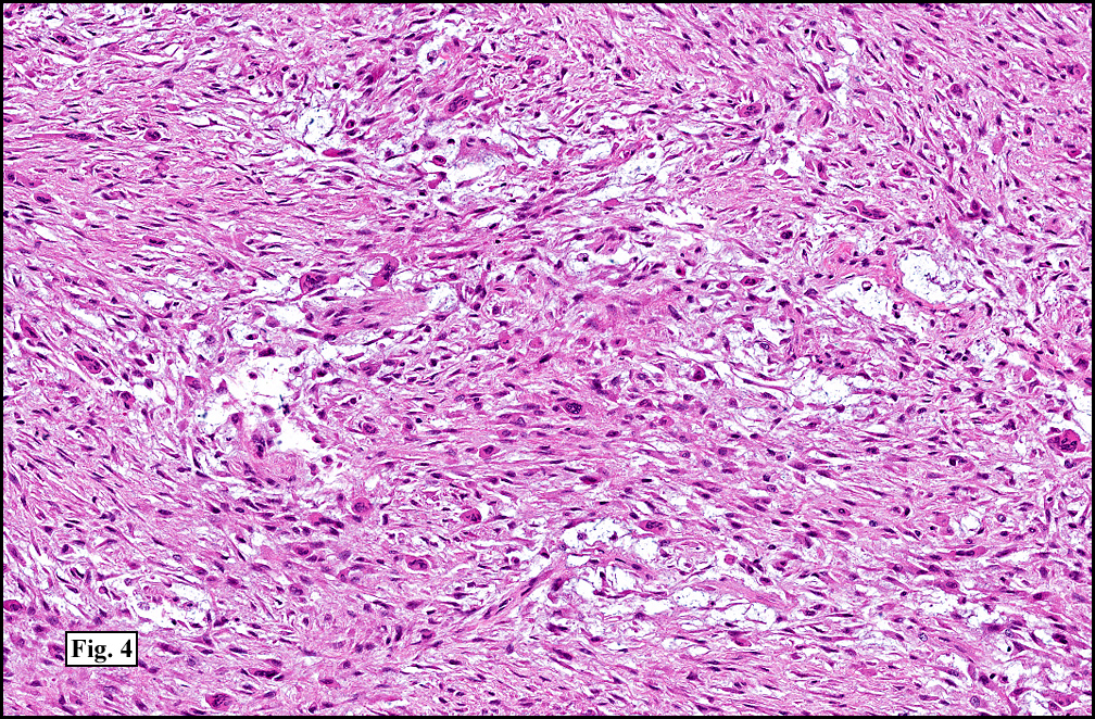
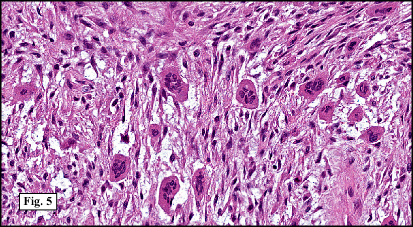
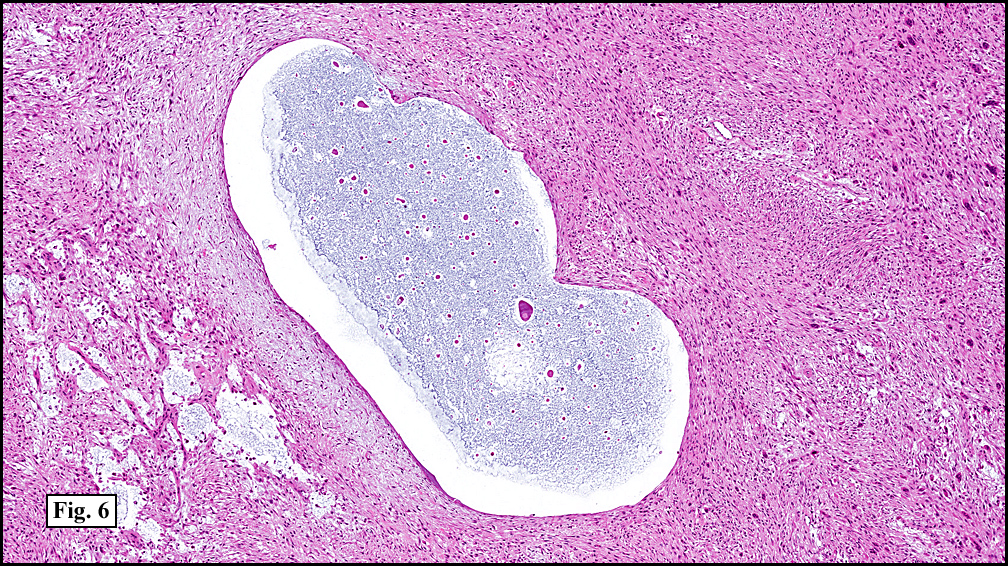
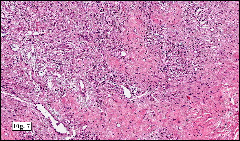
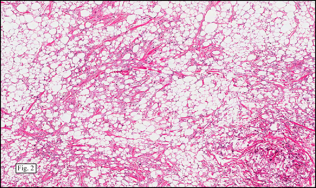
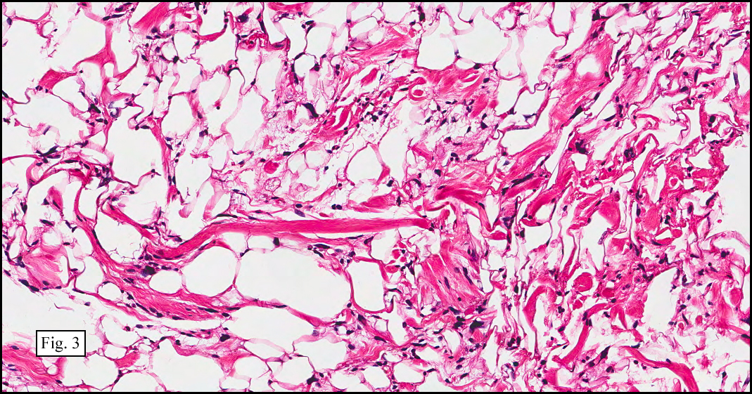
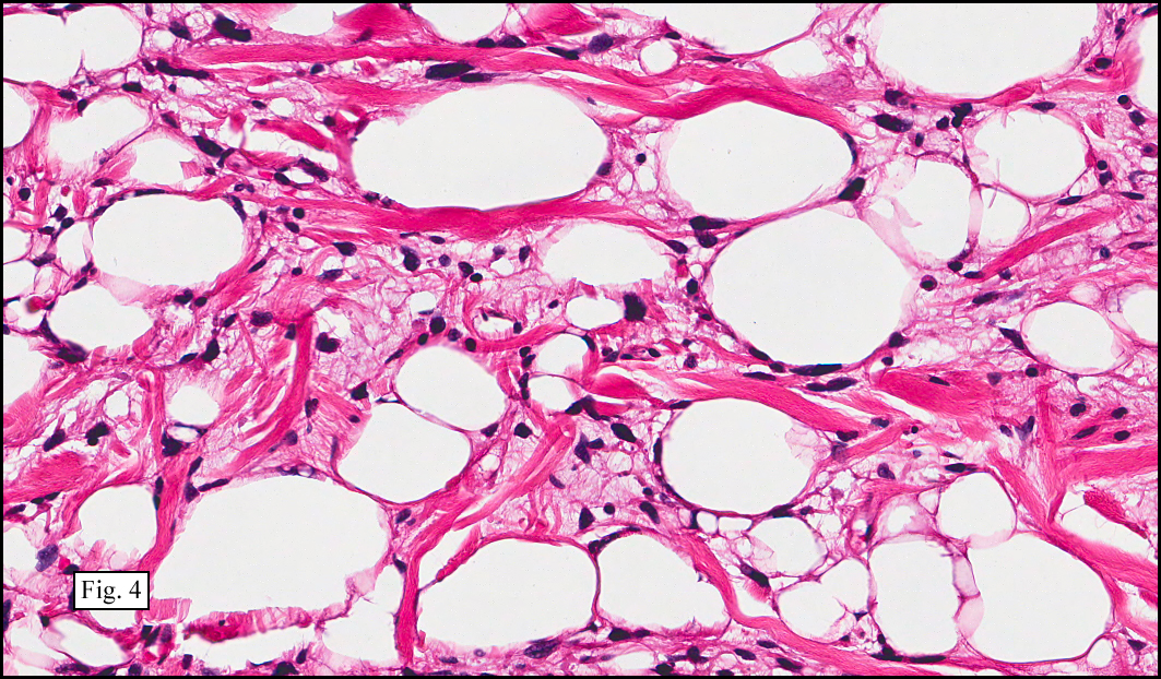
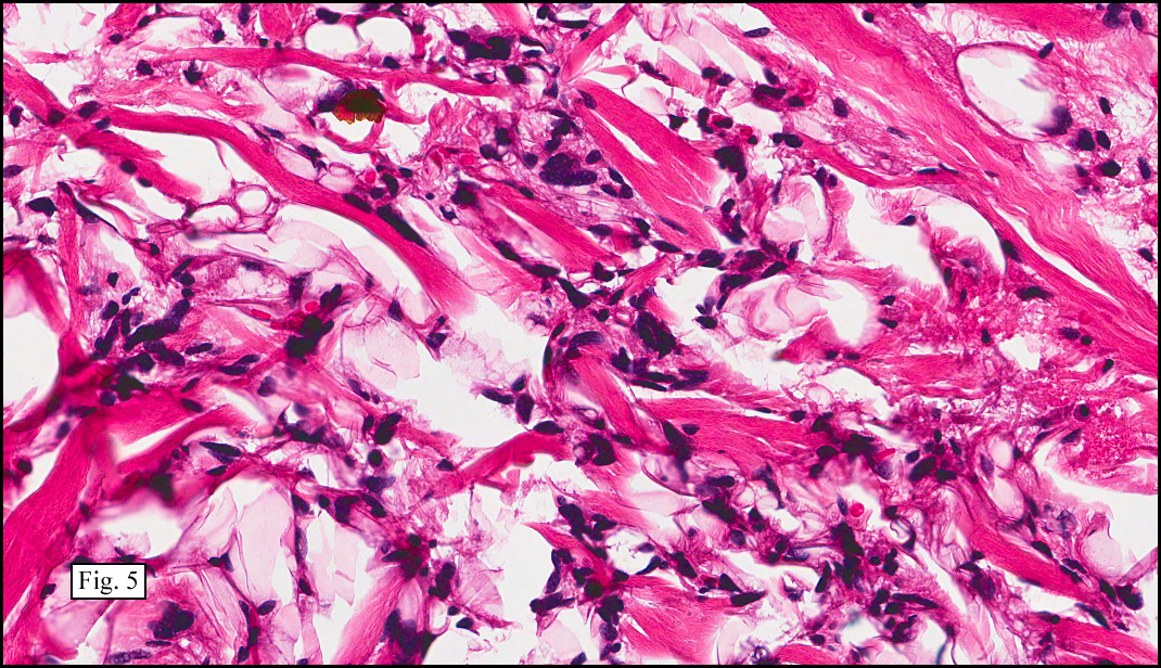
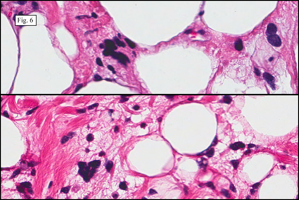
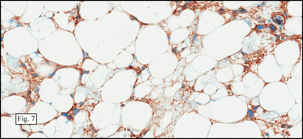
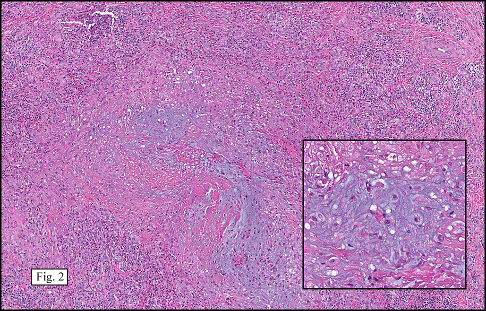
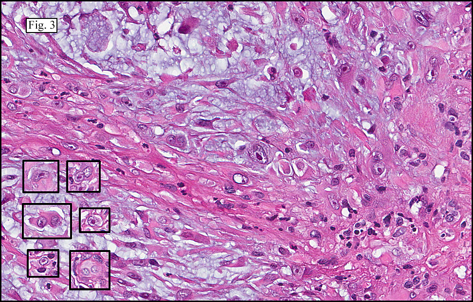
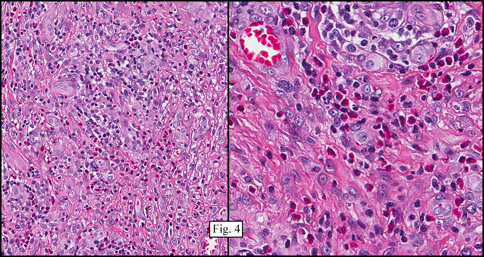
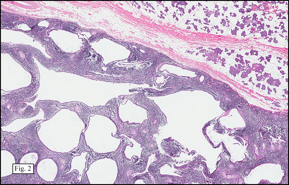
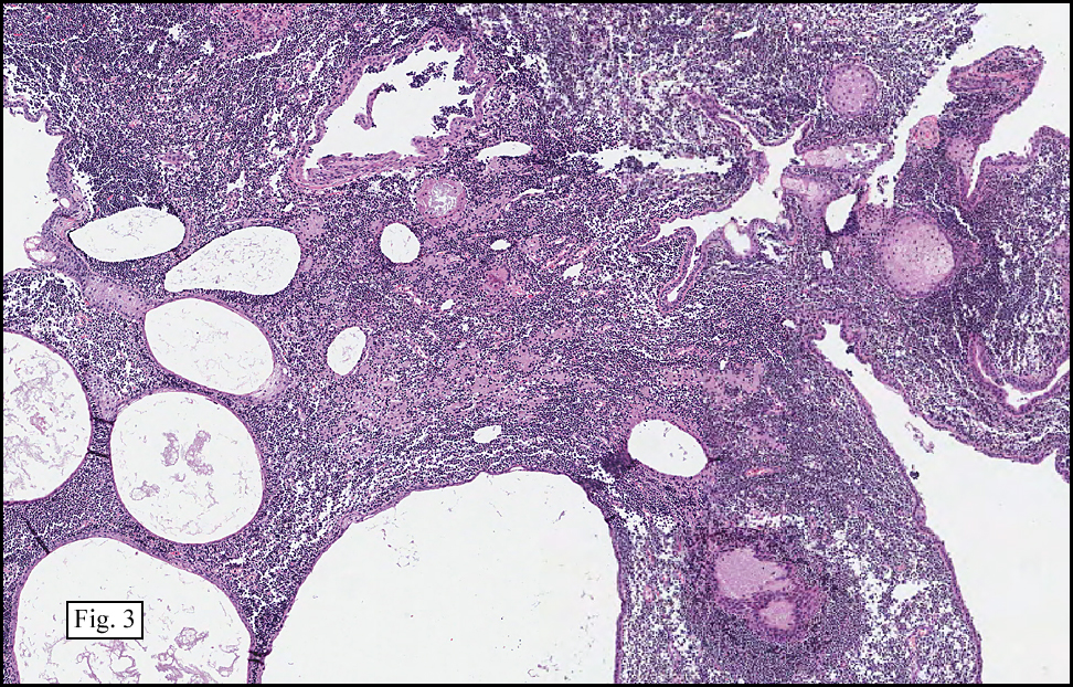
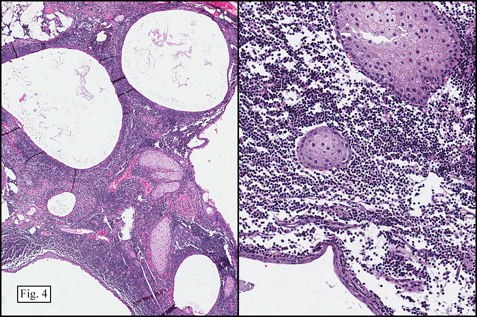
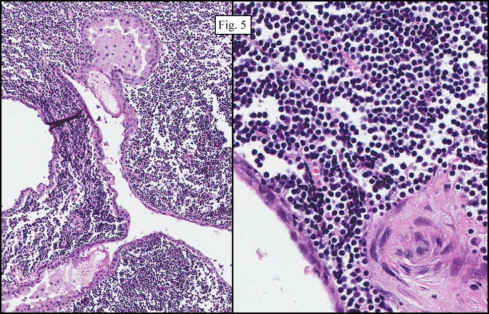
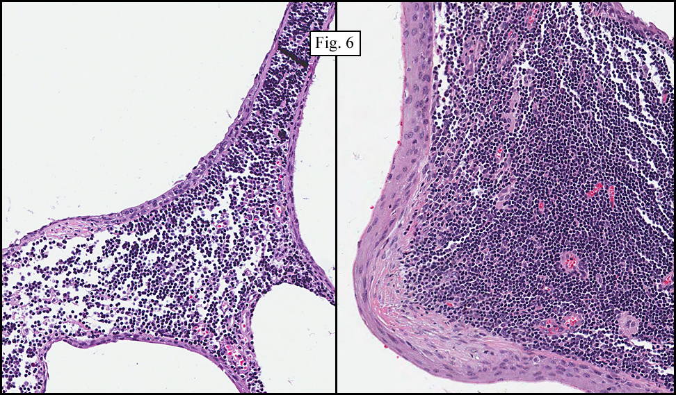
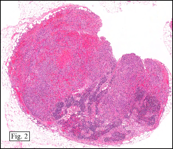
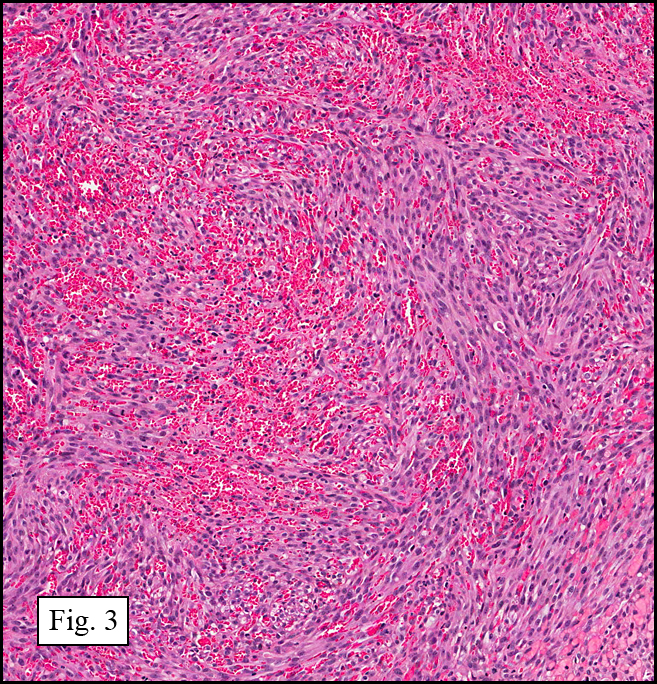
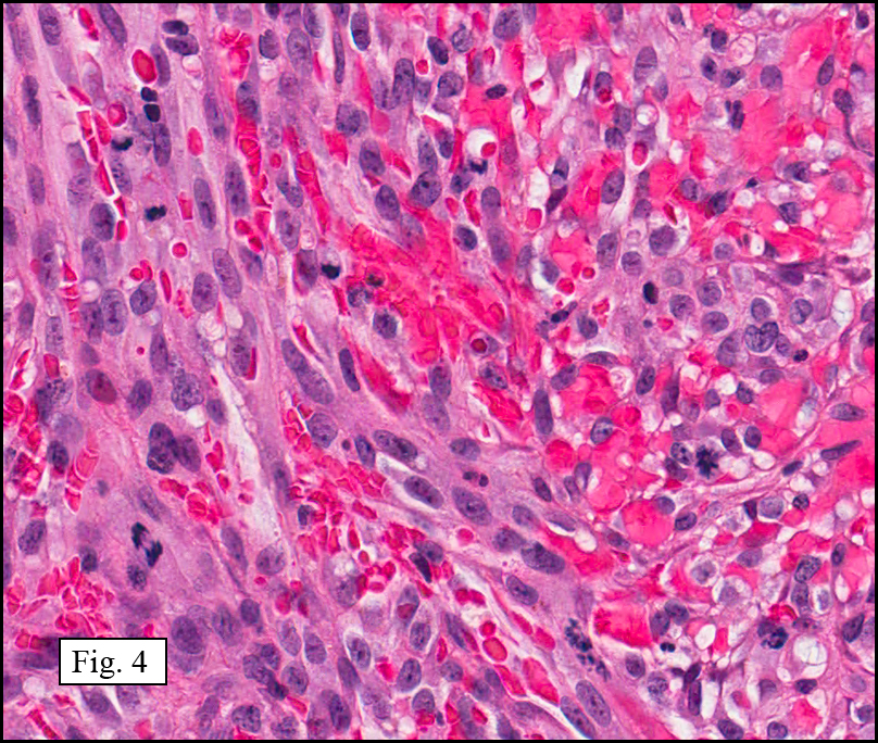
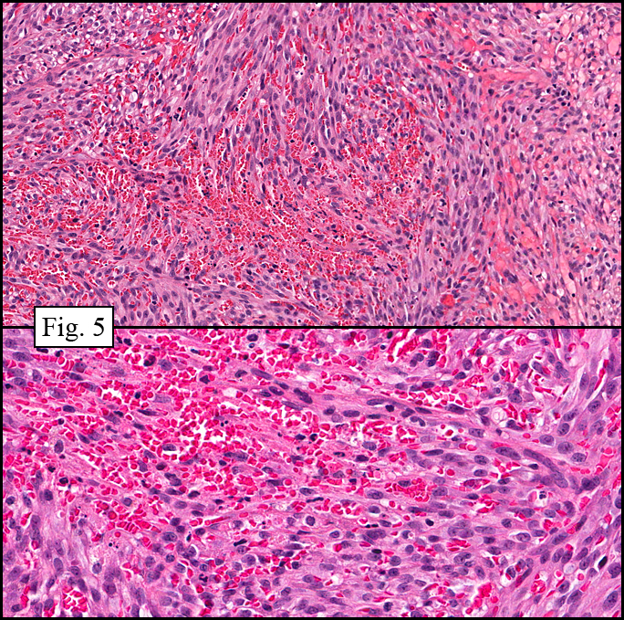
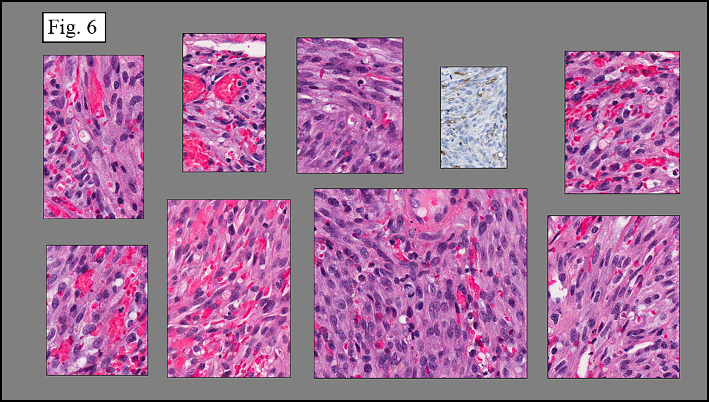
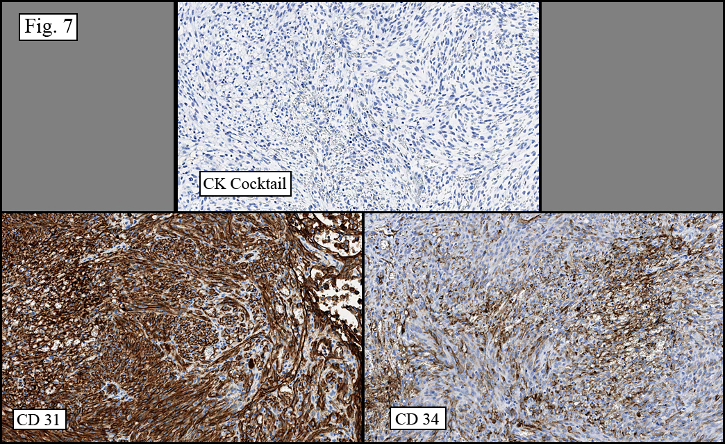
Microscopy showed spindled to stellate-shaped cells with a feathery or fibrillary appearance in a myxoid background (Fig. 2, 3, 4). Scattered giant cells were present, and mitotic figures were increased in number (Fig. 5). Also seen were occasional microcysts (Fig. 6), and areas of hyalinized fibrosis (Fig. 7). Immunohistochemistry positively marked the spindled cells for vimentin and actin. Stains were negative for desmin, S100 and cytokeratin.

Diagnosis: “Nodular Fasciitis with Giant Cells, Posterior Left Thigh”
Marnelli A. Bautista, MD, and Donald R. Chase, MD
Department of Pathology and Human Anatomy, Loma Linda University and Medical Center, Loma Linda, CA
California Tumor Tissue Registry, Loma Linda, CA

Discussion: Nodular fasciitis (NF) is a benign, reactive process consisting of myofibroblasts and fibroblasts. In 1955, Kornwaler and colleagues first described the lesion as “subcutaneous pseudosarcomatous fibromatosis” due to its rapid growth, areas with higher cellularity, and presence of mitotic figures, making it the most common benign mesenchymal lesion misdiagnosed as sarcoma. Commonly involved sites are the upper extremities, trunk, chest and back; however, it can also occur in the extremities, perhaps due to trauma. NF is hypothesized to be initiated by injury largely because it resembles organizing granulation tissue typically encountered in reactive or reparative processes. This lesion arises most commonly in adults 20-40 years of age, although the literature reports that about 10% of the cases are diagnosed in children and an additional 10% arise in patients more than 60 years.

Key features of NF include:
• Small size, generally less than 2 cm, with a well-circumscribed periphery
• Rapid initial growth, but regression over time
• Regional heterogeneity with prominent myxoid background and areas with hyalinized fibrosis
• Haphazardly arranged, uniform myofibroblasts and fibroblasts, with tissue culture growth-pattern or “torn tissue paper” appearance
• Increased mitotic figures

Correlation between the microscopic features and the duration of the lesion has been established. The acute phase usually has a mucoid appearance with less cellularity. Over a short period of time, an increasing myxoid matrix causes the lesion to expand. Then, a myofibroblastic proliferation occurs, which microscopically results in an appearance of increased cellularity. Long-standing processes sometimes referred to as the “resolving stage”, tend to have hyalinized fibrosis. This “sclerosis” seemingly reflects involution of the nodule. Microcysts may also be seen in this final stage. The eventual appearance may be that of a “dermatofibroma” or “fibroma.”

The myofibroblasts in NF are randomly arranged, being separated by the expanding mass of mucopolysaccharide matrix and the proliferation of capillaries. At this time the process may assume a feathery or fibrillary appearance, sometimes likened to as a “tissue culture-like appearance.” Reflecting the rapid expansion of the nodule, mitotic figures are numerous but atypical forms are rarely seen. Occasional red cells and lymphocytes can be identified. A few giant cells may also be present, but are very uncommon. Okoye and colleagues demonstrated that the giant cells in NF are likely related to myofibroblasts by virtue of their similarity in ultrastructural and immunohistochemical characteristics. This finding further supports the hypothesized reparative process in NF, similar to the response generated by injury.

Three described “classic” subtypes are:
• Subcutaneous NF – a well-defined nodule; most common among the three subtypes
• Intramuscular NF – slightly larger than the other two subtypes
• Fascial NF – less delineated due to its propensity to grow along septa producing a stellate pattern
Other benign variants of NF include:

• Intravascular fasciitis is a rare type (<3% of NF cases), most commonly found in younger patients. It is characterized by slow growth and mainly involves small and medium sized vessels, which could be mistaken for a thrombus. It has a less myxomatous background and tends to have more giant cells. • Cranial fasciitis primarily presents during the first year of life and is believed to be associated with birth trauma. This well-demarcated lesion is characterized by rapid growth, which may involve the dura & meninges. It consists of prominent myxoid and hyalinized matrix, and may occasionally have focal bone metaplasia. • Ossifying fasciitis is a less well-circumscribed nodule, with numerous foci of bone metaplasia. Differential diagnoses include: • Proliferative fasciitis (PF) - may be distinguished by characteristic large cells appearing similar to ganglion cells and having one or two large, basophilic, peripherally located nuclei. The clinical course, however, is the same as NF. • Giant Cell Tumor of Tendon Sheath (GCTTS) - mainly occurs adjacent to the interphalangeal joints of the fingers, a very unusual location for NF. Histologically, GCTTS is not characterized by a myxoid matrix, and has a lobular appearance. Giant cells are usually prominent, and have many more nuclei than are seen NF. • Fibromatosis - typically is a poorly demarcated lesion which infiltrates the surrounding subcutaneous tissue. It consists of long slender-shaped fibroblasts and lacks the characteristic mucoid background. Giant cells are never seen. • Myxoma - may mimic the acute phase of nodular fasciitis with expansion of myxoid matrix, but no proliferation of myofibroblasts is seen. The process does not regress. Unlike NF, the process is virtually non-vascular. • Fibrous histiocytoma - may resemble the cellular or early resolving phase of nodular fasciitis. This entity consists of spindle cells and polygonal histiocytes usually arranged in a cartwheel or storiform pattern. NF, however, may ultimately mimic this entity in its end “resolution” state, • Fibrosarcoma (FS) - can be differentiated from NF by the organized fibroblasts arranged in interweaving bundles resulting in a herringbone pattern. Although it shares with NF a high mitotic rate, FS does not show the tissue-paper myxoid phenomenon. In general, complete surgical excision is curative, and this lesion rarely recurs once removed. The literature reports about 1% recurrence rate. Reported exceptions include an article by Thompson who reported approximately 9% recurrence of external ear lesions. Another article documented 13% recurrence rate, although upon re-review, all recurrent lesions were later re-classified as entities other than NF. In general, NF is felt to be a non-recurrent reparative process. Suggested Reading:

Bernstein KE; Lattes R. Nodular (Pseudosarcomatous) Fasciitis, A Nonrecurrent Lesion: Clinicopathologic Study of 134 Cases. Cancer 49(8): 1668-78, 1982.

Okoye MI; Watanabe I. Ultrastructural and Immunohistochemical Investigations of the Giant Cells in Nodular Fasciitis. J Natl Med Assoc 80 (7): 770-5, 1988.

Akihiro Y; Hideki O. Nodular Fasciitis with Degeneration and Regression. J Craniofac Surg 19(4): 1167-70, 2008.

Lenyoun EH; Wu JK; Ebert B; Lieberman B. Rapidly Growing Nodular Fasciitis in the Cheek of an Infant: Case Report of a Rare Presentation. Eplasty 8: e30, 2008.

Thompson LD; Fanburg-Smith JC; Wenig BM. Nodular Fasciitis of the External Ear Region: A Clinicopathologic Study of 50 Cases. Ann Diagn Pathol 5(4): 191-8, 2001.

Weiss S, Goldblum J. Enzinger & Weiss’s Soft Tissue Tumors (5th ed). Philadelphia: Mosby/Elsevier Inc. 177-92, 2008.

December 2010: A 75 year old man with a large posterior neck mass

History: A 75-year-old man underwent surgical excision of a 4 x 3 cm yellow-tan, ovoid, lobular and encapsulated right posterior neck mass. It was superficial, approaching to within 1.2 cm of the skin surface. The mass had a homogeneous cut surface and was well circumbscribed (Fig. 1). Mature fat occupied about 50% of the tumor, with the remainder consisting of spindled and pleomorphic cells (Fig. 2). Conspicuous thick bands of eosinophilic, ropey collagen dissected throughout the fat and hyperchromatic spindled cells (Fig. 3, 4). Some areas showed clumping of the atypical spindled cells (Fig. 5). There were also scattered multinucleated giant cells with a “floret-like” appearance (Fig. 6). Neither necrosis nor hemorrhage was seen. Mitotic figures were absent. The tumor strongly decorated for CD34 in both the spindled cells, and the floret elements (Fig. 7).

Diagnosis: “Spindle Cell Lipoma/Pleomorphic Lipoma, Right Posterior Neck”

Marnelli A. Bautista, MD, and Donald R. Chase, MD
Department of Pathology & California Tumor Tissue Registry
Loma Linda University and Medical Center, Loma Linda, California

Discussion: Spindle Cell Lipoma (SCL) and Pleomorphic Lipoma (PL) have been recently re-classified as a single entity (SCL/PL) due to their overlapping clinical, cellular, immunoperoxidase and cytogenetic characteristics. They occur mostly in men (85-90%) between the ages of 45-60. Although SCL/PL can occur at other ages, they virtually do not occur before the age of 20. Approximately 80% arise in the posterior neck, shoulder or back, and are virtually always superficial, generally involving subcutaneous tissues. Deeper forms, however, have been described in a variety of sites.

The tumors show a wide histologic spectrum ranging from pure spindle cell lipoma to pure pleomorphic lipoma. Although the fatty component is mostly mature, occasional lipoblast-like cells may also be encountered. Problematically, cases with very minimal to absent adipose tissue have been reported, posing a diagnostic challenge.

In classic SCL, there is relatively equal distribution of mature fat and uniform, bland spindle cells, many of which have hyperchromatic nuclei. The spindle cells may be randomly arranged, clumped, or assembled in short parallel bundles. A consistent finding is eosinophilic, dense or ropey collagen strands admixed with the spindle cells. A pseudoangiomatous variant of SCL has also been described consisting of short bundles of spindle cells partitioned by dense connective tissue projections.

Pleomorphic lipoma also has ropey collagen bundles and occasional spindle cells, but is distinguished by the presence of peculiar giant cells with floret-like nuclear configurations, where the coalescing nuclei are organized into a wreath-like pattern. The spindle cells and floret giant cells stain strongly positive for CD34 but are negative for desmin, actin, keratin and S-100. Mast cells are seen in most cases. Some lesions may also display myxomatous change. On rare occasions, small foci of cartilaginous or osseous metaplasia may be identified. However, mitotic figures are not increased, and atypical forms are rarely present. SCL/PL behaves in a benign fashion, with a very low rate of recurrence once completely excised.

Cytogenetic findings: Most SC/PL show loss of 16q and occasional cases demonstrate loss of 13q, further highlighting the relationship between the two lesions. The absence of ring chromosomes and giant marker helps to separate SCL/PL from atypical lipomatous tumor/well-differentiated liposarcoma (ALT/WDL).

Differential Diagnosis:
• ALT/WDL, particularly the sclerosing type: These tumors are typically located in the deep muscles of the extremities and retroperitoneum. Floret-like giant cells may be encountered, with occasionally scattered atypical or pleomorphic lipoblasts, and dense fibrous zones, but the conspicuous ropey collagen bundles are not usually seen. Also, in contrast to SCL/PL, ALT/WDL is characterized by the presence of giant marker and ring chromosomes.

• Dedifferentiated Liposarcoma (DLS): Greater than 80% of DLS arises in the retroperitoneum. Most areas appear histologically similar to ALT/WDL, with focal overgrowth of pleomorphic sarcoma (dedifferentiation). An abrupt transition between the two regions is usually depicted. The dedifferentiated zone may show little pleomorphism mimicking fibrosarcoma or leiomyosarcoma, but much more commonly has the phenotype of so-called malignant fibrous histiocytoma (pleomorphic sarcoma). As in PL, scattered cells may have floret-like giant cells.

• Pleomorphic Liposarcoma: This least common type of liposarcoma (15%) is deeply located and consists of numerous pleomorphic lipoblasts and on occasion, large, bizarre giant cells. Unlike SCL/PL, the tumor is negative for CD34. Mitotic figures, including atypical forms, are commonly seen.

• Dermatofibrosarcoma Protuberans (DFSP) usually arises superficially within the dermis and infiltrates the underlying subcutaneous tissue. The uniform spindle cells are arranged in a storiform pattern. Giant cells may be encountered in some cases; however, ropey collagen bundles are typically absent, and the process is CD34 negative.

• Giant Cell Fibroblastoma (GCF) is commonly seen in children, usually 3-5 years old. It has been suggested that this lesion represents a juvenile form of DFSP. Multinucleated giant cells, which line the characteristic pseudovascular spaces, may emulate the floret-like giant cells of SCL/PL. Nevertheless, eosinophilic, ropey collagen bands are not seen.

Fine needle aspiration (FNA) may assist in categorizing SCL/PL, however, caution is urged as sampling limitation may lead to misdiagnosis of sarcoma. Definitive classification is best reserved for microscopic evaluation of the excised specimen.

Suggested Reading:

French CA; Mentzel T; Kutzner H; Fletcher C. Intradermal Spindle Cell/Pleomorphic Lipoma, A Distinct Subset. Am J Dermatopathol 22(6): 496-502, 2000.

Karacal N; Gulcelik N; Sapan LA; Kutlu N. Giant Pleomorphic Lipoma of the Neck. Plast Reconstr Surg 117(2): 692-693, 2006.

Sachdeva MP; Goldblum JR; Rubin BP; Billings SD. Low-Fat and Fat-Free Pleomorphic Lipomas: A Diagnostic Challenge. Am J Dermatopathol 33(5): 423-426, 2009.

Yong M; Raza AS; Greaves TS; Cobb CJ. Fine Needle Aspiration of a Pleomorphic Lipoma of the Head and Neck: A Case Report. Diagn Cytopathol 32(2): 110-113, 2005.

Thirumala S; Desai M; Kannan V. Diagnostic Pitfalls in Fine Needle Aspiration Cytology of Pleomorphic Lipoma: A Case Report. Acta Cytol 44: 653-656, 2000.

Weiss S, Goldblum J. Enzinger & Weiss’s Soft Tissue Tumors (5th ed). Philadelphia: Mosby/Elsevier Inc. 444-452, 2008.

November 2010: A 33 year old woman with a mass in a finger

History: A 33 year-old woman presented with a progressively enlarging, tender nodule in the palmar aspect of the right fourth finger. An approximately 1.0 cm diameter, circumscribed, nodular soft tissue mass was removed (Fig. 1).

Microscopically, the tumor was composed of spindled and epithelioid cells with oval to rounded nuclei and conspicuous nucleoli prominently supported by alternating undulations of myxoid and hyalinized matrix (Fig. 2). Several of the larger epithelioid cells had abundant eosinophilic cytoplasm, multilobulated nuclei and striking macro nucleoli reminiscent of Reed-Sternberg cells (Fig. 3). In addition, admixed within the mass was a dense chronic inflammatory infiltrate consisting mostly of lymphocytes and plasma cells with occasional areas of numerous eosinophils (Fig. 4). An infrequent multinucleated giant cell was also seen with intermingling round mononuclear cells with bland nuclei and amphophilic cytoplasm.

Immunohistochemical staining revealed limited focal positivity for CD34 and rare positivity for epithelial membrane antigen in the lesional cells. Other cytokeratin markers as well as CD31, or S-100 were negative.

Diagnosis: “Acral Myxoinflammatory Fibroblastic Sarcoma”

Brian Willis PSF and Donald Chase MD
Department of Pathology & California Tumor Tissue Registry
Loma Linda University and Medical Center, Loma Linda, California

Discussion: Acral myxoinflammatory fibroblastic sarcoma (AMIFS) is a rare, recently-described soft tissue tumor with just over one-hundred cases reported in the literature. First described in 49 patients by Montgomery and colleagues in 1998, it was originally entitled “Inflammatory myxohyaline tumor of the distal extremities with virocyte or Reed-Sternberg-like cells. The identical tumor was shortly later described by Meiss-Kindblom and Kindblom as “acral myxoinflammatory fibroblastic sarcoma”. Included in their presentation cases was a patient with biopsy-proven inguinal lymph node metastasis. The authors added the term “sarcoma” to emphasize the possibility of an aggressive clinical course, and AMIFS became the preferred term.

As reported, AMIFS occurs mostly in adulthood, generally within the fourth and fifth decades, but the age range is likely broader. Males and females are equally affected. The tumor presents as a slowly growing, multinodular, ill-defined mass usually in the fingers and/or hand. Magnetic resonance imaging shows a poorly circumscribed mass with involvement of the underlying tendon sheath; a finding that, when coupled with the location, leads to the preliminary diagnostic consideration of ganglion cyst or tenosynovitis.

Microscopically, there is usually a dense inflammatory infiltrate mostly composed of an admixture of lymphocytes and neutrophils, plasma cells, and eosinophils insinuated into a background of alternating undulations of myxoid and hyaline stroma. The more cellular zones may have bizarre cells that are spindled, epithelioid, or polygonal. The larger epithelioid cells frequently display prominent, sometimes multilobulated, vesicular nuclei with conspicuous macronucleoli and an abundant eosinophilic cytoplasm, strikingly reminiscent of Reed-Sternberg cells or virocytes. Despite the cytomorphology of these cells, they are negative for CD15/30, and are PCR negative for CMV and EBV. Occasional, ganglion-like cells, multinucleated giant cells, and lipoblast-like cells may also be found within the tumor. Mitotic figures are scarce, despite the relatively high cellularity and high degree of nuclear atypia.

Immunohistochemical findings are essentially non-contributory, with the larger bizarre cells showing consistent immunoreactivity for vimentin and focal, variable reactivity for CD68, CD34, and smooth muscle actin. There may be limited focal immunoreactivity for cytokeratins. S-100, HMB-45, desmin, CD15, and CD30 are virtually always negative. In one recent study by Kovarik and colleagues, diffuse, strong reactivity for CD163 and EGFR was demonstrated. KI-67 positivity is minimal, helping to confirm the low mitotic rate seen on H&E.

Ultrastructurally, the neoplastic cells have abundant endoplasmic reticulum and mitochondria along with large perinuclear whorled deposits of intermediate filaments without actin-type filaments, findings most consistent with a tumor of fibroblastic differentiation.

In the described cases of AMIFS, rates of local recurrence after surgical resection range from 20% to 70%, with approximately one-third of cases eventually necessitating digital amputation. Despite having a low metastatic rate (<2%), distant spread to lymph nodes and lung have been reported. The differential diagnosis of AMIFS includes infectious processes, lymphomas, and other tumors with myxoid or inflammatory characteristics, as well as tumors that arise in the distal extremities, such as giant cell tumor of tendon sheath. • Myxofibrosarcoma/myxoid malignant fibrous histiocytoma represents perhaps the most important distinction in the differential diagnosis. This much higher grade sarcoma shows pleomorphic cells of a degree not seen in AMIFS. Additionally, the myxoid stroma is more uniformly distributed than is seen in AMIFS, and there are not the unusual atypical cells which mimic Reed-Sternberg cells or virocytes. Also, the acral location argues strongly against the diagnosis of myxofibrosarcoma. • Giant cell tumor of the tendon sheath is the most common soft tissue tumor of the fingers. It usually shows numerous Touton-like giant cells along with a lympho-histiocytic cellular infiltrate; however it lacks the large, bizarre, epithelioid cells that characterize AMIFS. • Inflammatory myofibroblastic tumor may be considered based on the presence of a strong inflammatory component in conjunction with a spindle cell background. The presence of the ultrastructural features of myofibroblasts and the lack of bizarre cells precludes this diagnosis in cases of AMIFS. In addition, inflammatory myofibroblastic tumor is seldom encountered in acral locations. • Ganglion cysts and juxta-articular myxomas lack the bizarre, atypical cells of AMIFS. • The presence of Reed-Sternberg-like cells raises the question of Hodgkin’s disease; however, AMIFS does not show positivity for CD15/30. The origin of AMIFS is unknown. Only a few cytogenetic studies have been performed. They have demonstrated a t(1;10) switch with losses of chromosomes 3 and 13 as well as other complex non-specific chromosomal aberrations mostly involving shared gains at chromosome 7. Acral myxoinflammatory fibroblastic sarcoma is a unique slow-growing low-grade sarcoma that presents peculiarly in the distal extremities. Because the complex histology of this lesion may mimic benign inflammatory, reactive changes or even lymphoma careful evaluation is necessary to arrive at the correct diagnosis. Differences in reports of recurrence and metastases likely vary as a result of differences in initial diagnosis, underscoring the importance of proper diagnosis and primary surgical treatment by complete extirpation with clear margins to lower the likelihood of local recurrence or metastases. Suggested Reading

Kovarik CL, Barrett T, et al. Acral myxoinflammatory fibroblastic sarcoma: case series and immunohistochemical analysis. J Cutan Pathol; 2008; 35: 192-196.

Hassanein AM, Atkinson SP, Al-Quran SZ, et al. Acral myxoinflammatory fibroblastic sarcomas: are they all low-grade neoplasms? J Cutan Pathol 2008: 35:186-191.

Baumhoer D, Glatz K, Schulten HG, et al. Myxoinflammatory fibroblastic sarcoma: investigations by comparative genomic hybridization of two cases of and review of literature. Virchow Arch 2007; 451: 923-928.

Gonzalez- Campora R, Rios-Martin JJ, et al. Fine needle aspiration cytology of an acral myxoinflammatory fibroblastic sarcoma: case report with cytological and cytogenetic findings. Cytopathol 2008; 19: 118-123.

Alkuwari E, Gravel DH. A 30-year-old man with a soft tissue mass on the right elbow. Inflammatory myxohyaline tumor of the distal extremeties with prominent eosinophilic infiltrate. Arch Pathol Lab Med 2006; 130:e35

Montgomery EA, Devaney KO, Giordano TJ, et al. Inflammatory myxohyaline tumor of distal extremities with virocyte or Reed-Sternberg-like cells: A distinctive lesion with features simulating inflammatory conditions, Hodgkin’s disease, and various sarcomas. Mod Pathol 1998; 11:384.

Meis-Kindblom JM, Kindblom LG. Acral myxoinflammatory fibroblastic sarcoma: a low grade tumor of the hands and feet. Am J Surg Pathol 1998; 22:911.

October 2010: A 74 year old with a parotid gland mass

History: A 74 y/o man presented with a two year history of a left-sided facial mass. A CT scan identified a 4.0 cm mass in the parotid gland. Following a FNAB which showed only “reactive” lymphoid elements, a superficial parotidectomy found a well-circumscribed grey-tan cystic mass containing cheesy, grumous material.

Microscopically the tissue had a “pushing margin” abutting the parotid gland (Fig. 1, 2). It consisted of solid epithelial nests and small cysts or duct-like structures with focal sebaceous elements (Fig. 3). The background was of lymphocytes, without atypia (Fig. 4). In addition to the sebaceous elements, were scattered squamous pearls, and a yst lining of simple squamous to low cuboidal epithelium (Fig. 5,6).

Diagnosis: “Sebaceous Lymphadenoma”

Christina Martin MSIV, Donald R. Chase MD
Department of Pathology, Loma Linda University and Medical Center, Loma Linda California Tumor Tissue Registry, Loma Linda, California

Discussion: Sebaceous lymphadenoma (SL) is a rare benign epithelial tumor making up less than 0.1% of all salivary gland neoplasms. Most often occurring between the ages of 50-80, these tumors present as a progressively enlarging, painless mass. They are most commonly found in the parotid gland, but have also been reported in the oral region and the anterior midline of the neck. Some cases have been associated with a simultaneous Warthin tumor or primary squamous cell carcinoma.

Fine needle aspiration usually demonstrates aggregates of sebaceous cells with foamy cytoplasm and central crenated nuclei, as well as less mature squamoid forms with dense cytoplasm and rounded nuclei. A background of small mature lymphocytes may also be seen.

The excised specimen is usually described as solid to multicystic, and pinkish-grey to white or yellow. Sebum-filled spaces are sometimes present. Capsules are usually well-developed, but may be partially or completely absent. When absent, the periphery is still well-circumscribed, reflective of expansive compression of surrounding tissues.

The microscopic appearance of SL is usually of epithelial nests with peripheral basaloid cells maturing inwardly into sebaceous cells. Duct-like structures and small to medium sized cysts lined with squamous, cuboidal, columnar, and sebaceous cells are also seen. Many of the lumens contain secretions, and occasionally there are keratin-filled cysts. Oncocytic changes may also be present. The prominent lymphoid stroma is uniformly dense, but does not usually invade the epithelial elements. Lymphoid follicles may be absent or numerous, with mantle zones ranging from ill-defined to well-formed.

Immunohistochemical features include positive cytokeratin and epithelial membrane markers in the epithelial elements. S-100 and smooth muscle actin are generally negative. The lymphoid component has similar staining properties of a reactive lymph node.

The nature of the lymphoid tissue in sebaceous lymphadenoma is controversial, paralleling the controversy surrounding the lymphoid component of Warthin tumors. One theory is that the tumors arise from ectopic salivary gland tissue entrapped in lymph nodes during embryogenesis. Another suggests that the lymphoid component represents a secondary reactive response to epithelial proliferation (tumor-associated lymphoid proliferation) as seen in various other parotid gland tumors. The latter explanation is favored by current WHO and AFIP publications.

Treatment of this lesion is local excision. Recurrence is rare.

Some important entities to distinguish from Sebaceous Lymphadenoma include:

Lymphadenomas are identical tumors but lack sebaceous differentiation.

Mucoepidermoid carcinoma with tumor-associated lymphoid proliferation has a more haphazard distribution of cysts and ducts, and more variation in their size and shape than sebaceous lymphadenoma. Other distinguishing features pointing towards mucoepidermoid carcinoma include papillary growth and extensive, poorly organized intraluminal proliferation of the lining epithelium of the cysts, as well as the presence of clear and intermediate cells.

Sebaceous Adenocarcinoma forms irregular islands and narrow cords of tumor cells, but unlike sebaceous lymphadenoma, the cells infiltrate surrounding connective tissue and salivary gland parenchyma. The peripheries are infiltrative, not “pushing.”

Metastatic adenocarcinoma or squamous cell carcinoma can be a concern when seeing epithelial islands within a lymphoid stroma, however in sebaceous lymphadenoma they are morphologically bland and do not infiltrate surrounding tissue.

Suggested Reading:

Thompson’s: Head and Neck Pathology: a Volume in the Series Foundations in Diagnostic Pathology. Editors Thompson LDR, Goldblum JR. Elsevier, Inc. pp 312-15, 2006.

AFIP Atlas of Tumor Pathology: Tumors of the Salivary Glands. Editors Ellis GL, Auclair PL, pp 136-141, 2008

Sebaceous lymphadenoma of parotid gland in a child. Sun G, Hu Q, Huang X, Tang E. Oral Surg Oral Med Oral Pathol Oral Radiol Endod. 2009 Feb; 107 (2): 253-5.

Sebaceous lymphadenoma of the parotid gland. Majeed M, Murray B, Hume W, MakuraZ. Dentomaxillofac Radiol. 2008 Jul:37(5):300-4.

Lymphadenoma of parotid gland: Two additional cases and a literature review. Dardick I, Thomas MJ. Oral Surg Oral Med Oral Pathol Oral Radiol Endod. 2008 Apr;105(4):491-4.

Sebaceous lymphadenoma of salivary gland: a case report and a review of the literature. Maffini F, Fasani R, Petrella D, Maiorano E, Bruschini R, Pelosi G, Viale G. Acta Otorhinolaryngol Ital. 2007 Jun;27(3);147-50.

Sebaceous lymphadenoma of the parotid gland: report of two cases and review of the literature. Hayashi D, Tysome JR, Boyei E, Gluckman P, Barbaccia C. Acta Otorhinolaryngol Ital. 2007 Jun;27(3):144-6. Review.

Unilocular cystic sebaceous lymphadenoma: a rare tumor. Chandrasekar T, Ramani P, Anuja N, Karthikeyan R, Abhilash PR, Narayan V, Giri VV. Ann R Coll Surg Engl. 2007 May;89(4):1-3.

Sebaceous lymphadenoma identified by fine needle aspiration biopsy: a case report. Banich J, Reyes CV, Bier-Laning C. Acta Cytol. 2007 Mar-Apr;51(2):211-3.

September 2010: A 31 year old man with immunodeficiency and cervical lymphadenopathy

History: A 31 y/o physician with a retroviral disease and associated immunodeficiency was found to have cervical lymphadenopathy. He had a chronic cough, but sputum had been negative for mycobacterium and PCP. There was no evidence of fungal or acid fast bacilli. No skin lesions were found and there was no history of previous neoplasia. Excisional lymph node biopsy was performed. The specimen was received in multiple fragments (Fig. 1)

Microscopically the normal lymph node architecture was replaced by a disordered vascular proliferation (Fig. 2). The cells were spindled and showed a diffuse fascicular pattern (Fig. 3). Mitotic figures were increased (Fig. 4). Another characteristic was numerous slit-like spaces and regions containing many extravasated red blood cells (Fig. 5). Hyaline globules were also present (Fig. 6).

Immunostains for CD31 and CD34 were positive (Fig. 7). Markers for epithelial (Fig. 7), neural, melanocytic and lymphoid origins were negative.

Diagnosis: “Kaposi Sarcoma”

Christina Martin MSIV, Donald R. Chase MD
Department of Pathology, Loma Linda University and Medical Center, Loma Linda, California
California Tumor Tissue Registry, Loma Linda, California

Discussion: Kaposi Sarcoma (KS) is a locally aggressive endothelial tumor that classically presents with blue-red nodular skin lesions which spread and coalesce into plaques. Lesions may also involve mucosal sites, lymph nodes, and visceral organs. KS is uniformly associated with human herpes virus 8 (HHV-8), and may arise in the context of immunosuppression. It is believed that HHV-8 establishes a latent infection in a number of cell types including B lymphocytes and endothelium, and that reactivation of latent virus is pivotal in the development of KS.

Four epidemiological forms are recognized:

1. Chronic (Classic) KS occurs predominantly in men (90%) during late adult life (peak incidence in 6th and 7th decades) of Mediterranean/Eastern European descent with the classic presentation of purplish, reddish-blue or dark brown plaques, macules, and/or nodules found most often involving the skin of the lower extremities. It is prevalent in certain parts of the world including Poland, Russia, Italy, and equatorial Africa, but rare in the United States, and shows a statistically significant association with a second malignant tumor or altered immune state. The disease is usually indolent with a prolonged clinical course.

2. Endemic (African) KS arises in children and adults in Equatorial Africa who are not HIV infected. It presents primarily as localized or generalized lymphadenopathy, with sparse skin lesions which tend to be truncal. The course of the disease is fulminant, a feature attributed to internal tumor involvement.

3. Iatrogenic KS (i.e. transplant-associated) is well-established in renal transplant patients. Incidence varies based on patient population, suggesting genetic susceptibility. It usually develops several months to a few years after transplant (average 16 months). The extent of the disease correlates directly with the loss of cellular immunity as measured by response to skin testing with phytohemagglutinin (PHA), conconavalin A (Con A), pokeweed mitogen (PWM), and dinitrochlorobenzene (DNCB). Clinical course improves with the patient’s ability to tolerate reduction in immunosuppression and worsens with organ or internal involvement.

4. Acquired immunodeficiency syndrome-associated KS (AIDS KS) occurs in the setting of immunodeficiency caused by HIV-1 infection, which potentiates tumor growth. This form disproportionately involves the male homosexual population with other risk groups including intravenous drug users, sex trade workers and hemophiliacs receiving factor VIII-enriched blood fractions. Presentation is usually in young adults with initially small, flat, pink patches on the skin which later progress to classic blue violet papules, with a predilection for lines of cleavage and mucosal surfaces. It may also metastasize or synchronously involve deep viscera/organs.

Treatment for KS in limited mucocutaneous disease usually consists of local therapy including cryotherapy, intralesional injections and radiation therapy. Mortality is 10-20%. In patients with AIDS, KS responds to highly active antiretroviral therapy (HAART). The mortality rate in AIDS KS has decreased from 90% to less than 50%. AIDS patients with KS, usually die of secondary (opportunistic) infections many times due to pulmonary compromise.

The KS phenotype may be found in other tumors, generally as a minor component:

• Well-differentiated angiosarcoma (AS) has features virtually indistinguishable from early KS skin lesions, however a careful history (lifestyle, immunosuppression, etc) is of great importance. KS also tends to be HHV-8 positive while AS is generally negative.

• Fibrosarcoma can be confused with the advanced lesions of KS. The presence of ectatic slit-like spaces and inflammatory cells at the periphery of the lesions, and hyaline globules point towards KS, as does positivity for HHV-8.

• Arteriovenous malformations may duplicate the skin lesions of KS and have been termed “pseudo KS” but lack the marked spindling and formation of slit-like lumens of KS. They may present with bruits or by touch, “thrills”.

• Spindle-cell hemangioendothelioma (aka Kaposiform hemangioendothelioma) is often confused with KS. A younger population, without immunosuppression favors SCH, as does cavernous vessels and epithelioid endothelial cells. The preservation of lobules (not found in KS) is also helpful.

Suggested Reading:

Enzinger and Weiss’: Soft Tissue Tumors 5th Ed. Editors Weiss SW, Goldblum JB. Mosby, Inc. pp 722-730, 2008.

World Health Organization Classification of Tumours: Pathology & Genetics Tumours of Soft Tissue and Bone. Editors Fletcher CDM, Unni KK, Mertens F, pp 170-172, 2002.

An unusual cause of a parotid mass in an immunocompetent host: Classic Kaposi’s sarcoma. Pagani D, Bellinvia M, Capaccio P, Scoppio B, Brambilla L, Pignataro L. Tumori. 2009 Mar-Apr; 95(2);248-50.

Kaposi sarcoma in unusual locations. Pantanowitz L, Dezube BJ. BMC Cancer. 2008 Jul 7;8:190. Review.

Lymph nodes involved by multicentric Castleman disease among HIV-positive individuals are often involved by Kaposi sarcoma. Naresh KN, Rice AJ, Bower M. Am J Surg Pathol. 2008 Jul;32(7):1006-12.

Fine needle aspiration cytology determinants of the diagnosis of primary nodal Kaposi’s sarcoma as the first sign of unknown HIV infection: a case report. Morelli L, Pusiol T, Piscioli I, Del Nonno F, Brenna A, Licci S. Acta Cytol. 2007 July-Aug;51(4):602-4.

Scroll to top