August 2010: A 28 year old man with a long-standing epigastric mass

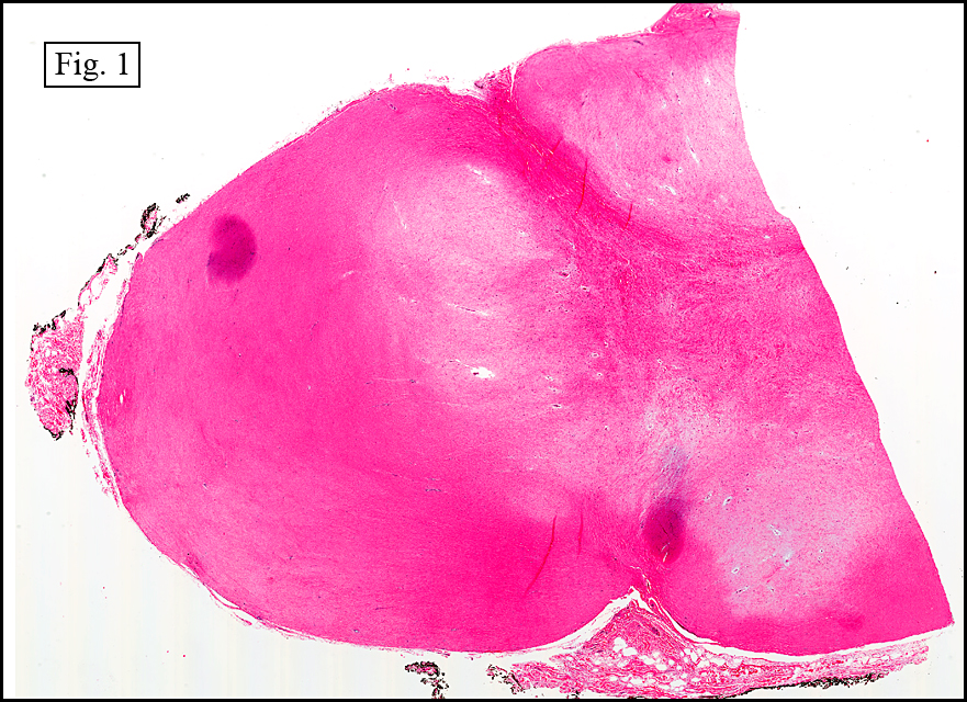
History: A 28-year-old male presented with a left epigastric mass that had been present for ten years. During exploratory surgery, a 15 cm mass was excised from the greater omentum. It was gray-tan with a heterogeneous surface.

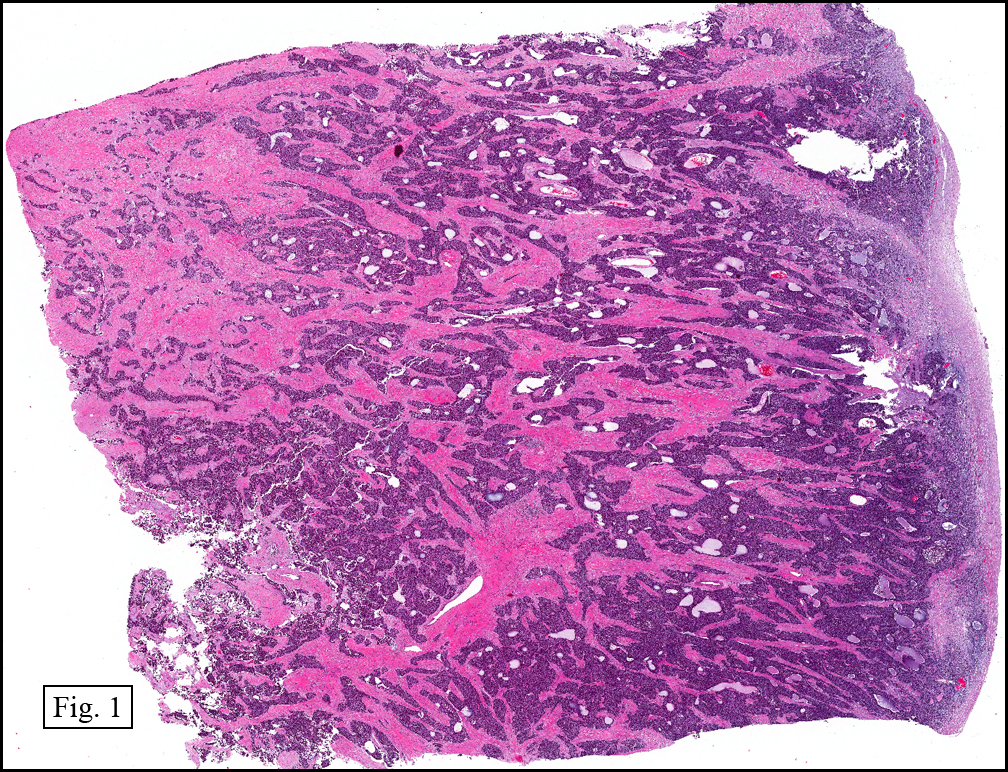
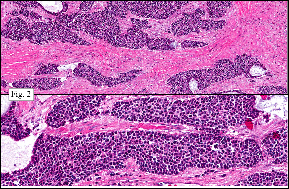
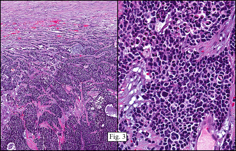
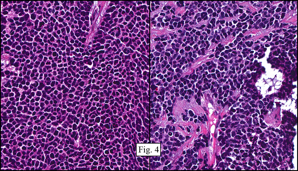
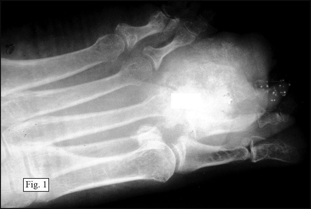
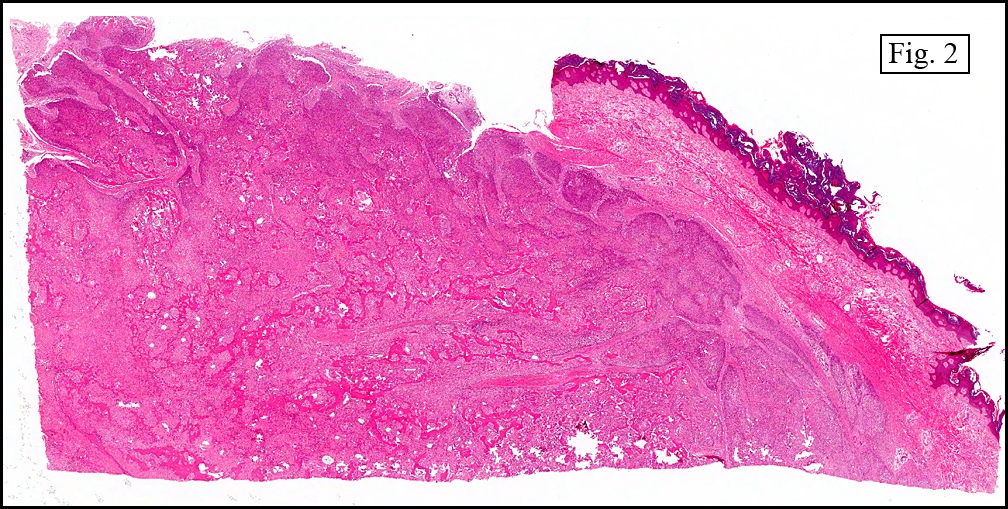
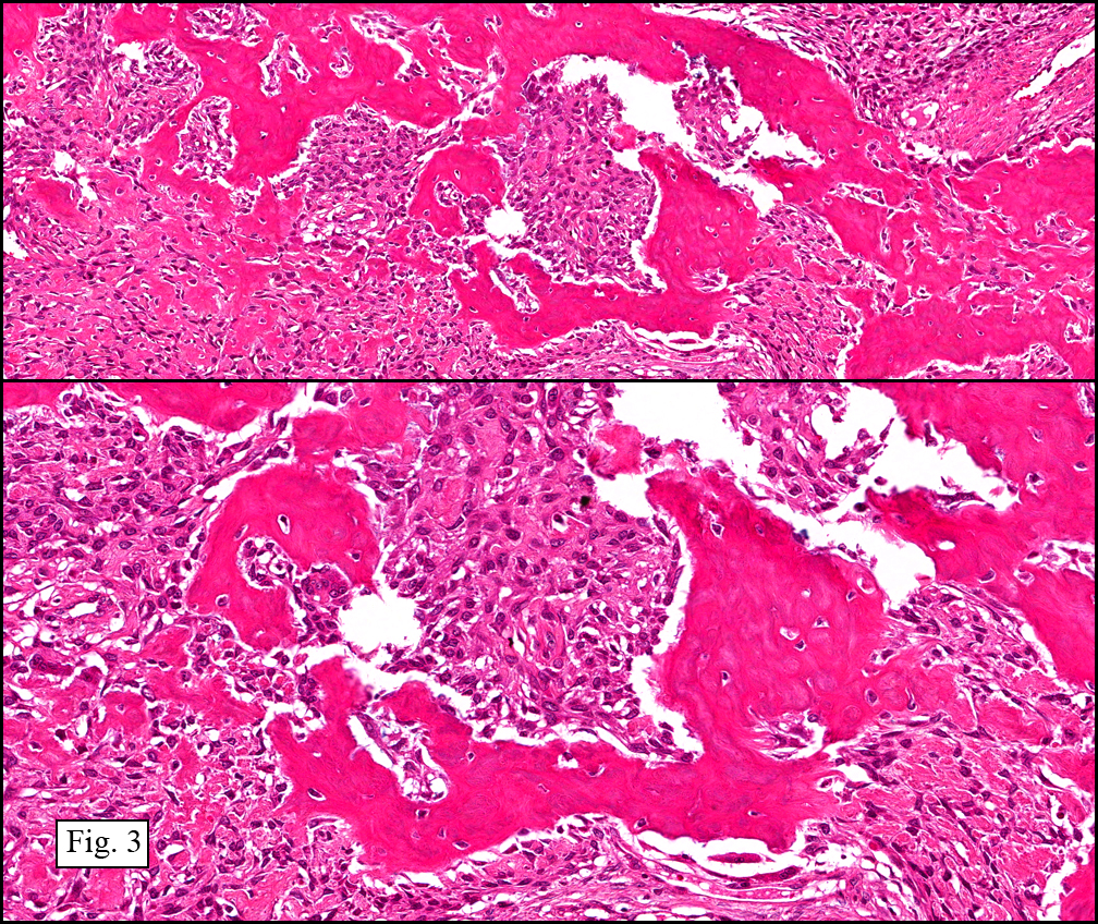
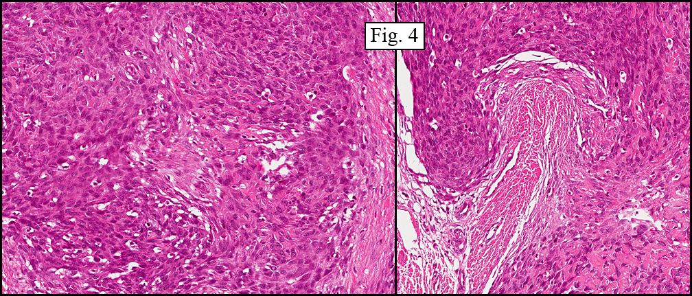
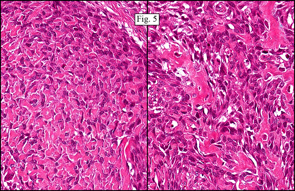
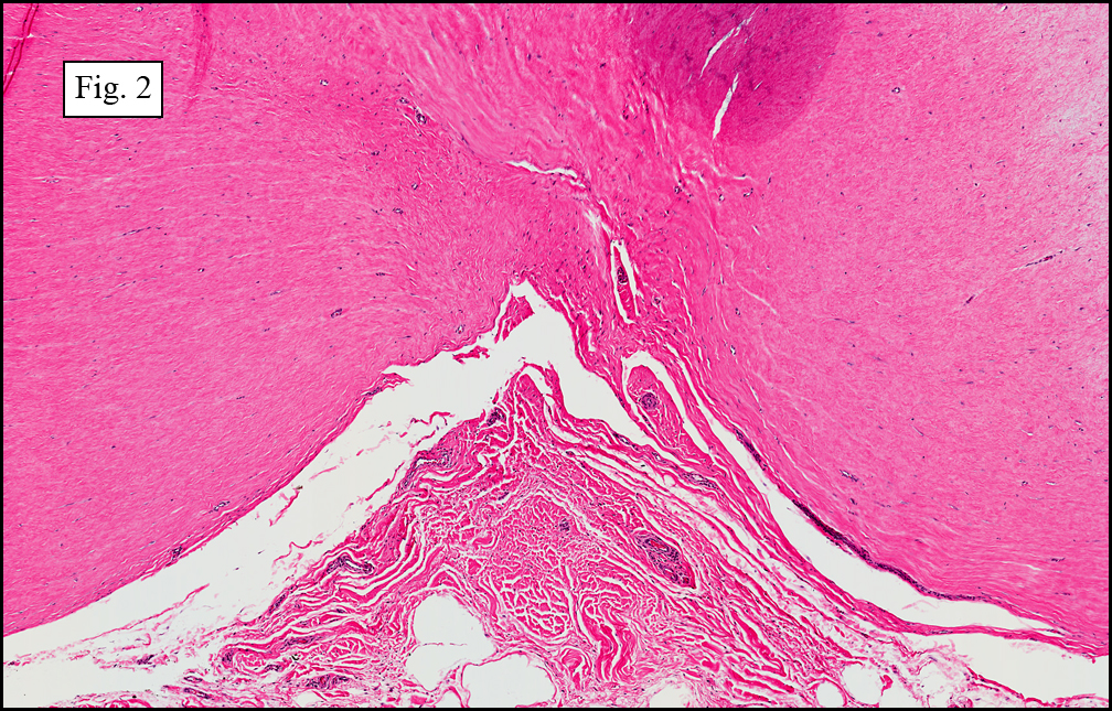
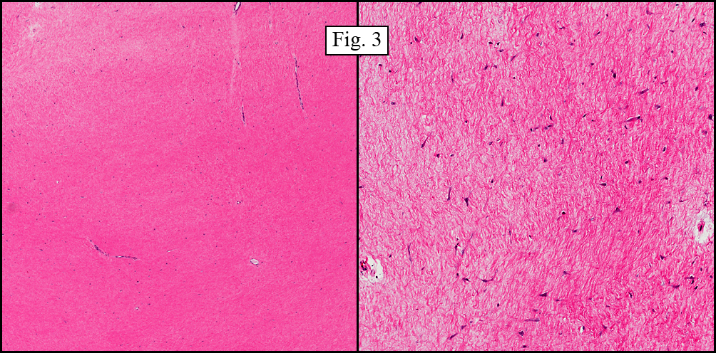
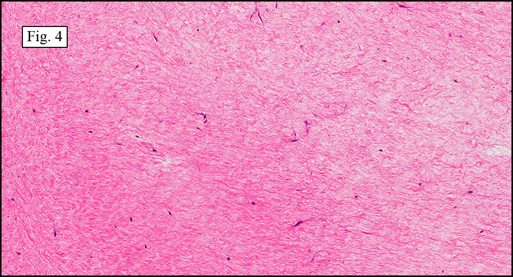
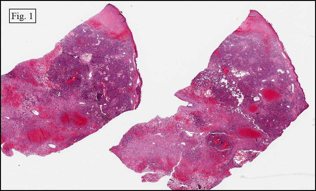
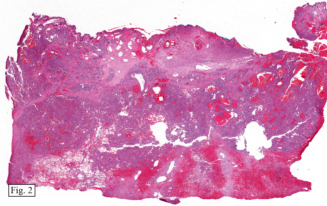
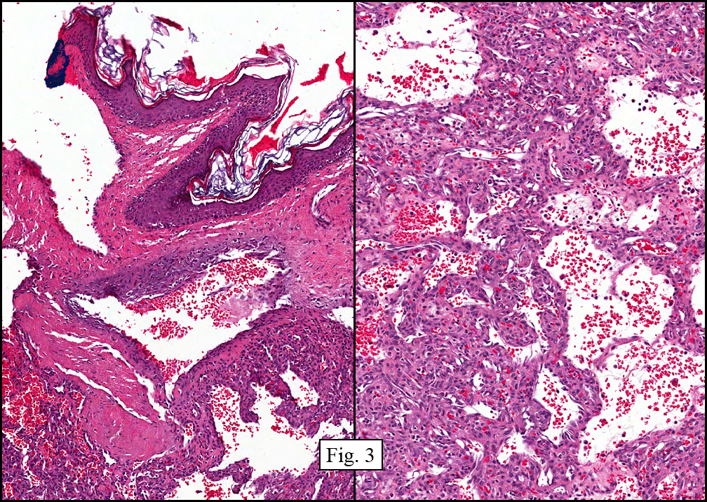
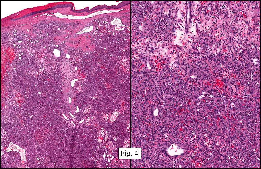
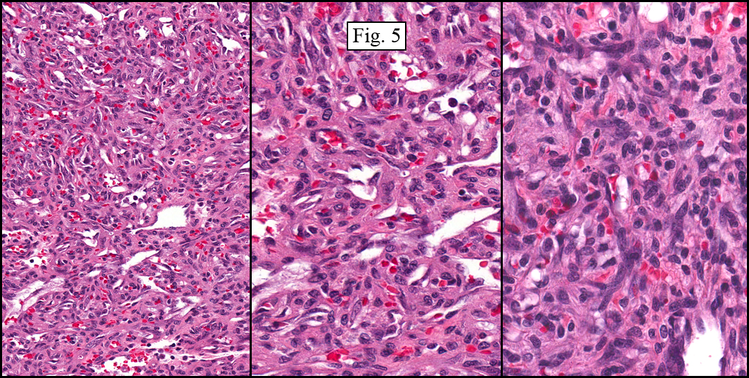
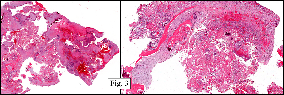
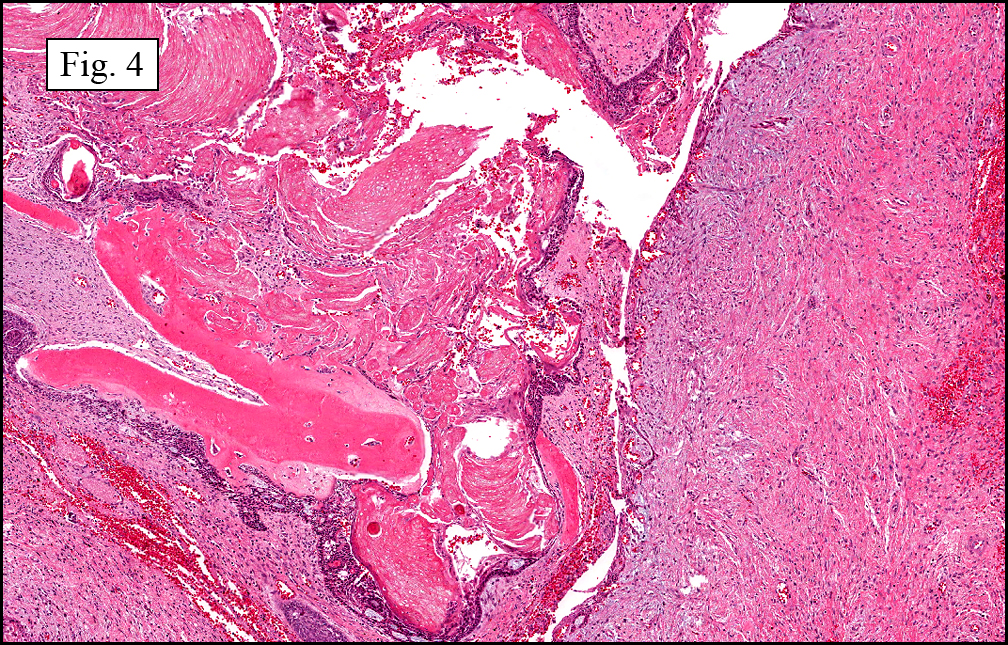
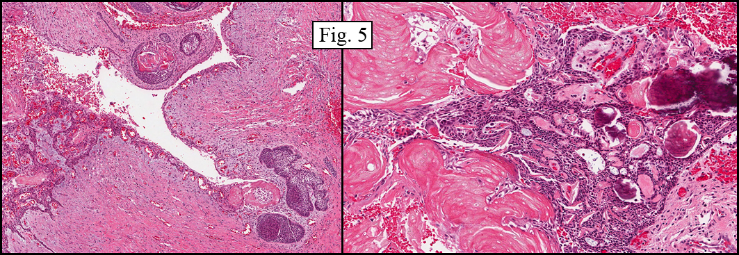
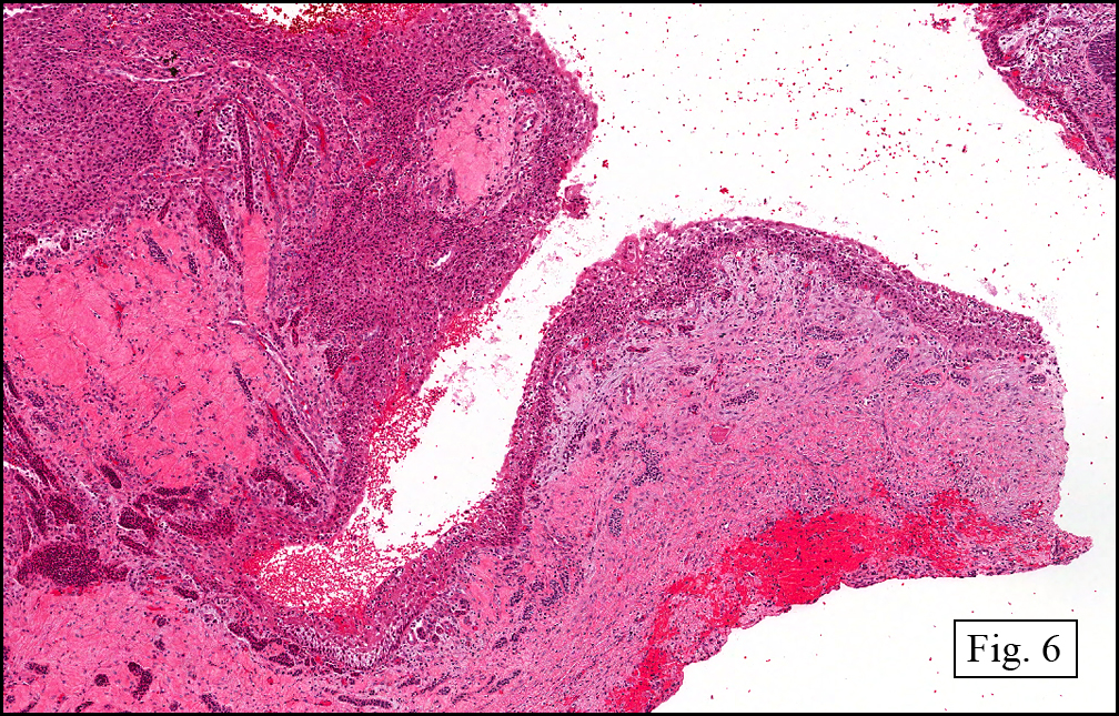
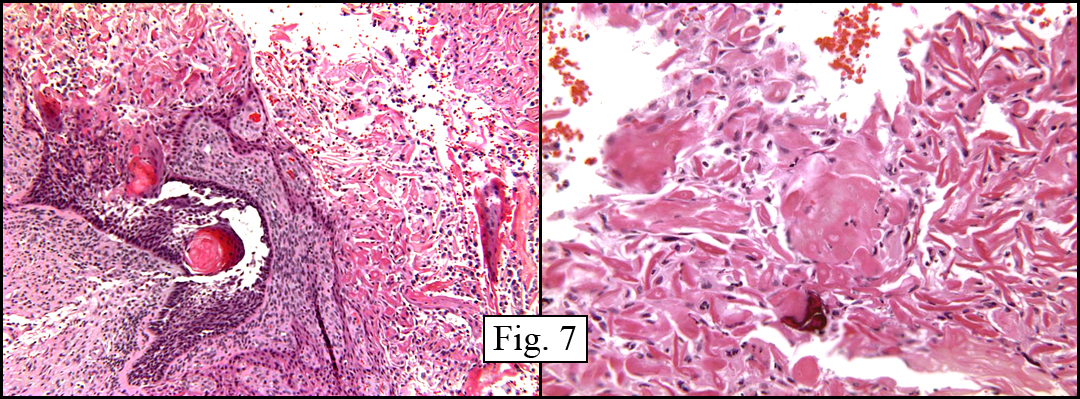
The tumor showed a peculiar trabecular growth pattern (Fig. 1), consisting of cohesive, small, polygonal to round blue cells, sometimes nested, with a prominent supporting desmoplastic stroma (Fig. 2). At the periphery of the tumor the tumor grew in nests and single files (Fig. 3). A subtle but substantial vascular network was seen (Fig. 3). There were scattered mitotic figures and mild pleomorphism (Fig. 3, 4). The tumor displayed a polyphenotypic immunoprofile and had positive staining for cytokeratin, EMA, CD99, Vimentin and NSE. It was negative for chromogranin, GFAP, S-100 and desmin (Fig. 5).

Diagnosis: “Desmoplastic Small Round Cell Tumor of Abdomen”

Miriam Peckham, PSF, and Donald R. Chase, MD
Department of Pathology and Human Anatomy, Loma Linda University and
Medical Center, Loma Linda, California
California Tumor Tissue Registry, Loma Linda, California

Discussion: First described about 20 years ago by Gerald and Rosai, desmoplastic small round cell tumor (DSRCT) is a rare malignant lesion presenting in the abdomen with distension and discomfort. It is usually peritoneal-based. DSRCT is typically found in young males (4:1) and occurs between the ages of 15 and 35. Though this tumor presents most commonly in the abdomen and pelvis, it has also been documented in various non-peritoneal sites including the parotid gland, CNS, pancreas, ovary, and lung.

Because of its lack of a definite visceral origin (though there is substantial evidence that it arises from mesothelial cells), this tumor is virtually undifferentiated and has a microscopic appearance of primitive, small, round, nested blue cells (Zellballen-like) within a desmoplastic stroma. Though this microscopic pattern is noted in a large portion of tumors, the histology may vary significantly. Some DSRCTs may show unusual cytology with spindle, rhabdoid, signet-ring, and pleomorphic cells.

Another diagnostic characteristic of DSRCT is its tendency to immuno-mark for neural, mesenchymal, and epithelial elements. The great majority of DSRCT cases stain positively for epithelial markers such as EMA and cytokeratins, as well as mesenchymal markers such as vimentin and desmin (usually in a dot-like, perinuclear pattern). Desmin positivity, which is frequently considered a diagnostic marker, was not noted in our presentation case. DSRCT has also been shown to have multiple neural antigens with neuron-specific enolase being the most common. Chromogranin and synaptophysin positivity, however, is not common. A more specific characteristic of this lesion is that it expresses WT1 – the Wilms’ tumor gene. This is believed to be a central component of DSRCT’s pathogenesis.

An important step in diagnosing DSRCT is separating it from similar-appearing small round blue cell tumors including rhabdomyosarcoma, PNET, Wilms tumor, lymphoma, neuroblastoma, small cell carcinoma, poorly differentiated carcinoma, malignant mesothelioma, and Merkel cell carcinoma. These tumors can be distinguished from DSRCT based on location (whether intra or extra-abdominal), age group (pediatric to young adult patients), and most convincingly, staining patterns via immunohistochemistry. Because of DSRCT’s characteristic polyphenotypia, an immunostain panel is needed to distinguish this entity from similar appearing tumors.

The most defining diagnostic feature of DSRCT is a reciprocal translocation of chromosomes 11 and 22, more specifically t(11;22)(p13;q12). This translocation exhibits the same genes found in Wilms tumor and Ewings sarcoma – fusion of the WT1 gene, from chromosome 11, and the EWS gene from chromosome 22. Other breakpoints for this translocation have been documented as well. Involvement of the WT1 protein has fueled speculation that its overexpression is related to the expression of both mesenchymal and epithelial markers. The EWS-WT1 chimeric protein has also been found to activate, through transcription factors, insulin-like growth factor 1. This is a possible mechanism for proliferation of tumor cells.

DSRCT behaves very aggressively and has a less than 2 year average survival rate. In the abdominal cavity it progressively fills the region with neoplastic nodules and accompanying ascites. These multiple implants frequently make complete excision impossible. A study done in 1998 followed up on 35 patients with DSRCT and found that 25 of those patients had already died, succumbing to widespread metastasis. The remaining patients lived on for an average of 25.2 months. Though the outlook of this disease is grim, the combination of surgical debulking and multimodality therapy can possibly improve survival. A study done by Lal & Sue, et al, showed a 55%, 3-year-survival in patients undergoing a combination of surgery, chemotherapy, and radiotherapy.

Suggested Reading:

1. Weiss SW, Goldblum JR. Soft Tissue Tumors. Mosby Inc. through Elsevier Inc. Philadelphia. 2008.
2. Neder L, Scheithauer BW, Turel KE, Arnesen MA, Ketterling RP, Jin L, Moynihan TJ, Giannini C, Meyer FB. Desmoplastic small round cell tumor of the central nervous system: report of two cases and review of the literature. Virchows Arch. 454:431-39, 2009.
3. Hiralal, Gamanagatti S, Thulkar S, Rao SK. Desmoplastic round cell tumour of the abdomen. Singapore Med J. 48(1): e19, 2007.
4. Lee Y, Hsiao C. Desmoplastic small round cell tumor: A clinicopathologic, immunohistochemical and molecular study of four patients. J Formos Med Assoc. 106(10):854-60, 2007.
5. Murphy AJ, Bishop K, et al. A new molecular variant of desmoplastic small round cell tumor: Significance of WT1 immunostaining in this entity. Hum Pathol. 39:1763-70, 2008.
6. Rosai J. Proceedings of the 107th California Tumor Tissue Registry semi-annual seminar, syllabus pages 32-33, December 5, 1999.
7. lal DR, Su WT, Wolden SL, et al. Results of the multimodal treatment for desmoplastic small round cell tumors. J Pediatr Surg. 40:251-5, 2005.

July 2010: A 50 year old woman with a mass in the toe

History: A 50-year-old female school teacher presented with a two year history of swelling of the right third toe. X-rays were read as being commensurate with a malignant tumor (Fig. 1), and an en-bloc resection of the third ray was performed. A hard, multinodular, tan-gray 5.0 cm mass was found to primarily involve the proximal phalangeal bone and adjacent soft tissue.

Microscopic sections showed the tumor to break out of bone and to involve surrounding tissues with extension up to the deep dermis (Fig. 2). The tumor displayed an osseous matrix consisting mostly of woven and trabecular bone often incorporating cells similar to those in the stroma (Fig. 3). In hypercellular regions that lacked bone formation, the tumor displayed both spindled and epithelioid features (Fig. 4) and had scattered mitotic figures. There were also areas which displayed a lace-like pattern of stromal cells intimately associated with, and incorporated into, osteoid (Fig. 5). Osteoclastic-like giant cells were not seen.

Diagnosis: “High-grade Conventional Osteosarcoma, Osteoblastic Type”

(Material donated by the Archdiocesan-Diocesan Health Services in Accra-Koforidua, Africa.)

Miriam Peckham, PSF, and Donald R. Chase, MD
Department of Pathology and Human Anatomy, Loma Linda University and
Medical Center, Loma Linda, California
California Tumor Tissue Registry, Loma Linda, California

Discussion: Osteosarcoma (OS) is a tumor of atypical mesenchymal tumor cells which are able to produce immature bone or osteoid. It is the most common malignant primary osseous neoplasm (20%) and has a bimodal age distribution. Most cases occur in the second decade of life. Less common are those which present over the age of 40.

Though the clinical course of this lesion is comparable in younger patients and patients over 40 years of age, there are different associations. In patients over 40, OS has been found to be secondary to radiation exposure, various chemical agents, viral infection or areas of pre-existing osseous Paget’s disease. In younger patients, no precursor conditions have been elucidated. There is, however, an association with previous trauma. OS has a male dominance (3:2).

Our presentation case is most unusual in that it occurred in the toe. Osteosarcoma arising in a small bone has only been documented five times in the literature, with the last case being reported in August of 2000. Although most cases of osteosarcoma present in the metaphyses of long bones (most commonly in the proximal tibia and distal femur), in patients over the age of 50, there is a higher occurrence in the flat bones and axial skeleton. In both age groups, however, occurrence in the digital bones of the hands and feet is fleetingly rare.

Osteosarcoma can be divided into three histologic subtypes based on the most prominent matrix material. These subtypes are “osteoblastic osteosarcoma,” “fibroblastic osteosarcoma,” and “chondroblastic osteosarcoma.” In rare cases with abundant giant cells, the tumor may be referred to as “giant cell osteosarcoma”.

Metastatic dissemination of OS is mostly hematogenous, with lung being involved in 80% of cases that metastasize. Standard treatment for OS has traditionally been amputation of the affected limb, however the success of pre-operative chemotherapy has enabled use of limb-salvage techniques. With amputation only, the 5 year survival rate is from 5-23%, but with the pre-surgical chemotherapy, the overall rate jumps to 50-65%. Differential diagnoses include osteoblastoma, chondrosarcoma, Ewing sarcoma, pleomorphic sarcoma (MFH), giant-cell tumor of bone, and exuberant fracture callus.

1. Osteoblastoma (OB) can resemble osteosarcoma due to the presence of bony trabeculae. However, the trabeculae in OB are more uniform and there is less nuclear atypia, unlike osteosarcoma which generally grows larger (>5cm) and has a destructive pattern of growth which can engulf preexisting trabeculae. Most helpful is that the sites of presentation for these two tumors are quite different with OB preferring the sacrum, vertebral column, and craniofacial bones, locations that are very rare in OS.

2. Chondrosarcoma (CS) is usually distinguished from OS through radiologic, clinical and pathologic studies. Its presenting location differs from OS by showing preference for the truncal bones. There may be morphologic overlap between variants of CS and OS, especially chondroblastic OS compared to a CS with enchondral ossification. When confronted with this dilemma, factoring in the location of the tumor, the age of the patient and any preexistent or concurrent conditions may be of help.

3. Ewing Sarcoma (ES) can be confused with an OS composed primarily of small cells. It may also be mimicked by other small, round-cell neoplasms including lymphoma, and even some rhabdomyosarcomas. However, none of these tumors form a stromal matrix and each has its own unique immunohistochemical profile.

4. Pleomorphic Sarcoma (PS, Malignant Fibrous Histiocytoma) has more atypism and pleomorphism than OS, and does not form an extra-cellular matrix. An OS with limited osteoid production has considerable morphologic overlap with PS, in fact, before PS was seen as an entity, it had been previously classified as “osteolytic, anaplastic type of osteosarcoma.”

5. Giant-cell tumor of bone may be confused with osteoclast-like giant cell osteosarcoma. However, giant-cell bone tumors most often occur in the epiphyses while osteosarcomas are most commonly found in the metaphyses. GCT usually has a uniform distribution of giant cells, while osteoclastic osteosarcoma shows region to region variation in the number of multinucleated cells.

6. Exuberant Fracture Callus may mimic OS by having a high mitotic rate, atypical osteoblasts and the production of osteoid. A distinguishing factor between the two entities is the more uniform histologic progression of healing that is present in callus but is lacking in OS. For example, the trabeculae in a callus are usually arranged in parallel, a pattern that differs from OS in which a haphazard deposition predominates. Also, the history of trauma and/or the presence of a hematoma is supportive of a reparative process.

Suggested Reading:

1. Gangadharan VP, Ramachandran K, Elizabeth SK, Preetha S, Chithrathara K, Elizabeth KA. Primary Osteosarcoma of Metatarsal Bone. Am J Clin Oncol-Canc; 23(4):429-30, 2000.
2. Dorfman HD, Czerniak B. Bone Tumors. Mosby, Inc. St. Louis,1998.
3. Rozeman LB, Cleton-Jansen AM, Hogendoorn PCW. Pathology of primary malignant bone and cartilage tumours. Int Orthop; 30(6):437-44, 2006.
4. Gassiamis A, Tsakonas G, Soukouli G, Mylonakis N, Karabelis A, Kosmas C. Diffuse calcification of metastases after intensive multiagent chemotherapy in widespread osteosarcoma leading to death in a 18-year-old male. Med Oncol; 23(4):455-62, 2006.
5. Marulanda GA, Henderson ER, Johnson DA, Letson GD, Cheong D. Orthopedic surgery options for the treatment of primary osteosarcoma. Cancer Control; 15(1):13-20, 2008.
6. Weiss SW, Goldblum JR. Soft Tissue Tumors. Mosby Inc. through Elsevier Inc. Philadelphia. 2008.
7. Wafaa E, Abdelhalim F, Chase DR. A 34 y/o male with a trauma-related mass in the right arm. California Tumor Tissue Registry, www.cttr.org, Case of the Month, vol 8(12), September, 2006.

June 2010: A 44 year old man with a right hip mass

History: An otherwise healthy 44-year-old man presented with a three month history of a painless lump over his right hip. Aspiration of the mass was dry, yielding no fluid. Surgical excision was performed, finding a 4.0 cm, smooth, pink, slightly glistening soft tissue tumor just proximal to the greater trochanteric region. The tumor was solid, without hemorrhage or necrosis.

Microscopically, the mass was composed mostly of thick, hypocellular collagenous tissue (Fig. 1). It was well-circumbscribed and minimally associated with adjacent soft tissue (Fig. 2). Widely interspaced within the collagen were stellate to spindle-shaped cells resembling fibroblasts and myofibroblasts (Fig. 3, 4). The tumor lacked pleomorphism and did not display mitotic figures.

Diagnosis: Desmoplastic fibroblastoma (Collagenous fibroma)

Miriam Peckham, PSF, and Donald R. Chase, MD
Department of Pathology and Human Anatomy, Loma Linda University and
Medical Center, Loma Linda, California
California Tumor Tissue Registry, Loma Linda, California

Discussion: Described first by Evans in 1995, desmoplastic fibroblastoma (DFB) is a rare, benign fibrous soft tissue mass that occurs most commonly in the skeletal muscle and subcutaneous tissue of adult males (5:1). This tumor presents most often in the fifth and sixth decades (mean 57 years) as a slow-growing, mobile, painless mass. The most common presentation sites include the upper and lower extremities, with the head and neck regions affected less frequently. The size of this tumor is normally less than 4 cm, but has been reported up to 20 cm.

Microscopically DFB is characterized by thick collagenous to myxocollagenous stroma with interspersed spindle and stellate-shaped fibroblasts/myofibroblasts. There are rare to absent mitotic figures and virtually no necrosis, hemorrhage or cellular atypia. Though DFB has the low-power appearance of a well-demarcated lesion, fat entrapment and infiltration of the surrounding soft tissues are commonly seen. Immunohisto- chemically, DFB positively marks for vimentin, and focally, expresses muscle-specific actin and alpha-smooth muscle actin. The tumor is generally negative for desmin, S-100 protein, and CD34.

Tumor behavior is usually indolent, with a slowly expanding soft tissue mass, which, when adequately excised, has limited capacity for local recurrence. To date, metastases have not been reported. This has led to question the nature of DFB and it has been reported that the tumor has a specific breakpoint at 11q12, supporting the notion that is a neoplastic process rather than a reactive one. Differential diagnoses include:

1. Nuchal fibroma: An uncommon lesion, nuchal fibroma most commonly occurs in the back of the neck of adult males. This lesion has a 30-40% association with diabetes and may recur after excision, most likely due to its persistent inciting cause. Histologically, its appearance is very similar to desmoplastic fibroblastoma in that it is made up of hypocellular collagen strands. Also seen are entrapped fat cells and an increased amount of small nerves. This morphology also overlaps with Gardner fibroma.

2. Gardner fibroma: Most commonly presenting in childhood, this lesion occurs mostly in the trunk with a predilection for the paraspinal region. The majority of patients with this lesion have familial adenomatous polyposis (FAP), with the fibroma preceding polyp formation. The correlation of this lesion with FAP makes the proper diagnosis of this fibroma of increased importance. These lesions are poorly demarcated and may recur as desmoid fibromatosis. Histologically, these fibromas show hypocellular collagen bands with artifactual cleft spaces. The cells within the mass have a bland, fibroblast-like appearance and there is frequently entrapment of surrounding soft tissue, all of which is similar to desmoplastic fibroblastoma. These masses stain positively for beta-catenin (similar as desmoid fibromatosis).

3. Neurofibroma: This lesion is characterized by wavy cells embedded within a myxocollagenous stroma. However, unlike desmoplastic fibroblastoma, they usually stain positive for S-100 protein.

4. Fibromatosis: This can be differentiated from desmoplastic fibroblastoma by its increased cellularity and its infiltrative nature. Another difference is this lesions characteristic cellular arrangement into broad fascicles.

5. Low-grade fibromyxoid sarcoma: This tumor is more cellular and more myxoid than DFB. It also tends to form whorls of spindled cells with a prominent fibromyxoid background.

6. Calcifying fibrous pseudotumor: This lesion affects a younger age group than DFB which is usually found in the fifth and sixth decades. This pseudotumor also shows a characteristic lymphoplasmacytic infiltrate, as well as multiple psammomatous calcifications located throughout its stroma.

7. Elastofibroma: This mass occurs mostly on the upper back in the region of the scapula. It is felt to be degenerative in nature, possibly due to repetitive motion. It diagnostically contains wavy elastic fibers which are absent in desmoplastic fibroblastoma.

8. Fibrous hyperplasia: Unlike desmoplastic fibroblastoma, this reparative inflammatory lesion presents with a history of trauma. The size of this lesion is also significantly smaller, ranging up to 1.5 cm.

9. Nodular fasciitis: This pseudosarcoma is more cellular, particularly in its early proliferative phase, and contains increased mitotic activity. It also shows rapid growth, contrasting with DFB which is indolent. A characteristic feature of NF is the mucinous separation of the tumor cells, resembling tissue paper that is being torn apart. The resolution stage of NF may mimic DFB, but is more cellular, mimicking benign fibrous histiocytoma.

Suggested Reading:

1. Watanabe H, Ishida Y, Nagashima K, Makino T, Norisugi O, Shimizu T. Desmoplastic fibroblastoma (collagenous fibroma). J Dermatol. 35(2):93-7, 2008.
2. Weiss SW, Goldblum JR. Soft Tissue Tumors, 5th Edition. Mosby Inc. through Elsevier Inc. Philadelphia. 2008.
3. Takahara M, Ichikawa R, Oda Y, Uchi H, Takeuchi S, Moroi Y, Kiryu H, Furue M. Desmoplastic fibroblastoma: a case presenting as a protruding nodule in the dermis. J Cutan Pathol. 35:70-3, 2008.
4. Walker KR, Bui-Mansfield LT, Gering SA, Ranlett RD. Collagenous fibroma (desmoplastic fibroblastoma) of the shoulder. AJR Am J Roentgenol. 183(6):1766, 2004.
5. Dagli M, Eryilmaz A, Acar A, Kulacoglu S, Akmansu H. Collagenous fibroma (desmoplastic fibroblastoma). Yonsei Med J. 45(5):941-3, 2004.
6. Shimoyama T, Horie N, Ide F. Collagenous fibroma (desmoplastic fibroblastoma): a new case originating in the palate. Dentomaxillofac Radiol. 34(2):117-9, 2005.
7. Fletcher, CDM. Diagnostic Histopathology of Tumors, 3rd Edition. Elsevier Limited. Philadelphia. 2007.

May 2010: A three month old boy with a rapidly growing mass in the eyelid

History: A three month old baby boy presented to plastic surgery clinic with a purple-red tumor on his right eyelid. Radiographs taken prior to surgery demonstrated a well-defined soft tissue mass without calcification.

The resected specimen microscopically showed a mildly hyperkeratotic epidermis undermined by a cellular, vascular tumor (Fig. 1, 2). The tumor showed cyst-like spaces filled with blood, similar to changes seen in an aneurysmal bone cyst (Fig. 3). Much of the tumor was solid, supported by ectatic blood vessels, focally resembling a hemangiopericytoma vascular pattern (Fig. 4). Closer evaluation of the cells showed a striking resemblance to pericytes, and smaller, ectatic vascular spaces which were sometimes slit-like (Fig. 5). Mitotic figures were rarely encountered, and no significant cellular pleomorphism was seen. Immunostains confirmed the vascular nature of the tumor and were also positive for GLUT-1 (see reference).

Diagnosis: Kaposiform Hemangioendothelioma of the Eyelid

Michael J. Matus PSF, Mia Perez MD, Donald R. Chase MD
Department of Pathology and Human Anatomy, Loma Linda University and
Medical Center, Loma Linda, California
California Tumor Tissue Registry, Loma Linda, California

Discussion: Kaposiform hemangioendothelioma (KHE) is a rare vascular tumor which can be locally aggressive and is most often found in skin, deep soft tissue, and/or bone in infants and children. Rarely does it occur in adults.

The term kaposiform refers to a morphologic resemblance to Kaposi sarcoma and the designation hemangioendothelioma implies its uncertain biologic behavior felt to lie between that of hemangioma and angiosarcoma.

Although KHE is often locally extensive and aggressive, it has not been shown to metastasize. Despite this, death has resulted due to severe coagulopathy related to the neoplasm. This association, termed Kasabach-Merritt syndrome (KMS), was described by Dr. Haig Haigouni Kasabach and Dr. Krom Merritt in 1940. Also known as Hemangioma with thrombocytopenia this syndrome occurs when a vascular tumor traps platelets, resulting in severe thrombocytopenia and consumptive coagulopathy in which clotting factors are activated and fibrinogen is diminished, worsening the bleeding tendency. This progress can progress to disseminated intravascular coagulation and death.

The differential diagnosis of Kaposiform hemangioendothelioma includes:
1) Kaposi sarcoma
2) Infantile (juvenile) hemangioma
3) Congenital non-progressive hemangioma
4) Tufted angioma

Unlike EHE, Kaposi sarcoma rarely occurs in children (except for the African lymphadenopathic form) and is usually associated with human herpesvirus 8 (HHV8), a result not found in EHE. There are several different clinical presentations, however all bare the same microscopic pattern. Most commonly occurring in adults, a flat red to purple lesion signals early tumor involvement, mirrored by a histologic proliferation of miniature vessels surrounding central ecstatic vessels. As vascular proliferation continues, bland spindle cells appear on the periphery. These spindle cells, devoid of significant pleomorphism, coalesce into distinct nodules and are eventually separated by slit-like spaces that contain erythrocytes.

Infantile hemangioma is the most common vascular tumor of infancy and occurs in nearly 4% of children. Histologically, tumor lobules are separated by normal appearing stromal elements composed of fibroblasts, pericytes, mast cells, and in some cases interstitial dendritic cells. The endothelial cells are prominent, and often contain significant rough endoplasmic reticulum and, by electron microscopy, Weibel-Palade bodies. This vascular lesion is immunoreactive to CD31, CD34, factor-VIII-related antigen as well as GLUT1 (see reference).

Congenital non-progressive hemangioma differs from EHE by being fully formed at birth with subsequent involution in most cases. A peripheral rim of pallor with a central depressed ulcer vividly portrays this change if the involution is rapid. The histological distinction from EHE is that the tumor lobules are surrounded by dense connective tissue. Also the clinical appearance of the overlying skin is often atrophic and microscopically, it lacks adnexal structures.

The term Tufted angioma (TA) was coined in 1989 by Wilson-Jones and Orkin for a tumor previously termed angioblastoma. Grossly the lesion presents as pink macules and plaques that grow slowly then stabilize. Histologically, there is an abundance of angiomatous lobules along the periphery of the lesion with fewer in the central region. In addition, they often include eccrine glands and venules surrounded by dense connective tissue. This appearance closely mimics EHE, and some authorities include TA in the EHE category, splitting them only by age of presentation. EHE appears to be the juvenile form, and TA, the adult form of what may actually be the same tumor. Both diagnoses are important, however, as each can give rise to Kasabach-Merritt syndrome, especially when deeply located in soft tissue or in the retroperitoneum.

The identification of EHE is important mostly for its association with KMS. Unlike congenital non-progressive hemangioma, it does not regress and therefore requires total surgical extirpation. To date, the separation of EHE from tufted angioma is mostly based upon patient age. Both, however, can give rise to KMS.

Suggested Reading:

Rastogi S, Banerjee S, et al. Glut-1 antibodies induce growth arrest and apoptosis in human cell lines. Cancer Letters, 253(2):244-251, 2007.

Lai, FMM, To, KF, Choi PCL, Leung PC, Kumta SM, Yuen PPM, Lam WY, Cheung ANY, Allen PW. Kaposiform Hemangioendothelioma: Five Patients with Cutaneous Lesion and Long Follow-Up. Mod Pathol 14(11):1087-1092, 2001.

Mulliken JB, Glowacki J. Hemangiomas and vascular malformations in infants and children: a classification based on endothelial characteristics. Plast Reconstr Surg 69:412-422, 1982.

North PE, Waner M, Beckmiller L, James CA, Mihm Jr MC. Vascular tumors of infancy and childhood: beyond capillary hemangioma. Cardiovascular Pathology 15:303-317, 2006.

North PE, Waner M, James CA, Mizeracki A, Frieden IJ, Mihm Jr MC. Congenital Nonprogressive Hemangioma. Arch Dermatol 137:1607-1620, 2001.

Weiss, SW, Goldblum, JR. Enzinger & Weiss’s Soft Tissue Tumors. 5th Ed. Mosby Inc.

April 2010: A 16 year old boy with a left mandibular mass

History: A 16 year-old boy presented to his orthodontist with a two-year history of left mandibular swelling. An x-ray showed a well-circumbscribed radiolucent lesion of the left body of mandible extending from teeth #18-22 (Fig. 1). The left mandibulectomy specimen contained a 4.2 x 2.2 cm cystic lesion (Fig. 2). Tissue removed from the cyst consisted of loosely adherent pink-brown soft-tissue fragments with focal areas of necrosis.

The tumor consisted of an admixture of trabecular bone and hemorrhagic fibrous tissue (Fig. 3). A cystic lining was composed of squamoid elements with a prominent basal component (Fig. 4, 5, 6). Aggregates of “ghosted epithelium” were present throughout (Fig. 4, 5, 7) as were areas of ameloblastic-type epithelium (Fig. 5). Focally calcified keratinaceous debris was seen, sometimes associated with basaloid epithelium (Fig. 7). Adjacent soft tissues showed regions of tooth germinal epithelium with tooth-like pulp stroma, with occasional amounts of eosinophilic material consistent with dentin.

Diagnosis: “Calcifying Odontogenic Cyst (‘Keratinizing and Calcifying Odontogenic Cyst’, ‘Gorlin Cyst’) with associated early odontoma formation”

Justin Kerstetter, MD, Craig Zuppan, MD, and Donald R. Chase, MD
Department of Pathology and Human Anatomy, Loma Linda University and Medical Center, Loma Linda, California
California Tumor Tissue Registry, Loma Linda, California

Discussion: Calcifying odontogenic cyst (COC), classified as an odontogenic tumor by the World Health Organization (WHO), was first identified as a distinct entity by Gorlin et al. in 1962, and now is felt to be a true neoplasm. These lesions arise from a more mature enamel epithelium than ameloblastoma and accordingly appear to have less growth potential. After Gorlin’s first description, COC was re-labeled as a “calcifying ghost cell odontogenic tumor” by Fejerskov and Krogh in 1972, and now is sometimes referred to as a “Keratinizing and Calcifying Odontogenic Cyst”. The tumor usually arises in younger adults with most cases diagnosed in the second to third decades, but patients may range in age from infant to adult. COC is predominantly an intraosseous tumor although approximately 13-30% of the cases occur as extraosseous lesions. Both intraosseous and extraosseous lesions occur with equal frequency in the maxilla and mandible with about 65% of the cases being found in the incisor and canine areas.

In addition to the histologic features described in this case, calcifying odontogenic cysts are well-defined with a fibrous capsule and a lining of odontogenic epithelium of 4 to 10 cells in thickness. The basal cells may be cuboidal or columnar and resemble ameloblasts. The most characteristic feature of this tumor is variable numbers of ghost cells within the epithelial component. These cells are altered epithelial cells showing loss of their nuclei with preservation of basic cell outline. Finally, COC may be associated with other odontogenic tumors, with odontoma being the most common. However, other tumors reported in association with COC include ameloblastomas and adenomatoid odontogenic tumors.

The differential diagnosis of COC includes:

• Dentigerous cysts may be associated with unerupted teeth. They originate from the separation of the follicle from around the crown of the unerupted tooth and remain attached to the tooth which remains in the alveolar bone. Histologically, they demonstrate a fibrous capsule with a thin, non-keratinized epithelial lining. These cysts may demonstrate an inflamed or non-inflamed appearance based on the presence of a chronic inflammatory cell infiltrate.

• Eruption cysts are the soft-tissue variant of the dentigerous cyst. Their origin is also from the separation of the dental follicle from the crown of an erupting tooth that is within the soft tissue overlying the alveolar bone. Histologically there is the presence of oral epithelium overlying the roof of the cyst which is comprised of a thin layer of nonkeratinizing squamous epithelium.

• Odontogenic keratocysts arise from cell rests of the dental lamina and are associated with Gorlin syndrome when they are multiple. The luminal surface of the cyst demonstrates flattened, parakeratotic epithelium which is wavy or corrugated in appearance. The basal epithelial layer is composed of a palisaded layer of cuboidal or columnar epithelial cells which are hyperchromatic. These have a tendency to recur.

• Glandular odontogenic cysts occur mainly in the anterior portion of the mandible. They are lined by squamous epithelium of varying thickness. The superficial epithelial cells lining the cyst cavity are cuboidal to columnar and have an irregular, sometimes papillary surface. Pools of mucinous material are often present within the epithelium.

Suggested Reading:

Calcifying odontogenic cyst – a possible analogue of the cutaneous calcifying epithelioma of Malherbe (an analysis of fifteen cases). Gorlin RJ, Pindborg JJ, Clausen FlP et al. Oral Surg, 15:1235-1240, 1962.

Syndromes of the Head & Neck, by Gorlin RJ and Pindborg, McGraw-Hill, New York, 1964.

The calcifying ghost cell odontogenic – or the calcifying odontogenic cyst. Fejerskov O, Krogh J. J Oral Pathol, :273-287, 1972.

Calcifying odontogenic cyst associated with odontoma: report of two cases. Keszler A, Gugliemontti NB. J Oral Surg, 30:893-897, 1972.

Calcifying odontogenic cyst. A review and analysis of seventy cases. Freedman PD, Lumerman H, Gee JK. Oral Surg, 40:93-106, 1975.

The keratinizing and calcifying odontogenic cyst (Gorlin cyst). Clark MJ, Alley RS, Steed DL, Tilson HB. J Am Dent Assoc. 100(4):563-564, 1980.

Calcifying odontogenic cyst: case report and review of literature. el-Beialy RR, el-Mofty S, Refai H. J. Oral Maxillofac Surg, 48(6)637-40, 1990.

Scroll to top