History: A 36 year-old man underwent surgical resection of a 10.0 x 5.5 x 4.0 cm ill-defined left anterior leg mass The excised specimen had a tan, vaguely lobulated cut surface with central hemorrhage It grossly appeared to abut the deep fascial resection margin.
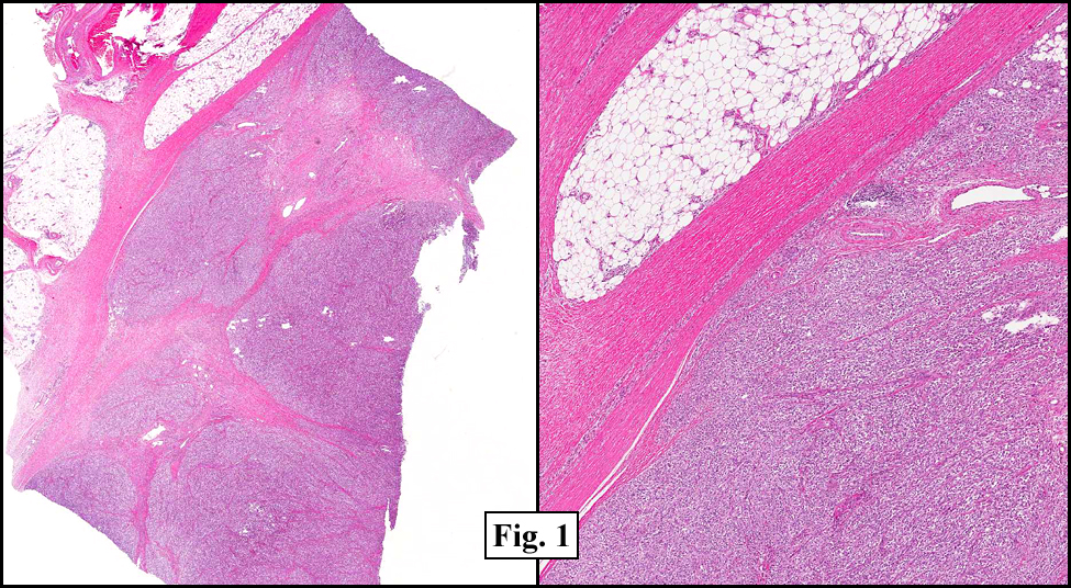
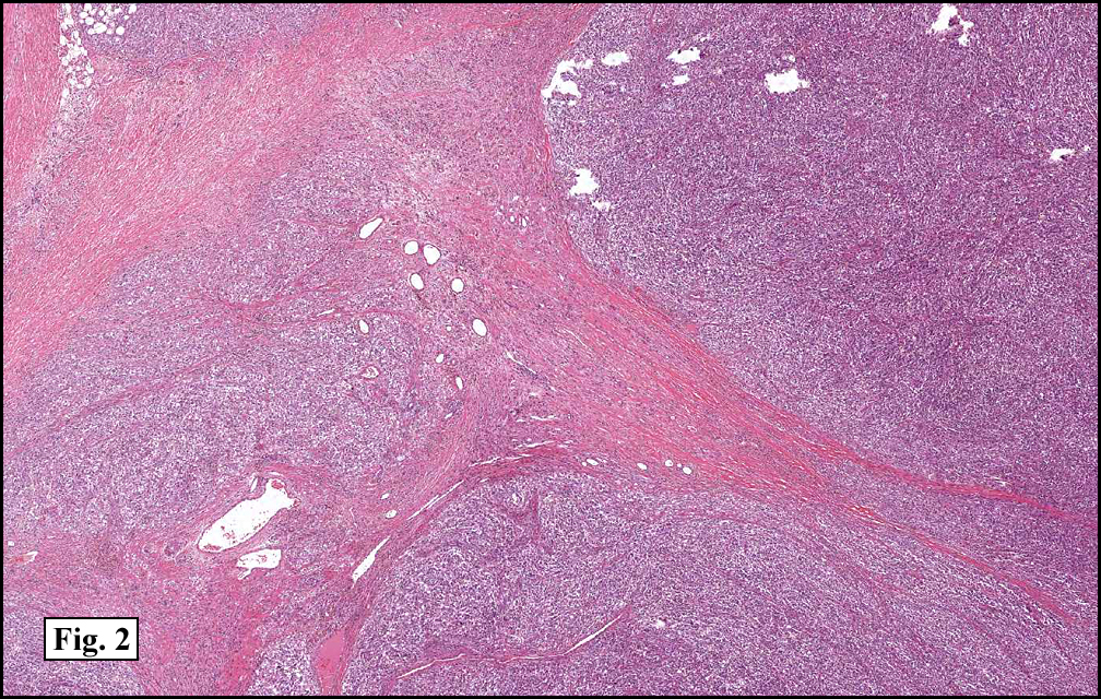
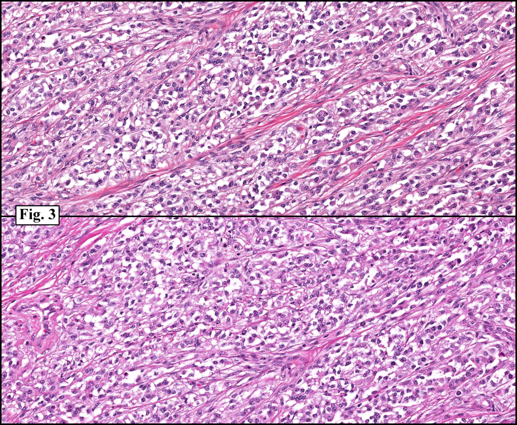
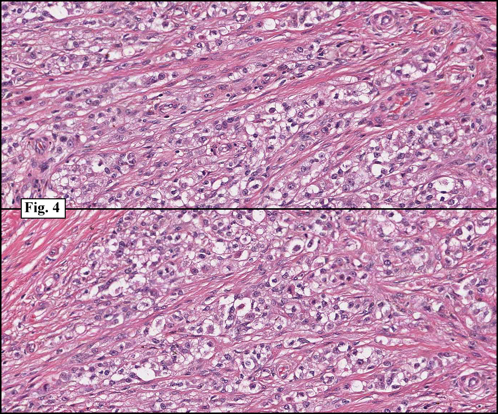
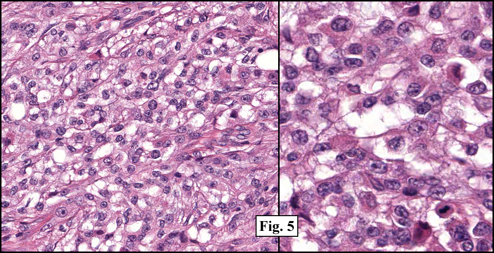
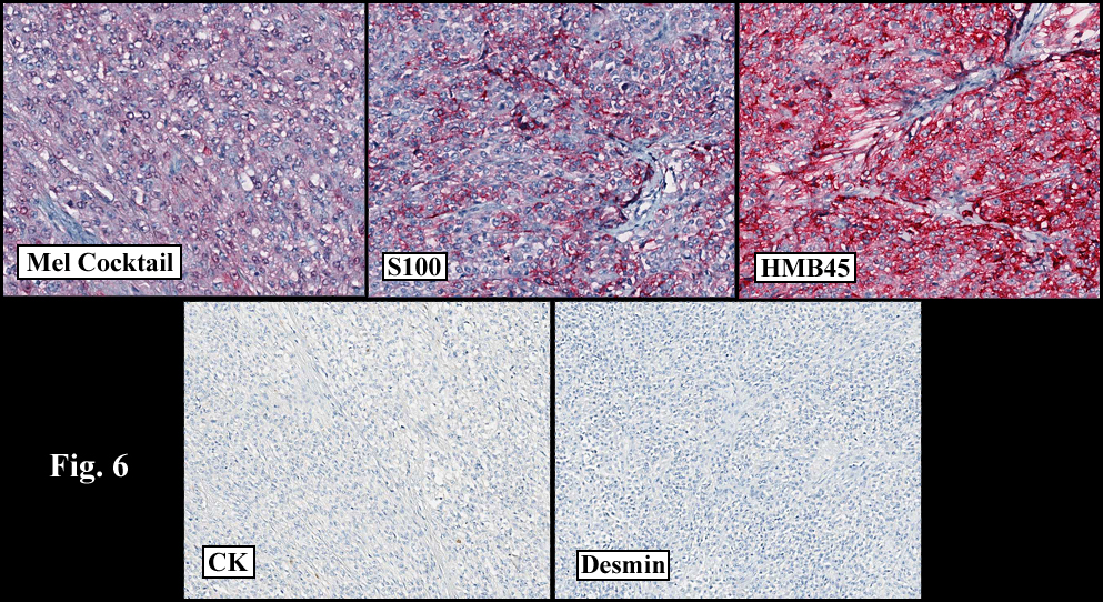
Microscopically, the mass was well-circumbscribed (Fig. 1). It had lobules of nested spindled cells separated by fibrous septae (Fig. 2, 3). The cytoplasm was generally clear and the nuclei were vesicular, many times with prominent nucleoli (Fig. 4). Rare mitotic figures were seen (Fig. 5, right). Immunohistochemistry revealed that the tumor cells were positive for melanoma cocktail, S100, and HMB45, and were negative for cytokeratin and desmin (Fig. 6).
Diagnosis: Clear cell sarcoma
Kate Grogan, MD, and Donald R. Chase, MD
Department of Pathology & California Tumor Tissue Registry
Loma Linda University and Medical Center, Loma Linda, California
Discussion: Initially described by Franz Enzinger in 1965, clear cell sarcoma (CCS or “Malignant melanoma of soft partsâ€) is a rare aggressive neoplasm of adolescents and young adults (with a mean age of 31 years). It typically presents as a mass in the deep tissues of the lower extremities, usually adjacent to tendons, fascia or aponeuroses.
Histologically, the CCS consists of nests or fascicles of spindled cells with a vesicular chromatin pattern, prominent nucleoli and abundant clear to weakly eosinophilic cytoplasm. In general, CCS tends not to be highly pleomorphic. Mitotic figures are scarce, but multinucleated tumor giant cells are frequently identified. Melanin is present in approximately 50% of cases.
The great majority of cases show diffuse staining for S-100 and frequently also express HMB-45, MiTF and Melan-A.
Cytogenetic findings: One of the most defining diagnostic features of clear cell sarcoma is a reciprocal translocation of chromosomes 12 and 22, more specifically t(12;22)(q13;q12). This translocation results in the fusion of EWS with ATF-1, with the resultant protein mimicking the action of melanocyte stimulating hormone (MSH).
Differential Diagnosis:
- Malignant melanomas typically involve the dermis, whereas clear cell sarcomas traditionally originate in deep structures, with rare dermal involvement. In ambiguous cases, FISH analysis and RT-PCR can be performed to evaluate for the hallmark EWS-ATF1 gene fusion, which is not seen in malignant melanoma.
- Cellular Blue Nevi often occur in similar age groups and locations. The blue nevi cells tend to be smaller and nuclei have a less vesicular pattern. Often recurrent cases have pronounced cytologic atypia, and molecular genetic analysis may be indicated to differentiate atypical blue nevus from clear cell sarcoma.
- Fibrosarcoma, synovial sarcoma and peripheral nerve sheath tumors also have a fascicular growth pattern, however are usually easily distinguished from CCS with immunohistochemistry.
- Poorly preserved or degenerating specimens may be confused with a round cell sarcoma, particularly alveolar rhabdomyosarcoma, but immunohistochemistry should help delineate them.
In general, complete surgical excision with generous tumor-free margins is the treatment of choice. Chemotherapy is typically ineffective in these patients. Recurrences, which reflect the adequacy of initial excision, range from 14% to 39%. Up to one half of these patients will develop lung or lymph node metastases within 2-8 years. Late metastases (up to 20 years post surgical intervention) have been reported. Prognostic factors include size, necrosis, and nodal metastases.
Suggested Reading:
Chase DR, Rosai J, Argani, P. Clear Cell Sarcoma of Tendon Sheath. CTTR 129th Semi-Annual Slide Seminar. 37-9, 2010.
Antonescu CR, Dal Cin P, Nafa K, Teot LA, Surti U. Fletcher CD, Ladanyi M. EWSR1-CREB1 Is the Predominant Gene Fusion in Angiomatoid Fibrous Histiocytoma. Genes, Chromosomes & Cancer 2001; 46: 1051-1060.
Antonescu CR, Tschernyavsky SJ, Woodruff JM, Jungbluth AA, Brennan MF, Ladanyi M. Molecular Diagnosis of Clear Cell Sarcoma. Detection of EWS-ATF1 and MITF-M Transcripts and Histopathological and Ultrastructural Analysis of 12 cases. Journal of Molecular Diagnostics 2002 4: 44-52.
Enzinger FM, Clear Cell Sarcoma of Tendons and Aponeuroses. An Analysis of 21 cases. Cancer 1965; 18: 1163-74.
Chung EB, Enzinger FM. Malignant melanoma of soft parts. A reassessment of clear cell sarcoma. Am J Surg Pathol 1983; 7: 405-13.
Meis-Kindblom JM. Clear Cell Sarcoma of Tendons and Aponeuroses: A historical Perspective and Tribute to the Man Behind the Entity. Adv Anat Pathol 2006; 13: 286-292.
Weiss S, Goldblum J. Enzinger and Weiss’s Soft Tissue Tumors (5th edition). Philadelphia: Mosby/Elsevier Inc. 926-34, 2008.
Zucman J, Delattre O, Desmaze C, et al. EWS and ATF-1 gene fusion induced by t(12:22) translocation in malignant melanoma of soft parts. Nature Genetics 1993; 4: 341-345.