January 2010: A 45 year old woman with a large cystic ovarian mass and multiple uterine nodules

History: A 45-year-old gravida 0 woman presented with frequent menometrorrhagia. Transabdominal sonography and computerized tomography revealed a large multi-loculated cystic mass in the left ovary, multiple nodules in the myometrium, and pelvic lymphadenopathy. The patient was treated with total abdominal hysterectomy, bilateral salphingo-oophorectomy, and limited pelvic lymph node dissection.

The excised uterus and adnexa weighed 2450 grams and contained a multicystic 34.5 x 21.0 x 9.5 cm left ovarian mass dominated by thin and thick-walled cysts filled with mucoid material. The outer surface was smooth. The uterus was 8.0 × 4.5 × 2.5 cm and had unremarkable endometrium and multiple intramural leiomyomata up to 0.8 cm. Both oviducts were normal as was the right ovary. Multiple enlarged pelvic lymph nodes up to 1.5 cm in diameter were present having grey and pink homogeneous cut surfaces. The omentum was unremarkable.

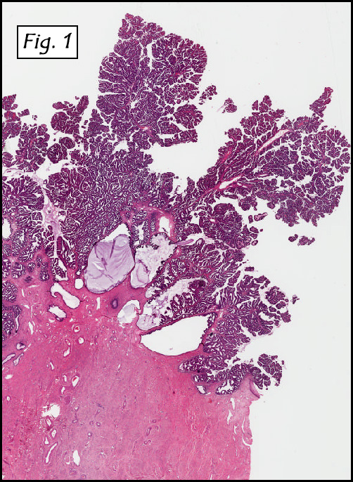
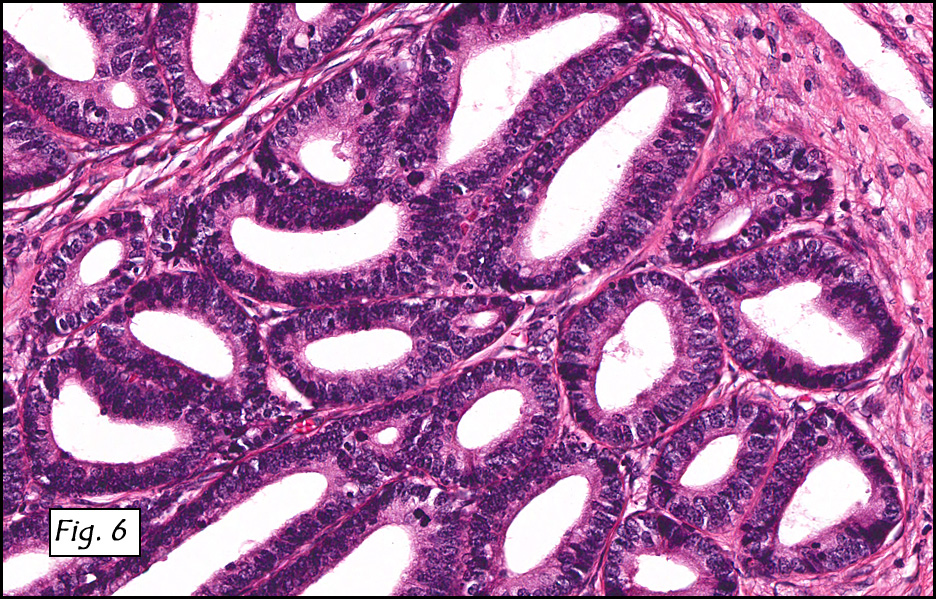
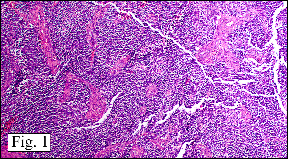
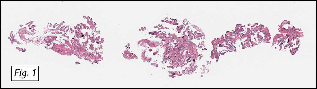
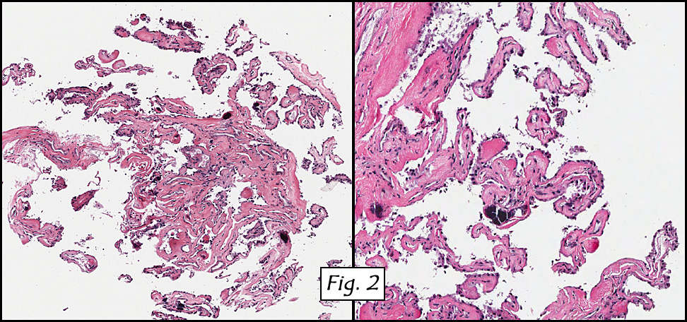
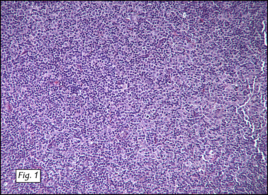
Microscopically, the dominant left ovarian mass had cystic spaces lined by mucin-secreting columnar calls (Fig. 1). Minimal stratification was seen and neither complex microvilli nor marked nuclear pleomorphism were encountered. Stromal invasion was not seen.

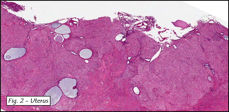
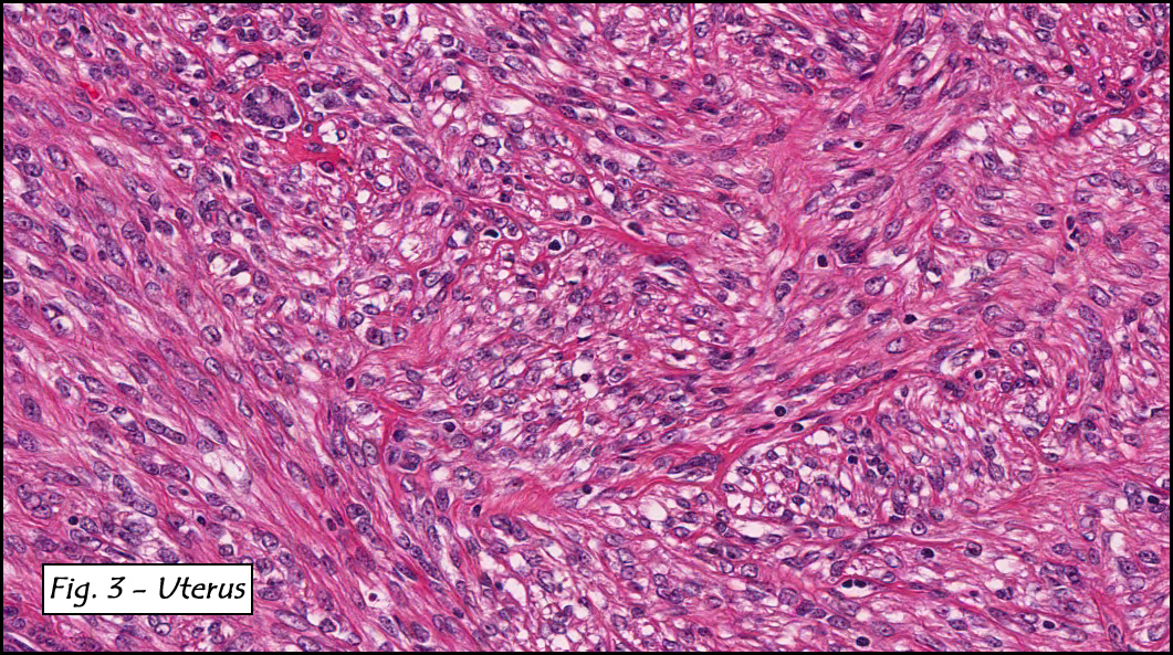
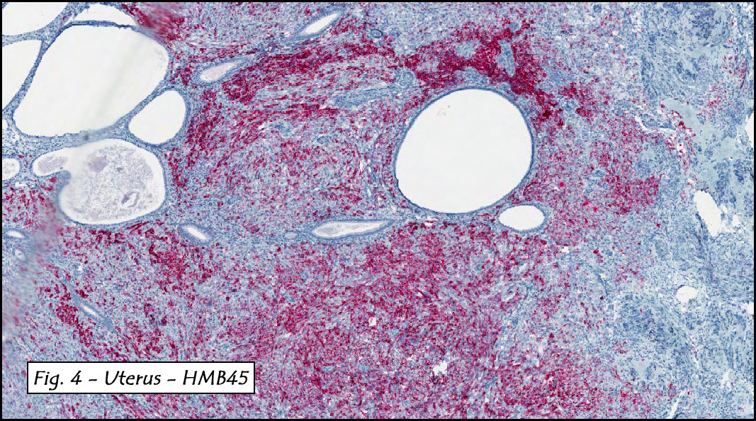
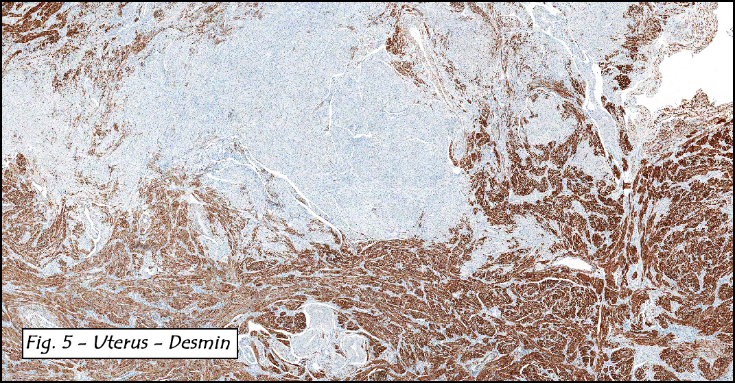
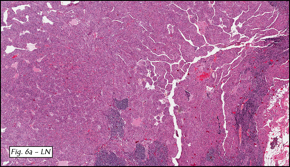
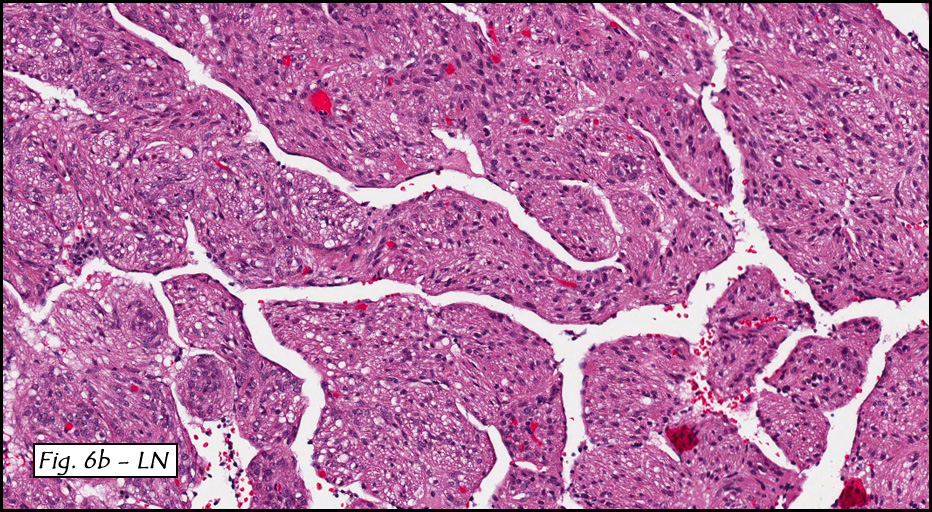
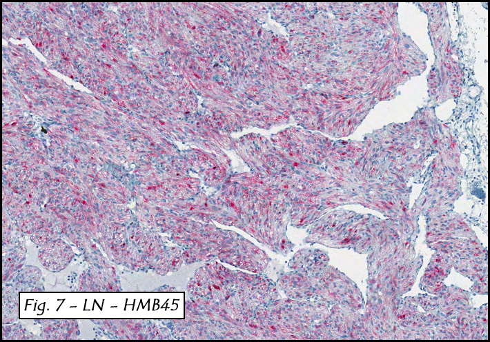
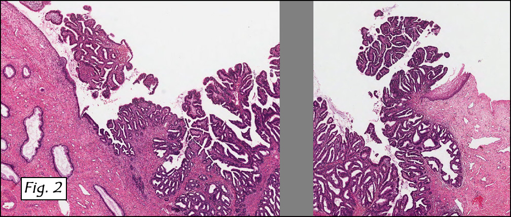
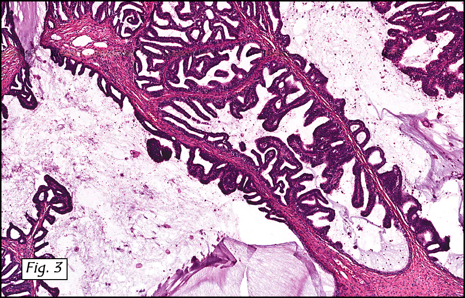
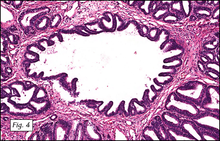
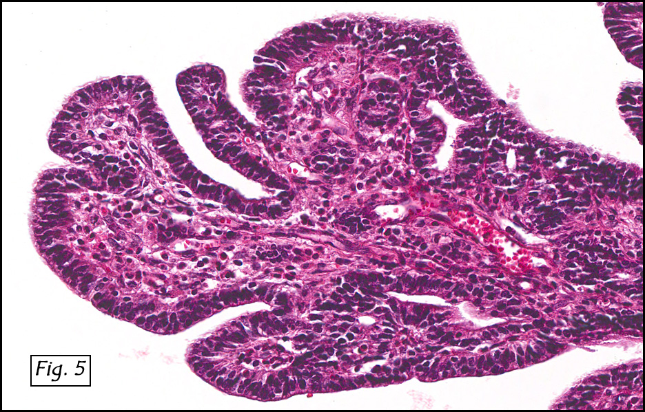
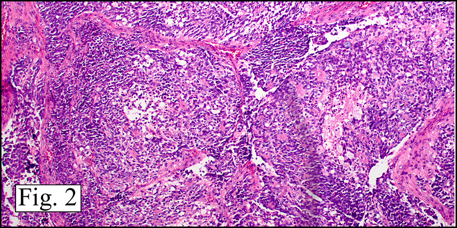
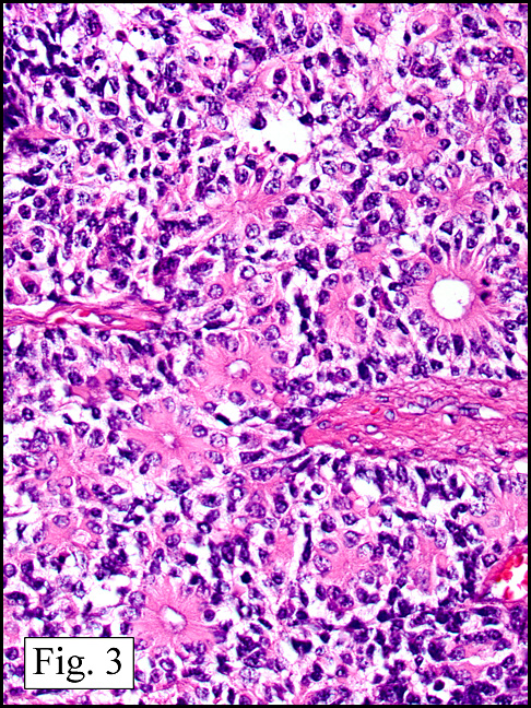
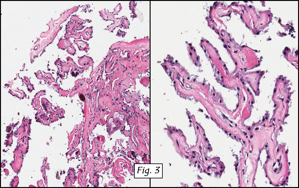
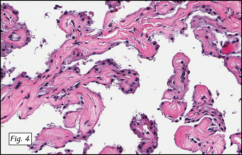
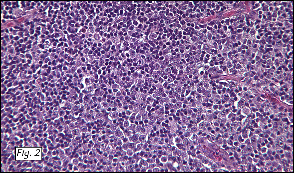
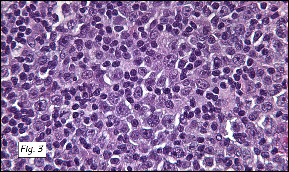
In addition to focal adenomyosis and benign leiomyomata, the uterus was found to have a 3-4 mm superficial tumor in the myometrium consisting of confluent nodules of blunt-ended spindled and epithelioid cells growing in short fascicles around mildly dilated entrapped endometrial glands (Fig. 2). The neoplastic nuclei were round or oval, mildly hyperchromatic and displayed small nucleoli (Fig. 3). Tumor marked for HMB-45 (Fig. 4) as well as for actin, but was negative for CD10. A desmin stain highlighted the surrounding myometrium but was negative in the tumor (Fig. 5). Mitotic figures were only rarely seen (<1/10 HPF). Two pelvic lymph nodes were involved by a proliferation of bland spindled cells surrounding endothelium-lined spaces reminiscent of a pericytoma pattern. Most of the cells had moderate clear to eosinophilic with partially vacuolated cytoplasm (Figs. 6a & 6b). The cells were HMB-45 positive (Fig. 7) and also marked for smooth muscle actin.

The patient had an uneventful postoperative recovery. Additional clinical investigation uncovered stigmata of the tuberous sclerosis complex (TSC). A postoperative Tc99m bone scan showed a solitary focus of increased activity in the upper pole of the left kidney suspicious for angiomyolipoma (AML).

Diagnosis: “Perivascular Epithelioid Cell Tumor (‘PEComa’) of
the Uterus Associated with Lymphangioleiomyoma of the Pelvic Lymph Nodes (another ‘PEComa’) and Cystic Mucinous Tumor of the Ovary”

Mingyi Chen MD, Craig Zuppan MD, and Donald R. Chase MD
Department of Pathology, Loma Linda University and Medical Center, and
California Tumor Tissue Registry, Loma Linda, California

Discussion: The histogenesis of angiomyolipoma, lymphangioleiomyomatosis, and clear cell sugar tumor has long been controversial. The concept of a common cell origin for this collection of neoplasms was first advanced by Bonetti et al. in a letter published in the American Journal of Surgical Pathology in 1992, in which the authors noted that both angiomyolipoma (AML) and clear cell ‘‘sugar’’ tumor of the lung (CCST) were immunoreactive with melanocytic markers and exhibited an epithelioid appearance with clear-acidophilic cytoplasm, usually in a perivascular distribution. In 1996, Pea et al proposed the perivascular epithelioid cell (PEC) as the cell of origin of these tumors and noted that due to the pleuri-immunohistochemical potential of the PEC, that tumors of this type may express varying degrees of melanocytic, muscle, pericytic, and perivascular epithelioid differentiation. Since then, the World Health Organization has broadly defined PEComas as ‘‘mesenchymal tumors composed of histologically and immunohistochemically distinctive perivascular epithelioid cells.’’

The PEComa family of tumors is now recognized to include angiomyolipoma (AML), lymphangioleiomyomatosis (LAM), clear-cell “sugar” tumor of the lung (CCST), and clear-cell myomelanocytic tumor of the falciform ligament/ligamentum teres. The PEC is also found in other clear-cell tumors such as “abdominopelvic sarcoma of perivascular epithelioid cells”, “primary extrapulmonary sugar tumor,” and “extra-renal epithelioid AML”.

PEComas have been reported in almost every body site, and the growing list includes gynecological sites, genitourinary and gastrointestinal locations, extremities, skin, as well as isolated reports in the heart, breast, oral cavity, orbit, and skull base. The uterus is the most commonly reported site of a PEComa.

Although the clinical significance is unclear, Vang and Kempson have divided uterine PEComas into two types based on morphology and immunohistochemical characteristics:
• Type A tumors show a tongue-like growth pattern and are composed of cells with clear to eosinophilic pale granular cytoplasm, diffuse HMB-45 expression, and focal smooth muscle reactivity.
• Type B tumors are composed of epithelioid cells with less prominent clear cell features, only a few of which are HMB-45 positive. In addition, these cells demonstrate extensive muscle marker expression and a lesser degree of tongue-like growth pattern.

The PEC does not have a known normal histologic counterpart. Morphologically it is somewhat spindled, yet epithelioid with clear to granular cytoplasm, a round to oval centrally located nucleus and an inconspicuous nucleolus. Immunohistochemically, PEC proliferations co-express both myogenic markers (actin and desmin) and melanocytic markers such as HMB45, HMSA-1, MelanA/Mart1, and microophthalmia transcription factor (MiTF). It minimally decorates for vimentin. Ultrastructure shows PEC tumors to contain microfilament bundles with electron-dense condensations, numerous mitochondria and membrane-bound dense granules.

Many PEComas have been reportedly associated with the tuberous sclerosis complex (TSC). TSC is an inherited disorder characterized by mutations of TSC1 and TSC2, two tumor suppressor genes located on chromosomes 9q34 and 16p13.3, respectively. This chromosomal imbalance has been demonstrated both in TSC and in PEComa. The TSC1 gene produces hamartin, whereas the TSC2 gene produces tuberin, these two proteins being responsible for the regulation of multiple intracellular signaling pathways of cell growth control. Inherited or acquired loss of heterozygosity at the TSC1 and TSC2 loci leads to uncontrolled cell proliferation and the development of hamartomas and tumors. Activation of the mTOR signal pathway through loss of inhibition by TSC1/2 likely occurs in most, if not all PEComas.

PEComas Versus Uterine Smooth Muscle Cell Tumors (U-SMTs). PEComas may be confused with true smooth muscle neoplasms with both spindled and epithelioid morphology. On microscopic examination, PEComas show overlapping features with epithelioid U-SMTs, as they have cells with abundant clear or eosinophilic cytoplasm and round to oval nuclei arranged in sheets, small solid nests, or cords separated by scant hyalinized stroma. Features that will favor the diagnosis of PEComa over an epithelioid U-SMT include the association with tuberous sclerosis and lymphangiomyomatosis, the presence of multinucleated giant cells and ‘‘spiderlike’’ cells in PEComas, and the expression of melanocytic markers such as HMB-45, Melan A, and MiTF. Other helpful clues include the absence of delicate capillary networks in epithelioid SMT and the frequent keratin and EMA expression in epithelioid U-SMTs but none in PEComas.

PEComas Versus Uterine Tumor Resembling an Ovarian Sex Cord Tumors (UTROSCT). UTROSCTs are rare stromal tumors showing prominent sex cord–like differentiation. On microscopic examination, the neoplastic cells show an epithelioid appearance with indistinct eosinophilic (sometimes vacuolated) cytoplasm and oval to round nuclei and grow in tight nests, cords, and sheets. Sometimes, spindle-shaped cells are present, and the stroma is generally sparse with some hyaline strands. The epithelial differentiation in UTROSCTs is more pronounced than in PEComas, with tubular formation, retiform differentiation, or prominent vacuolated cytoplasm, as seen in the luteinized cells of sex cord stromal tumors of the ovary. The presence of sex cord stromal markers (inhibin and calretinin) in UTROSCTs is helpful in this differential diagnosis.

PEComas Versus Placental Site Trophoblastic Tumor and Epithelioid
Trophoblastic Tumor. Microscopically, placental site trophoblastic tumors (PSTTs) and epithelioid trophoblastic tumors (ETTs) are composed of mononucleated round or polyhedral intermediate trophoblastic cells with abundant eosinophilic to clear cytoplasm frequently associated with a diffuse or nested growth. Features favoring a diagnosis of trophoblastic tumor include history of recent pregnancy or abortion and an elevated serum human chorionic gonadotropin level, an infiltrative growth of single cells or small aggregates of cells dissecting individual muscle fibers (PSTT), prominent vascular involvement with associated fibrinoid change (PSTT), islands or nests of cells surrounded by extensive necrosis or a hyaline-like matrix (ETT), as well as immunoreactivity for inhibin, human placental lactogen, and p63 (ETT), with negativity for myomelanocytic markers.

PEComas Versus Endometrial Stromal Sarcoma. Cells with abundant dense eosinophilic cytoplasm have been rarely described in endometrial stromal tumors, and this finding may lead to a differential diagnosis of a PEComa, especially in small samples. In these cases, the distinct immunohistochemical features (CD10 vs HMB-45) are helpful clues for the diagnosis.

PEComas Versus Melanoma. Although unusual, conventional clear cell sarcoma (melanoma of soft tissue) or metastatic melanoma from other genital or extragenital sites could involve the uterus and should always be in the differential diagnosis of PEComas. Diffuse positivity for S100, HMB-45, and Mart-1, and negative expression of smooth muscle markers will favor the diagnosis of melanoma. Identification of the t(12; 22) (EWS-ATF1) translocation is diagnostic for CCS.

Our presentation case shows a most interesting combination of two PEComas in the setting of the tuberous sclerosis complex and points to the possibility of “PEComatosis”, a condition that is likely under-reported and therefore under-studied. Literature supports the existence of malignant PEComas that have metastasized, but to date, no uterine primary tumor has been described with this behavior, and virtually all metastasizing lesions have been malignant angiomyolipomas (Pea M, et. al, and Vip SK, et. al). For this reason, we interpret the multicentricity of these two PEComas as being synchronous primaries.

It has been suggested that HMB-45 immunostaining should be performed on uterine mesenchymal tumors with lymphangiomyomatous pattern or with clear and epithelioid features to identify PEC tumors, and that the diagnosis of a PEComa warrants the examination of the patient for TSC.

Although most uterine PEComas follow a benign course, they should be viewed as having uncertain malignant potential particularly those larger than 8 cm with marked hypercellularity, cytological atypia, high mitotic activity (including atypical forms), coagulative necrosis and/or an infiltrative growth pattern. The mainstay of treatment of conventional uterine PEComas is wide excision or hysterectomy. A recent pilot study from the Dana-Farber Cancer Institute suggests that malignant PEComas may show responses to mTOR Inhibitor (Sirolimus) treatment.

Suggested Reading:

Martignoni G, Pea M, Reghellin D, Zamboni G, Bonetti F. PEComas: the past, the present and the future. Virchows Arch. 2008 Feb;452(2):119-32.

Folpe AL, Mentzel T, Lehr HA, Fisher C, Balzer BL, Weiss SW. Perivascular epithelioid cell neoplasms (PEComas) of soft tissue and gynecologic origin: a clinicopathologic study of 24 cases. Mod Pathol 2005;18:48A.

Folpe AL. Neoplasms with perivascular epithelioid cell differentiation (PEComas) In: Fletcher CDM, Unni KK, Epstein J, Mertens F (eds). Pathology and Genetics of Tumours of Soft Tissue and Bone. Series: WHO Classification of tumours. IARC Press: Lyon, 2002, pp. 221-222.

Vang R, Kempson RL. Perivascular epithelioid cell tumor (‘PEComa’) of the uterus: a subset of HMB-45-positive epithelioid mesenchymal neoplasms with uncertain relationship to pure smooth muscle tumors. Am J Surg Pathol 2002;26:1-13.

Pea M, Bonetti F, et. al. Apparent renal cell carcinomas in tuberous sclerosis are heterogeneous; the identification of malignant epithelioid angiomyolipoma. Am J Surg Pathol 1998:22:180-187.

Vip SK, Sim CS, Tan BS. Liver metastasis and local recurrence after radical nephrectomy for an atypical angiomyolipoma. 2001:J Urol 165:898-899.

December 2009: A 35 year old woman with a mass in the cervix

History: A 35-year-old woman presented with persistent post-coital bleeding. Examination revealed a coarsely granular, tan mass protruding from the cervical os. Following multiple cervical biopsies, the patient underwent a hysterectomy with bilateral salpingo-oophorectomy. A 6.6 x 2.5 x 0.8 cm well-circumbscribed, exophytic tumor was removed from the region of the transition zone.

Microscopically, the mass was mostly exophytic (Fig. 1), involved the squamo-columnar junction (Fig. 2), and only superficially invaded the underlining cervical tissue. It was uniformly papillary with long thin branching papillae that were lined by endocervical-type epithelium. Occasional mucin-filled cysts were present at the base (Fig. 3). The lining cells showed minimal cytologic atypia and displayed mild nuclear stratification. Although papillary and micropapillary regions dominated (fig. 4), some areas were “adenomatous” resembling tubular adenomas of the colon. Most of the cells had abundant eosinophilic cytoplasm and had nuclei with finely granular chromatin and small but distinct nucleoli (Fig. 5, 6). Occasional cells showed discrete vacuoles, suggestive of some degree of mucinous differentiation. Mitotic figures were rarely seen. Neither lymphovascular nor perineural invasion was present.

Diagnosis: “Well Differentiated Papillary Adenocarcinoma of Cervix / Villoglandular Adenocarcinoma”

Jin Guo, MD, and Donald R. Chase, MD
Department of Pathology, Loma Linda University and Medical Center,
Loma Linda, California
California Tumor Tissue Registry, Loma Linda, California

Discussion: First reported in 1989, villoglandular adenocarcinoma (VGA) is a rare tumor of the uterine cervix with just over 100 cases in the literature. It mostly arises in young women (often <40 years) without known risk factors. Patients usually present with vaginal bleeding and/or abnormal cervical smears. Although VGA generally carries a good long-term prognosis, it may be locally invasive. There are also scattered reports of metastases and rarely death from the tumor. VGA may be grossly visualized during pelvic examination as an exophytic polypoid cervical mass. It grows as complex papillae varying from long, delicate, fingerlike projections to short, broad, complex and branching excrescences. The fibrovascular cores are covered with stratified endocervical, endometrial or intestinal-type tall columnar epithelium. Generally, the cells have abundant eosinophilic cytoplasm and elongated hyperchromatic mildly atypical nuclei. Mucin production is variable. The deeper regions of the papillae may show adenocarcinoma in situ or superficial invasion. Interpretation of an initial, superficial biopsy is often challenging largely because the tumor usually lacks host desmoplasia, shows minimal atypia, and only occasionally shows invasion. For these reasons deferment of the diagnosis is suggested pending full examination of a cone biopsy or hysterectomy specimen. VGA, having a relatively good prognosis, needs to be distinguished from several conditions including cervical implant(s) from conventional or papillary endometrial adenocarcinoma, serous papillary adenocarcinoma of the cervix, as well as hyperplastic reactive glands. Unlike most endometrial adenocarcinomas, VGA occurs mainly in young women at the cervical transitional zone, and may decorate for HPV. Traditional endometrial adenocarcinomas usually arise in the uterine fundus or ovaries of postmenopausal women and tend to be HPV negative. Serous papillary adenocarcinoma (SPA) of the cervix is rare and resembles serous papillary carcinoma of the ovary or endometrium. Although the growth patterns of SPA and VGA are similar, SPA is less glandular, rarely displays mucinous differentiation, and is highly invasive of both stroma and lymphatics. Unlike VGA, SPA may present with distant metastasis to pelvic and/or periaortic lymph nodes. There is also more cytologic atypia and increased mitotic activity. Reactive, hyperplastic glands do not show the profound villoglandular architecture and lack the degree of cellular atypia of VGA or SPA. VGA usually marks positively for B72.3, Ca-125, carcinoembryonic antigen, keratin 7, and p16 protein. It is usually negative for vimentin, P53, and estrogen and progesterone receptors. Molecular studies with PCR amplification of tumor DNA may reveal a positive signal for HPV-DNA but not for HSV-DNA. Villoglandular adenocarcinoma of the cervix is usually suspected by microscopic examination of tissues from a cervical biopsy and/or a cervical cone. Careful review of this material will show if any further surgery is needed. A completion hysterectomy is usually reserved for residual/recurrent disease, or in those cases showing unequivocal adenocarcinoma, significant invasion, atypical squamous features, and/or high grade glandular dysplasia. Cone biopsy is curative in most cases and also gives the potential benefit of preserving fertility. Suggested reading:

R.H. Young and R.E. Scully. Villoglandular papillary adenocarcinoma of the uterine cervix. A clinicopathologic analysis of 13 cases, Cancer, 63:1773–1779, 1989.

Heather Stanley-Christian, Bradley K. Heim, Jeffrey F. Hines, Kevin L. Hall, Gerald D. Willett and Willard A. Barnes. Villoglandular Adenocarcinoma of the Cervix: A Report of Three Cases and Review of the Literature. Gyn Oncol, 66(2):327-330, 1997.

R.D. Macdonald, J. Kirwan, K. Hayat, C.S. Herrington and H. Shawki. Villoglandular adenocarcinoma of the cervix: Clarity is needed on the histological definition for this difficult diagnosis. Gyn Oncol 100(1):192-194, 2006.

Michael S. Ballo, M.D., Steven G. Silverberg, M.D., and Mary K. Sidawy, M.D. Cytologic Features of Well-Differentiated Villoglandular Adenocarcinoma of the Cervix. Acta Cytol, 40:536-540, 1996.

Fadare Oluwole, Wenxin Zheng. Well-differentiated papillary villoglandular adenocarcinoma of the uterine cervix with a focal high-grade component: is there a need for reassessment? Virch Archiv, 447:883-887, 2005.

Giovanna Giordano, Tizsiana D’Adda, Letizia Gnetti, Carla Merisio, Marzio Gabrielli, Mauro Melpignano. Villoglandular adenocarcinoma of the cervix: two new cases with morphological and molecular study. Int J Gynecol Pathol, 26(2):199-204, 2007.

Richard J Zaino M.D. Glandular Lesions of the Uterine Cervix. Mod Pathol, 13(3):261-274, 2000

Young RH, Scully RE. Villoglandular papillary adenocarcinoma of the uterine cervix: a clinicopathologic analysis of 13 cases. Cancer, 63(9):1773–1779, 1989.

Al-Nafussi A, Obafunwa J, Jordan LB, Fulton I, Martin C, Beattie G. Cervical implant from villoglandular endometrial adenocarcinoma masquerading as cervical villoglandular adenocarcinoma. Int J Gynecol Cancer, 12(3):308-11, 2002.

November 2009: A 70 year old man with a mass in the left frontal lobe

History: A 70-year-old male presented with a one year history of nasal obstruction, facial pain, occasional epistaxis, and headache. A CT scan of the head found a 5.0 x 3.0 cm heterogeneous mass in the left frontal lobe. A biopsy was performed.

Microscopically, the lesion consisted of irregular nests and sheets with fibrovascular stroma (Fig. 1) consisting of uniform small cells containing scant cytoplasm and round to ovoid nuclei with indistinct nuclear membranes, punctuate chromatin and indistinct nucleoli. A fibrillary or reticular background was seen in some areas with others demonstrating more diffuse sheets of cells with a little intervening stroma (Fig. 2). Rosettes of variable size with delicate neurofibrillary stroma and palisading tumor cells were identified with and without lumens (Fig. 3). Immunohistochemically, the tumor cells were positive for S100 and NSE and negative for chromogranin. The rosettes stained positively for keratin but were GFAP negative, while occasional tumor cells and adjacent glial cells were positive for GFAP.

Diagnosis: “Olfactory Neuroblastoma”

Jin Guo, MD, Amita Mistry MD and Donald R. Chase, MD
Department of Pathology, Loma Linda University and Medical Center,
Loma Linda, California
California Tumor Tissue Registry, Loma Linda, California

Discussion: Olfactory neuroblastoma (esthesioneuroblastoma) is a rare malignant tumor of neuroectodermal origin accounting for 1 to 5% of malignant neoplasms in the nasal cavity. Occurring in all age groups (3-88 years) with bimodal peaks in the second and fifth decades, there is no gender predilection. Fewer than 1,000 cases have been reported in the literature since the tumor was first described more than 75 years ago. The tumor is thought to arise from the specialized sensory neuroepithelial (neuroectodermal) olfactory cells in the superior one third of the nasal cavity, including the cribriform plate of the ethmoid sinus as well as the superior turbinate. Almost all olfactory neuroblastomas involve the cribriform plate to some degree, and even when the bulk of a tumor lies intracranially, as in the current case, it is often still attached to the cribriform plate. Patients often present with nonspecific symptoms such as unilateral nasal congestion or obstruction (70%), recurrent epistaxis (50%) and, less commonly, facial pain, headache, anosmia, visual disturbances and/or diplopia. Because of the nonspecific nature of the presenting symptoms, the diagnosis is often missed during its early stages and may not be made until an advanced stage.

Morphologically, olfactory neuroblastoma is characterized by lobular architecture with circumscribed islands or nests of primitive neuroblastoma cells in a vascularized fibrous and fibrillary stroma. The tumor cells are small and round with a blue appearance and a high nucleus-to-cytoplasm ratio. Small and uniform nuclei have an even distribution of granular chromatin and often contain inconspicuous nucleoli. Nuclear pleomorphism, increased mitotic figures and necrosis are uncommon, but may be present in the high grade tumors. Tumor cells are often arranged in a syncytial pattern forming epithelial tubules or rosettes which sometimes resemble that of an ependymoblastoma. Immunohistochemically, the tumor cells stain positive for neuron-specific enolase, synaptophysin, chromogranin, neurofilament, catecholamines and S100 (sustentacular distribution), while staining negatively for EWS-FL1, CD99, EMA and desmin. Since chromogranin can be negative, alone it neither proves nor disproves the diagnosis of neuroblastoma. GFAP positivity is often restricted through active gliosis and occasional GFAP positivity in the tumor cells may indicate the presence of some degree of glial differentiation. The diagnosis of olfactory neuroblastoma is often established through histopathology accompanied by clinical and demographic findings, and then confirmed with pertinent immunohistochemical studies.

The degree of differentiation combined with the presence or absence of a neural stroma, mitotic figures and necrosis determines the grade (I through IV) of an olfactory neuroblastoma. Higher-grade tumors are poorly differentiated and characterized by more pleomorphic nuclei with coarser chromatin, increased mitoses, and necrosis. Pseudorosettes and fibrillar stroma are less commonly seen. Histological grade correlates with the prognosis, although not as closely as tumor stage. Stage A tumors are limited to the nasal cavity, stage B tumors involve the nasal cavity and paranasal sinuses, and stage C tumors extend beyond these structures. At presentation, approximately one-half of all olfactory neuroblastomas are stage C tumors.

Owing to the “small, round, blue-cell” morphology of this neoplasm, the differential diagnosis is quite extensive:

  • Neuroendocrine carcinomas of the nasal cavity and nasal sinuses are especially uncommon and thought to originate from the glandular epithelium of the exocrine glands found in normal olfactory mucosa. Often occurring in older patients (mean age 50 years), these tumors seldom involve the cribriform plate and are composed of sheets and nests of small to intermediate-sized cells with a high nuclear-cytoplasmic ratio, hyperchromatic nuclei, and high mitotic rates.
  • Sinonasal undifferentiated carcinomas were first recognized as a distinct entity in 1986, and are comprised of medium-sized cells arranged in nests and sheets with wide trabeculae, extensive necrosis and vascular permeation. Prognosis is considerably less favorable than for olfactory neuroblastoma.
  • Pituitary tumors involving the paranasal sinuses frequently arise secondary to invasion from an intrasellar tumor rather than from an ectopic focus. These neoplasms share the wide histological spectrum of other pituitary adenomas, ranging from lesions with neuroendocrine features to those resembling poorly differentiated carcinomas. Sinonasal pituitary adenomas in general may be more aggressive than regular pituitary adenomas because they are commonly more invasive macroadenomas.
  • Lymphomas arising from the paranasal sinuses are distinctly less common in Western than in Asian populations, accounting for less than 1% of all extranodal malignant lymphomas. Virtually every subtype of lymphoma can occur in the paranasal sinuses with B-cell lymphomas and natural killer/T-cell lymphomas more regularly encountered. There is an increased male to female ratio with most cases occurring in the sixth to eighth decades of life.
  • Sinonasal melanoma, which migrates from the neuroectoderm, is an uncommon tumor constituting 4.8% of all neoplasms in this region. It is composed of a variable morphology of cells that may be round, oval, polygonal, epithelioid, or spindle shaped. Predominantly developing in patients in their fifth through eighth decades of life, these tumors are aggressive with a 5-year survival rate of 36%.
  • Despite that facial rhabdomyosarcoma is relatively uncommon, it should be considered in the presence of a rapidly growing facial swelling which is unresponsive to antibiotics. Histologically, the tumor consists of solid sheets of small cells with scattered foci lining alveola-like spaces. Immunohistochemical studies demonstrate the presence of desmin and myoglobin, and absence of pre-keratin, neuron-specific enolase, and leukocyte common antigen (LCA).
  • Ewing sarcoma and primitive neuroectodermal tumors (PNET) have a characteristic profile with diastase sensitive PAS positivity, and positive staining for CD99, FLI1 protein NSE, and CD57. Negative staining is seen for S100, LCA, muscle and vascular markers. These tumors routinely share the cytogenetic translocation t (11;22) (q24;q12). Though morphologically similar, it is controversial whether olfactory neuroblastoma and Ewing’s sarcoma/PNET carry the same cytogenetic translocations.
  • Plasmacytomas and paragangliomas should also be considered as part of the differential investigation. The distinction of high-grade olfactory neuroblastoma from other poorly-differentiated neoplasms arising in the nasal cavity is challenging and essential to determining patient management and prognosis. A future potential diagnostic marker could be based upon the human achaete-scute homologue (hASH1) gene which is critical in olfactory neuronal differentiation and expressed in immature olfactory cells.

Even though diagnostic and treatment modalities have improved over the past two decades, limited data exists with respect to optimum management strategies due to the rare nature of olfactory neuroblastomas. The natural history of the disease, which may range from slow progression to aggressive local recurrence and distant metastasis, has yielded various treatment protocols and recommendations. Current consensus recommendation for treatment of primary low to moderate-grade tumors is radical craniofacial resection followed by radiotherapy with the addition of chemotherapy for patients with advanced, recurrent, or metastatic disease. Approximately 15% of patients acquire cervical lymph node metastasis, and 10% develop a distant metastasis at some point during the course of their disease. The 5-year survival rate varies from 45 to 80% based on different studies with a local recurrence rate of 30%. Overall negative prognostic factors include age (greater than 50 years at presentation), advanced stage or grade, tumor recurrence, and metastasis.

Suggested reading:

Porter AB, Bernold DM, Giannini C, Foote RL, Link MJ, Olsen KD, Moynihan TJ, Buckner JC. Retrospective review of adjuvant chemotherapy for esthesioneuroblastoma. J Neurooncol. 90(2):201-4. 2008.

Bumm K, Grizzi F, Franceschini B, Koch M, Iro H, Wurm J, Ceva-Grimaldi G, Dimmler A, Cobos E, Dioguardi N, Sinha UK, Kast WM, Chiriva-Internati M. Sperm protein 17 expression defines 2 subsets of primary esthesioneuroblastoma. Hum Pathol. 36(12):1289-93, 2005.

Zvi R. Cohen, M.D., Eric Marmor, M.D., Gregory N. Fuller, M.D., Ph.D., and Franco Demonte, M.D. Misdiagnosis of Olfactory Neuroblastoma. Neurosurg Focus. 2(5). 2002.

Dulguerov P, Allal AS, Calcaterra TC. Esthesioneuroblastoma: A metaanalysis and review. Lancet Oncol. 2:683-90. 2001.

Miyamoto RC, Gleich LL, Biddinger PW, Gluckman JL. Esthesioneuroblastoma and sinonasal undifferentiated carcinoma: impact of histological grading and clinical staging on survival and prognosis. Laryngoscope. 110:1262-1265. 2000.

Eriksen JG, Bastholt L, Krogdahl AS, et al. Esthesioneuroblastoma — what is the optimal treatment? Acta Oncol. 39:231-235. 2000.

Mezzelani A, Tornielli S, Minoletti F, Pierotti MA, Sozzi G, Pilotti S. Esthesioneuroblastoma is not a member of the primitive peripheral neuroectodermal tumour-Ewing’s group. Br J Cancer. 81:586-591. 1999.

Broich G, Pagliari A, Ottaviani F. Esthesioneuroblastoma: a general review of the cases published since the discovery of the tumour in 1924. Anticancer Re. 17:2683-2706. 1997.

Hirose T; Scheithauer BW; Lopes MB; Gerber HA; Altermatt HJ; Harner SG; VandenBerg SR. Olfactory neuroblastoma. An immunohistochemical, ultrastructural, and flow cytometric study. Cancer. 1;76(1):4-19. 1995.

Whang-Penn J. Translocation t (11:22) in esthesioneuroblastoma. Cancer Genet Cytogenet. 1:155-159. 1987.

Thompson L. Olfactory neuroblastoma. Ear Nose Throat J. 85(9):569-70. 2006.

October 2009: A 76 y.o. man with progressive right-sided hearing loss and frequent falls

History: A 76 year old man with a one year history of progressive right-sided hearing loss and tinnitus presented with a four month history of frequent falls.  A physical examination was essentially normal.  The tympanic membranes were clear, without drainage.  An MRI of the head revealed a 1.2 x 1.1 x 1.0 cm peripherally-enhancing lobulated mass involving the right internal auditory canal with extension into the right cerebellopontine angle.  The clinical impression was schwannoma vs. meningioma.

At surgery, two 0.2 cm fragments of red-tan tissue were removed from the right cerebellopontine angle.  They exuded “cheesy-white” material.

Microscopic sections revealed a papillary tumor (Fig. 1) with spaces lined by a single layer of bland cuboidal cells with acidophilic cytoplasm.  The supporting stroma was fibrovascular and showed focal calcifications and perivascular hyalinization (Figs. 2,3,4).  Neither significant pleomorphism nor mitotic activity was seen.

Diagnosis: “Low grade adenocarcinoma of endolymphatic sac origin (Heffner tumor)”

Hannah Wong MD, Norman Peckham MD, Donald Chase MD
Department of Pathology, Loma Linda University and Medical Center, Loma
Linda, California
California Tumor Tissue Registry, Loma Linda, California

Discussion: Low grade adenocarcinoma of endolymphatic sac origin (LGAE) is also referred to as endolymphatic sac papillary tumor, aggressive papillary tumor of temporal bone, endolymphatic sac adenoma, temporal-mastoid bone adenoma/adenocarcinoma, low grade adenocarcinoma of potential endolymphatic sac origin, and aggressive papillary tumor of the temporal bone.

LGAE is a rare tumor first described by Heffner in 1989 as an aggressive neoplasm arising in the region of the temporal bone.  There is no apparent gender predilection.  Of curiosity is that 11-30% of patients with LGAE also have von Hippel-Lindau (VHL) disease.  The average age of patients with LGAE (alone) is 52.5 years, but is younger (31.3 years) when associated with VHL disease.  Most tumors are unilateral, but patients with VHL have a higher incidence of bilateral disease.

Clinically, patients may present with sensorineural hearing loss, tinnitus, vertigo, serous otitis media, cranial nerve paralysis and/or jugular foramen syndrome.  The tumor may be found on physical examination as a blue or red mass behind an intact or ruptured tympanic membrane.  Imaging studies generally show soft tissue involvement with varying degrees of local destruction usually at the posterior aspect of the petrous portion of the temporal bone.  More advanced tumors may show intraosseous extension.

Histologically, the tumor is predominantly papillary with a variable degree of cystic change.  The papillae have fibrovascular cores lined by a single layer of cuboidal to columnar cells.  They may have a follicular appearance or be “crowded” into a solid mass.  Individual cells may show clear, vacuolated or acidophilic cytoplasmic features and usually have distinct cell borders.  The nuclei are oval, mildly irregular, centrally to apically located and have inconspicuous nucleoli.  Mitotic activity is generally absent.  The surrounding stroma is hypocellular with focal areas of fibrosis, hemorrhage, and/or cholesterol clefting.  PAS-positive proteinaceous material similar to colloid may be present.

LGAE generally displays one or both of two main histological patterns:
•    Follicular pattern consisting of colloid filled follicles.   This pattern is believed to be secondary to cystic dilation of the glands, resulting in a follicle-like appearance similar to that seen in thyroid tumors.
•    Papillary pattern consisting of a more cellular lesion made of papillary and solid areas.

Although histogenesis is still debated, most studies suggest that LGAE originates from the endolymphatic sac, which in turn was derived from neuroectoderm (otocyst) between the dura and the posterior surface of the petrous portion of the temporal bone.  The tumor strongly resembles normal endolymphatic sac tissue consisting of papillary epithelium composed of cuboidal or columnar cells arranged in villous folds overlying loose connective tissue.

LGAE expresses several cytokeratins including CAM5.2, 34βE-12, CK7, CK8 and CK19.  It also usually marks for epithelial membrane antigen and vimentin.  Expression of vascular endothelial growth factor and neuron specific enolase may also occur and increased Ki-67 expression has been reported.

The differential diagnoses for LGAE includes middle ear adenoma, middle ear carcinoma, paraganglioma, meningioma, squamous cell carcinoma and primary and secondary bone lesions.  Of these lesions, middle ear adenoma is the most common tumor with which LGAE can be confused with.  Middle ear adenomas are less aggressive neoplasms than LGAE and are typically confined to the middle ear.  Unlike LGAE they usually show glandular-trabecular growth patterns.

Despite local aggressiveness, clinical progression of LGAE is generally slow, allowing for initial local surgical resection without use of chemotherapy or radiotherapy.  Postoperative radiotherapy is generally reserved for recurrent disease or for cases of persistent disease due to previous incomplete excision.

Suggested reading:

1.    Mills SE, Faggey MJ and HF Frierson, Jr. Aggressive papillary tumor of temporal bone and endolymphatic sac (low-grade adenocarcinoma of endolymphatic sac origin). Tumors of the upper aerodigestive tract and ear. AFIP. Third series, fascicle 26; 436-438.

2.    Horiguchi H, Sano T, Toi H, Kageji T, Hirokawa M, Nagahiro S. Endolymphatic sac tumor associated with a von Hippel-Lindau disease patient: an immunohistochemical study. Mod Pathol. 14(7):727-732, 2001.

3.    Yilmaz I, Bolat F, Demirhan B, Aydin V, Ozluoglu LN. Endolymphatic sac papillary tumor: a case report and review. Auris Nasus Larynx. 35:276-281, 2007.

4.    Delisle MB, Uro E, Rouquette I, Yardeni E, Rumeau JL. Papillary neoplasm of the endolymphatic sac in a patient with von Hippel-Lindau disease. J Clin Pathol. 47:959-961, 1994.

5.    Ho VT, Rao VM, Doan HT, Mikaelian DO. Low-grade adenocarcinoma of probable endolymphatic sac origin: CT and MR appearance. AJNR. 17:168-170, 1995.

6.    Heffner DK. Low-grade adenocarcinoma of probable endolymphatic sac origin: a clinicopathologic study of 20 cases. Cancer. 64(11):2292-2302, 1989.

September 2009: A previously healthy 80 y.o. male with right axillary lymphadenopathy

History: A previously healthy 80 year-old man presented with a one-month history of right axillary lymphadenopathy.  Physical examination revealed a mildly tender, firm, two cm lymph node in the right axilla.  No other lesions identified.
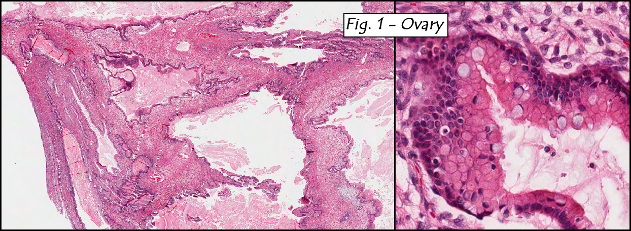
Imaging studies confirmed the presence of an enlarged lymph node in the right axilla.  There were no other radiological abnormalities.

Microscopic sections revealed complete effacement of lymph node architecture by a polymorphous lymphoid infiltrate consisting of large atypical lymphoid cells with open chromatin, prominent nucleoli, and moderate amounts of cytoplasm against a background of predominately small lymphocytes (Figs. 1,2,3).  Large atypical lymphoid cells were positive for CD20 and CD79a, while the smaller background lymphocytes were CD3, CD5 and PAX5 positive (Fig. 4).  Epstein-Barr virus genomes were detected in the large atypical lymphoid cells by EBER in situ hybridization.

Diagnosis: “EBV positive diffuse large B-cell lymphoma of the elderly”

Hannah Wong MD, Jun Wang MD, Donald Chase MD
Department of Pathology, Loma Linda University and Medical Center, Loma
Linda, California
California Tumor Tissue Registry, Loma Linda, California

Discussion: EBV positive diffuse large B-cell lymphoma of the elderly (EBV+ DLBCL, aka “age-related EBV-associated lymphoproliferative disorder”), is a rare B-cell lymphoproliferative disorder that occurs in elderly patients with no known history of immunodeficiency or lymphoma.  The entity was first described by Oyama et al. in 2003 and was added as a distinct entity in the newly published WHO Classification of Tumours of Haematopoietic and Lymphoid Tissues.

As the name suggests, EBV+ DLBCL of the elderly is a disease affecting patients over the age of 50 years.  There is a slight male predominance of 1.4:1.  Typically there is no history of immunodeficiency or lymphoma.  Presentation symptoms and signs include fever, night sweats, weight loss and lymphadenopathy.  Extranodal sites found at presentation include skin, lung, tonsil and stomach.

Histologically, the nodal or native tissue architecture is completely effaced by large atypical lymphoid cells/immunoblasts as well as Reed-Sternberg-like giant cells.  Accompanying these elements are variable numbers of small lymphocytes, plasma cells, histiocytes, centroblasts and immunoblasts.  Although EBV+ DLBCL of the elderly was originally separated into two subtypes; polymorphic lymphoproliferative disorder (PLPD) and large cell lymphoma (LCL) recent studies have shown the sub-classification to be clinically irrelevant.

The neoplastic large lymphoid cells show expression of pan-B cell markers (CD20, CD79a and PAX5), with variable expression of CD30, LMP-1 and EBNA-2.  Light chain restriction may be present but has been difficult to demonstrate in most cases. CD15, CD10 and Bcl-6 are generally negative.  Neoplastic cells showing EBER positivity and Ki-67 expression is generally high.

Differential diagnoses of EBV+ DLBCL of the elderly include other B-cell lymphoproliferative disorders such as Burkitt lymphoma, post-transplant lymphoproliferative disorder (PTLD), HIV-associated lymphoproliferative disorder, EBV- DLBCL, and EBV+ classical Hodgkin lymphoma (cHL).  It is important to differentiate these entities in order to provide the most targeted and effective treatment for the patients.

•    Burkitt lymphoma (BL) is a highly aggressive lymphoma with three clinical variants:

  1. Endemic type mostly occurs in children between the ages of 4 – 7 in equatorial Africa.
  2. Sporadic type  is more common in the United States and Western Europe, where the median age is 30 years.
  3. Immunodeficiency-associated BL patients are virtually all immunocompromised, mostly due to HIV infection.

BL has a variable degree of EBV involvement with endemic types having the highest association.  Although the morphology of BL varies slightly in each type, the most common pattern is of medium-sized cells in a diffuse monotonous pattern with multiple benign macrophages having ingested apoptotic tumor cells resulting in the classic “starry-sky” pattern.  Multiple mitotic figures may be present.  Immunohistochemical stains can be helpful in differentiating BL from EBV+ DLBCL of the elderly since the malignant cells in BL are usually CD10 and BCL-6 positive while the malignant cells of EBV+ DLBCL are negative for those two markers.  BL is also unique in that t(8;22) involving the c-myc oncogene is a constant feature; however this translocation has not been investigated in EBV+ DLBCL of the elderly.

•    Post-transplant lymphoproliferative disorder (PTLD) is a lymphoid proliferation that arises in immunosuppressed recipients usually involving a solid organ or bone marrow allograft.  EBV has been found to be the driving force in PTLD, leading to early polyclonal proliferations and ultimately the development of lymphomas.  Four morphologic categories of PTLD are:

  1. Early lesions resemble reactive hyperplasia and/or infectious mononucleosis. These lesions show an expanded paracortex filled with plasma cells and immunoblasts with a background of small T-cells with partial preservation of the underlying normal tissue architecture.
  2. Polymorphic PTLD shows effacement of the normal nodal/extranodal architecture by a mixture of large immunoblasts and plasma cells with smaller lymphocytes.  Necrosis, mitoses and atypical immunoblasts may also be present.
  3. Monomorphic PTLD has a monomorphic infiltrate effacing nodal/extranodal tissues with confluent sheets of atypical B-cells or plasma cells.
    The Hodgkin lymphoma (HL) variant is similar in morphology and immunophenotype to classical Hodgkin lymphoma.  It displays Reed-Sternberg (HRS)-like cells and variants, and are positive for CD30 and CD15, but not CD20.

Despite splitting PTLD variants into four morphologies, the distinction is of limited value.  The clinical history currently appears to be the most helpful criterion for differentiating PTLD from EBV+ DLBCL of the elderly.

•    HIV-related lymphomas display EBV presence about 60% of the time.  They are generally aggressive B-cell lymphomas related to other lymphoproliferative disorders which are associated with HIV infection such as BL, DLBCL, primary effusion lymphoma, plasmablastic lymphoma of the oral cavity and others.  Interestingly, other viruses such as Kaposi’s sarcoma-associated herpes virus/human herpes virus 8 (KSHV/HHV8) have also been found in several categories of immunodeficiency-associated lymphoma.  But despite the similar morphological and immunohistochemical features of HIV- associated LPD and EBV+ DLBCL of the elderly, a positive HIV serology usually separates the two.

•    EBV+ classical Hodgkin lymphoma (cHL).  Classical Hodgkin lymphoma accounts for 95% of all Hodgkin lymphomas.  cHL has a bimodal age distribution with the first peak at 15 to 35 years and a second peak later in life.  Approximately 30-50% of cHL have been found to be EBV positive.  Patients with EBV+ cHL have a more aggressive clinical course and worse prognosis in comparison to those with EBV- cHL.  Morphologically, EBV+ cHL has the classic morphology of cHL with HRS cells in a background of inflammatory cells.  The large HRS cells have abundant cytoplasm and at least two large round nuclei with prominent nucleoli.  These HRS cells are positive for CD30 and mostly positive for CD15 but are negative for CD45.  This is in comparison to EBV+ DLBCL of the elderly which is usually positive for CD45, CD20/CD79a, variably positive for CD30 but negative for CD15.  EBER has been found to be positive in the neoplastic cells of EBV+ DLBCL of the elderly more often than in the neoplastic HRS cells and their variants of EBV+ cHL.  Thus, immunohistochemical studies and EBER are the most useful tools to distinguish EBV+ cHL from EBV+ DLBCL of the elderly.

•    EBV- DLBCL is virtually the same as EBV+ DLBCL of the elderly except that it may occur in patients of any age and is not EBV+.  Morphologically, these two entities are virtually indistinguishable.  However, EBER positivity would help separate it from the EBV+ DLBCL of the elderly.

EBV+ DLBCL of the elderly has a highly aggressive behavior; the median survival for this disease is approximately two years.  Patients with EBV+ DLBCL of the elderly have been found to be less responsive to the standard chemotherapy regimens for large B-cell lymphoma compared with other forms of B-cell lymphoproliferative disorders.

Suggested reading:

1.    Nakamura S, Jaffe E.S, Swerdlow S.H.EBV+ Diffuse large B-cell of the elderly.. In: Swerdlow S.H., Campo E., Harris N.L., Jaffe E.S., Pileri S.A., Stein H., Thiele J., Vardiman J.W. (Eds.): WHO Classification of Tumours of Haematopoietic and Lymphoid Tissues. IARC: Lyon, 243-244 2008.
2.    Shimoyama Y., Yamamoto K., Asano N., Oyama T., Kinoshita T., Nakamura S. Age-related Epstein-Barr virus-associated B-cell lymphoproliferative disorders: Special references in lymphomas surrounding this newly recognized clinicopathologic disease.  Cancer Sci  9:1085-1091, 2008.
3.    Shimoyama Y., Oyama T., Asano Net al. Senile Epstein-barr virus-associated B-cell lymphoproliferative disorders: a mini review. J Clin Exp Hematopathol  46:1-4, 2006.
4.    Oyama T., Ichimura K., Suzuki R., et al . Senile EBV+ B-cell lymphoproliferative disorders: a clinicopathologic study of 22 patients. Am J Surg Pathol  27;16-26. 2003.
5.    Mueller S., Aigner T., Haag J., Schwartz R., Niedobitek G. Senile EBV-associated B-cell lymphoproliferative disorder of prepatellar bursa in elderly patient with multifocal urate arthropathy. Hematol Oncol  25:140-142, 2007.
6.    Oyama T., Yamamoto K., Asano N., et al . Age-related EBV-associated B-cell lymphoproliferative disorders constitute a distinct clinicopathologic group: A study of 96 patients. Clin Cancer Res  13:5124-5132,  2007.

Scroll to top