June 2013

Critical Pathology Diagnoses and Differentials of the Head and Neck
June 2, 2013
Costa Mesa, CA
Lester D.R. Thompson, M.D.


Objectives: At the conclusion of the seminar, attendees will be able to recognize a variety of reactive and neoplastic lesions that occur in the Head & Neck, formulate a differential diagnosis, and identify important histologic criteria to separate them.  Select and integrate immunohistochemical, molecular, and/or genetic procedures that aid in diagnosis or have prognostic significance. Understand their clinical behavior and be able to communicate effectively with clinicians about their management.

Materials: 1) Glass slides and/or digital images representative of 20 head and neck disorders, 2) Correlating clinical histories, 3) Six hour lecture including images of each case and other illustrative materials, 4) Comprehensive printed color syllabus, including diagnosis, discussion, and appropriate literature references. CME credit applies only to the day of the seminar. Full Package can earn up to 10 hrs CME, Attendance only can earn up to 6 hrs CME.

June 2012: A 59 year old man with an enlarging abdominal scar

History: A 59-year-old man had a cutaneous melanoma of his right abdomen excised three years earlier. He presented with an ulcerated, enlarging scar at the site of previous excision.

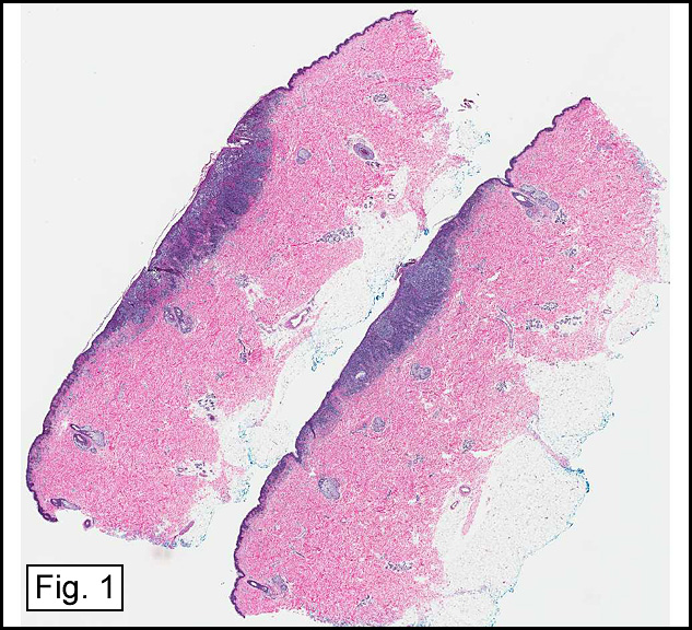
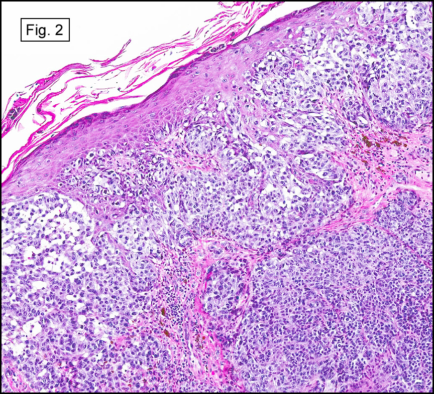
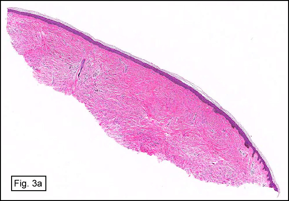
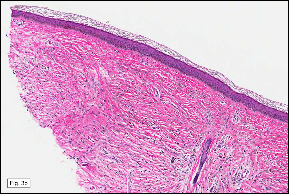
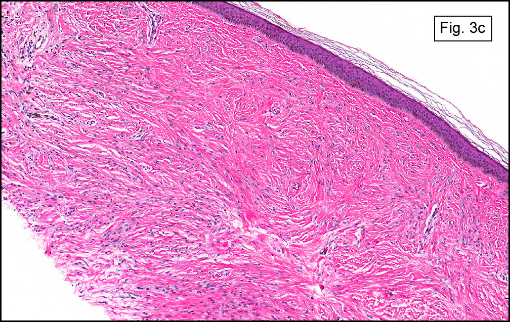
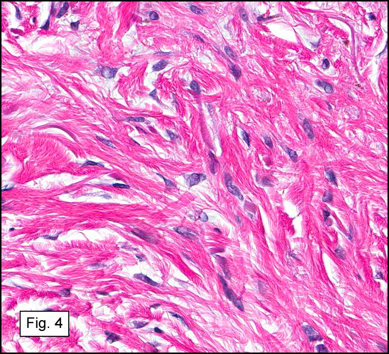
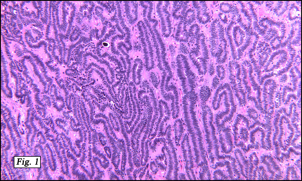
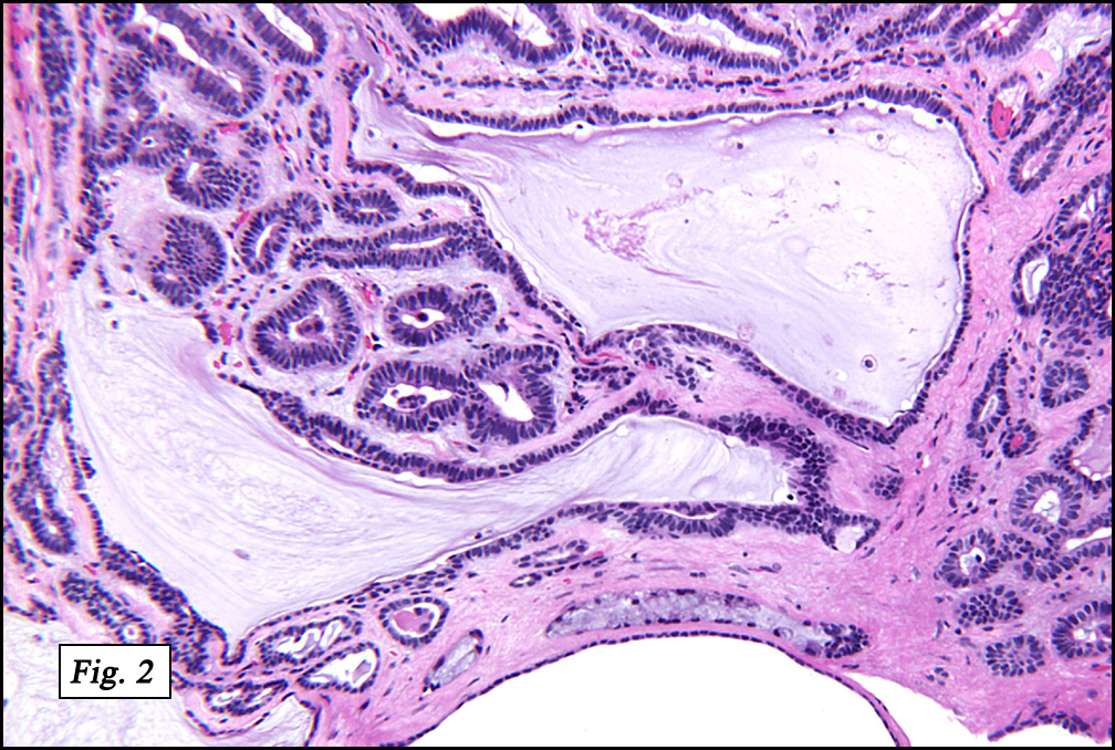
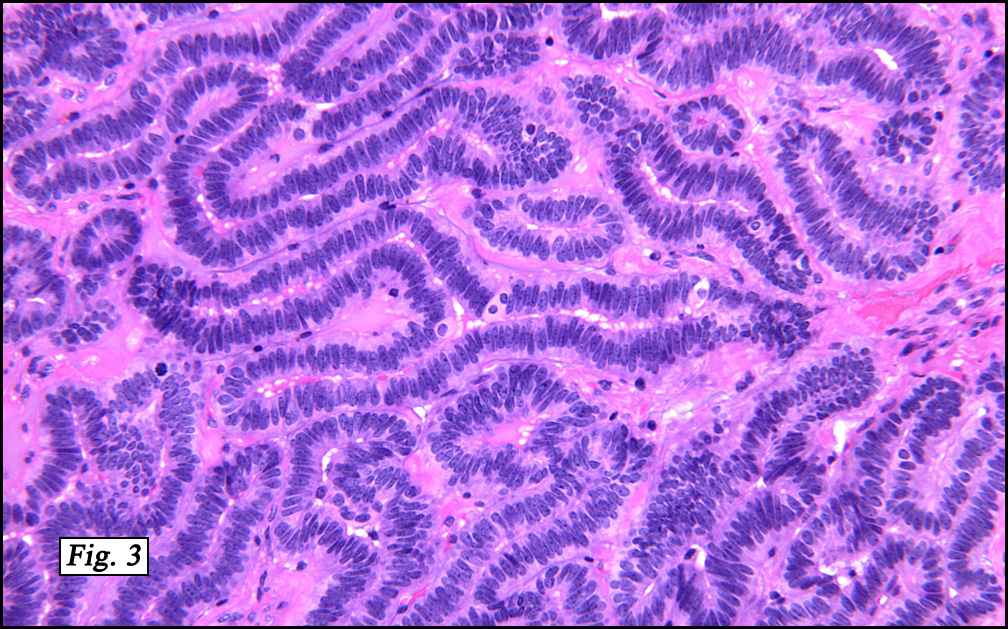
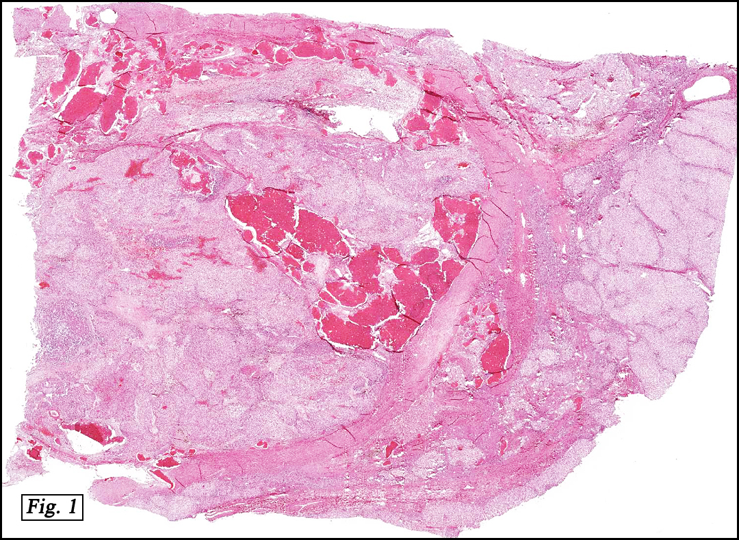
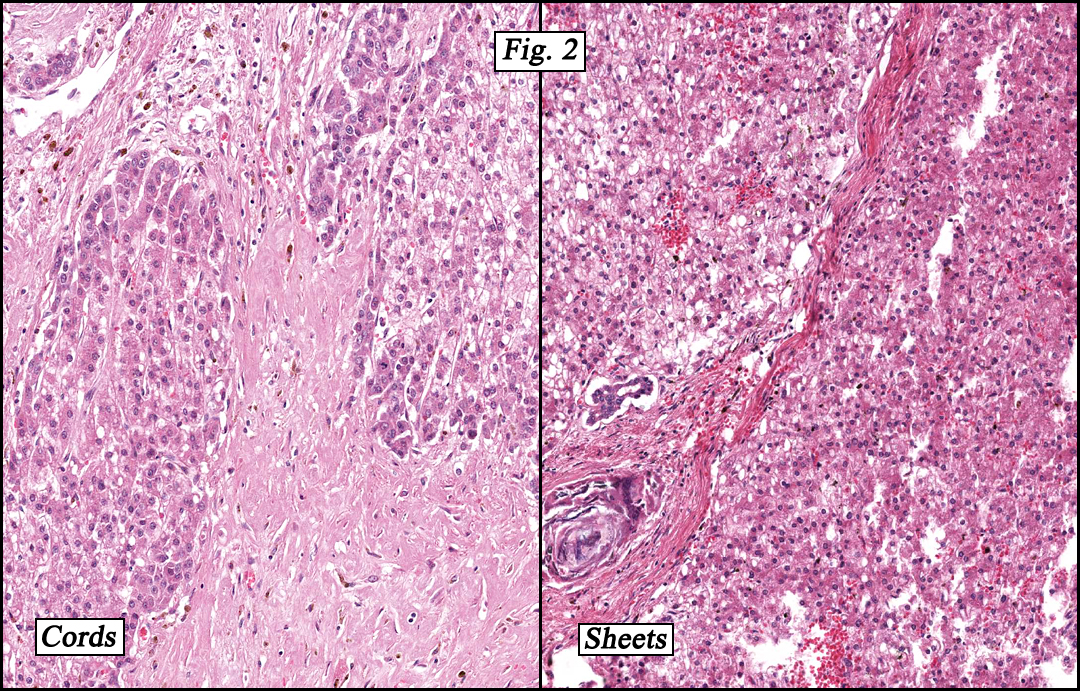
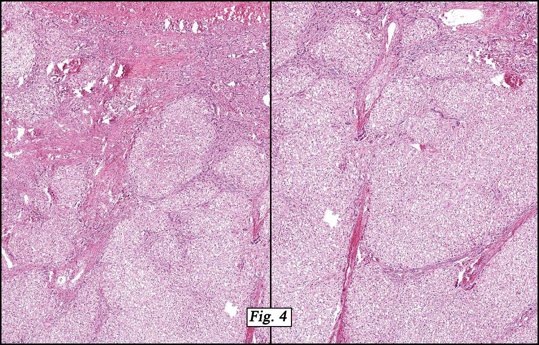
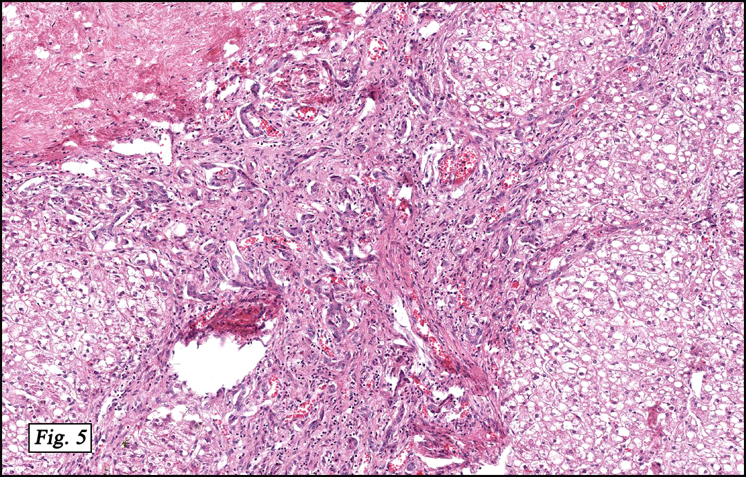
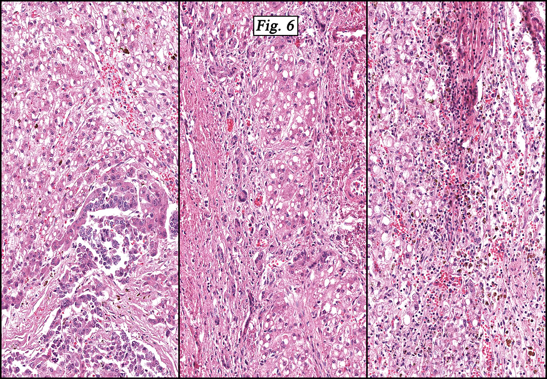
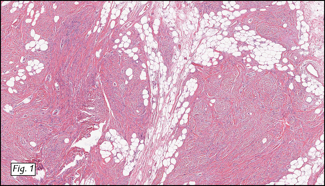
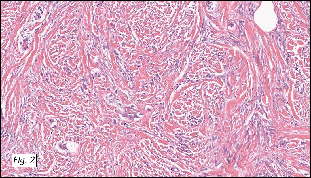
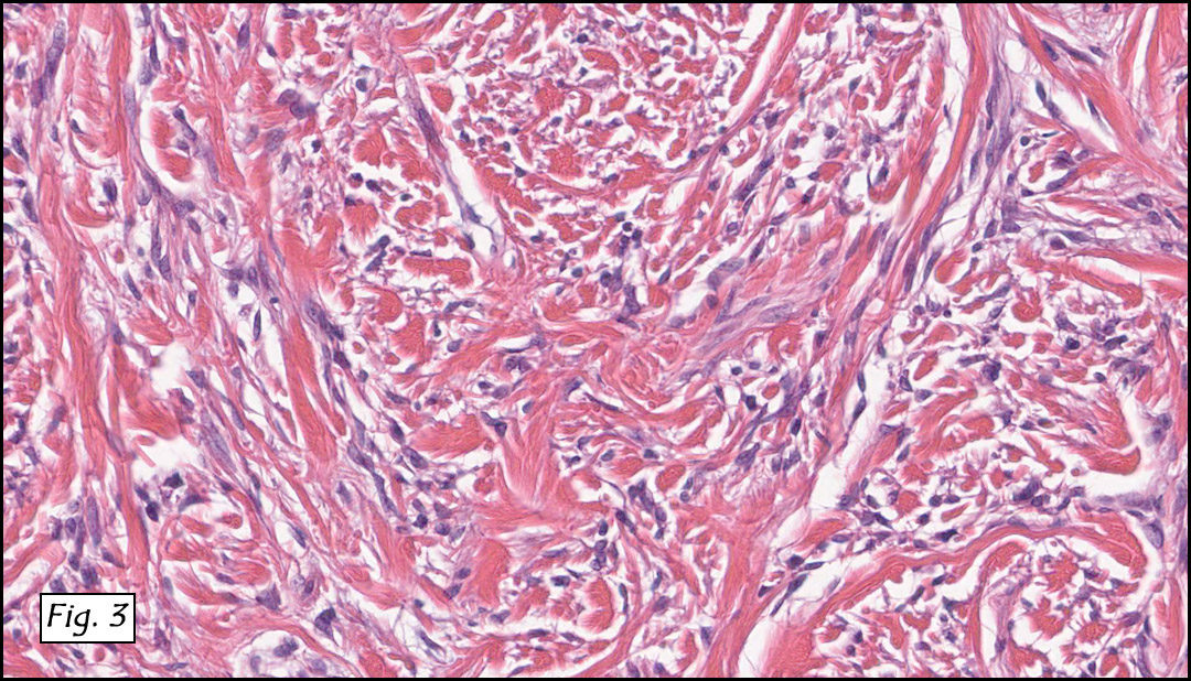
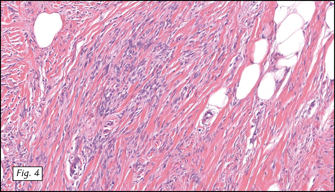
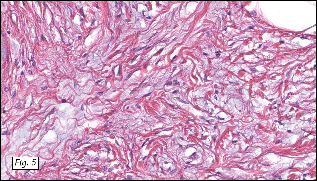
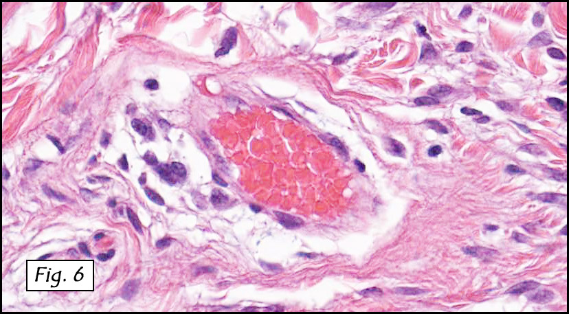
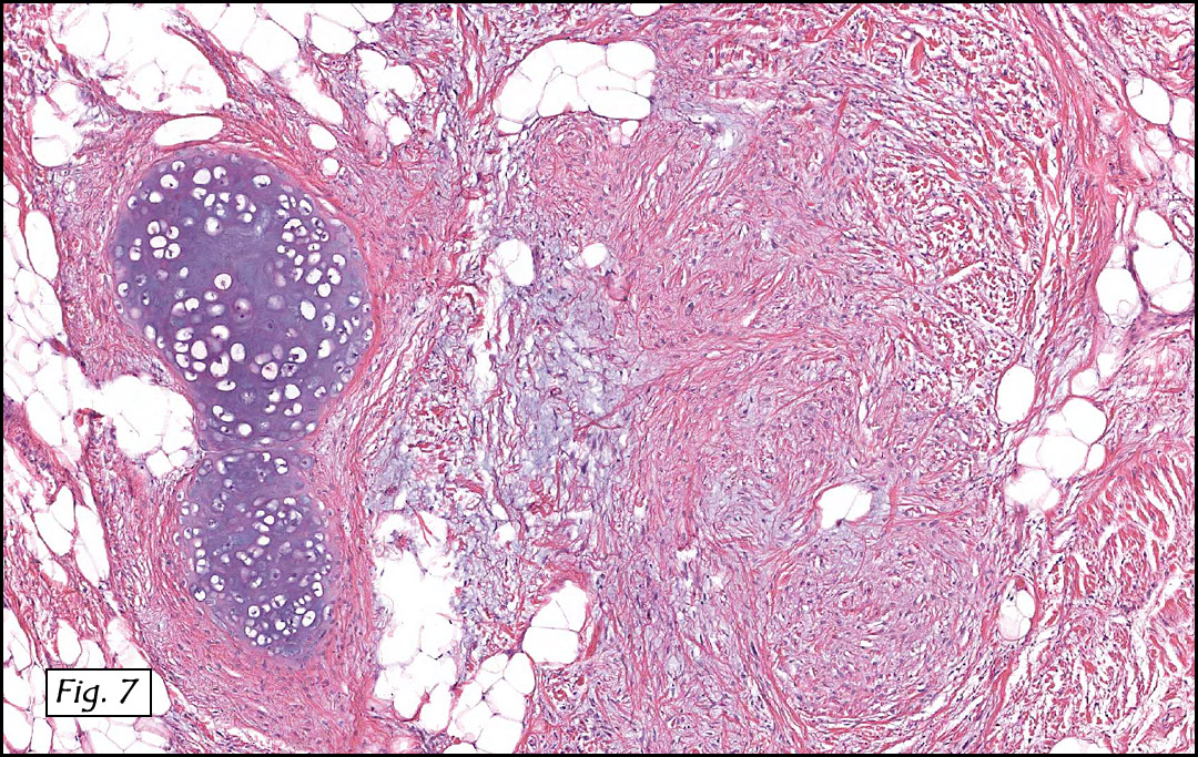
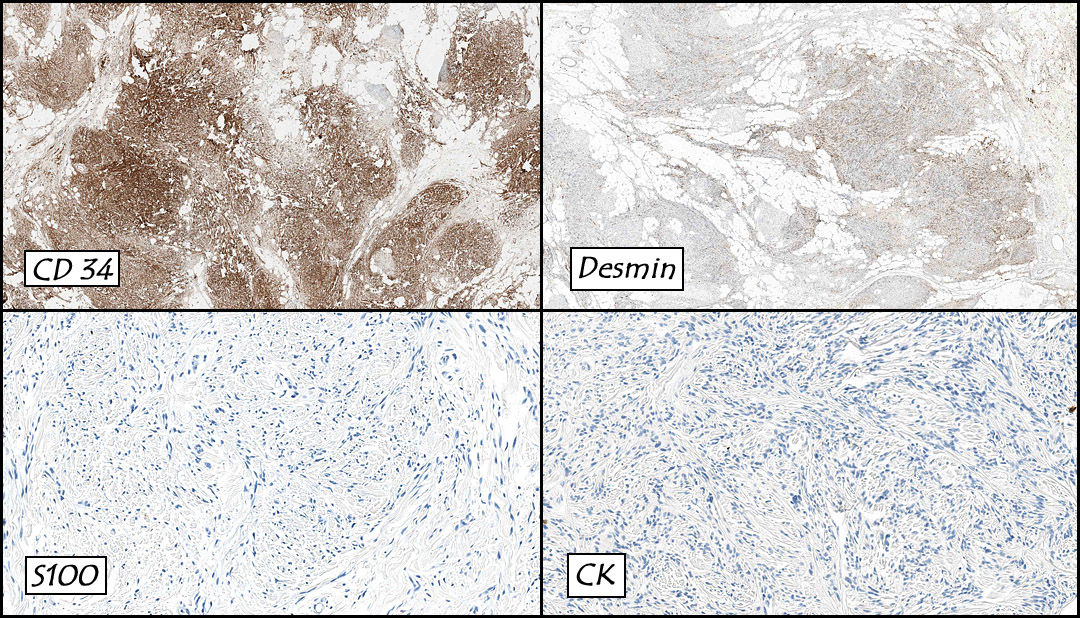
The original melanoma was classic, composed mostly of polygonal melanocytes, without spindled elements (Figs. 1, 2). The peripheral margins were microscopically free of tumor (Fig. 1). The more recent excision had a cicatrix appearance (Figs. 3a,3b,3c). Upon closer examination, the tumor had an infiltrative pattern consisting most of fibrosing spindle cells (Fig. 4). The cytology of these spindle cells was rather bland, resembling fibroblasts. Interspersed between the spindle cells were (rarely) macrophages filled with dark pigment (Fig. 5). Polygonal cells, which dominated in the original tumor, were not present. Immunohistochemical stains showed the spindled cells to be positive for both HMB-45 (Fig. 6 left) and Melan A (Fig. 6 right).

Diagnosis: “Recurrent Melanoma with Desmoplastic Phenotype (“Desmoplastic Melanoma”)

Jonathan Zumwalt, MSIV, Resa L. Chase, MD, and Donald R. Chase, MD
Department of Pathology and Human Anatomy, Loma Linda University and Medical
Center, Loma Linda, California
California Tumor Tissue Registry, Loma Linda, California

Discussion: In 1812, Rene Laennec first described the disease entity currently known as “melanoma”, in a paper entitled “The Melanoses”. Today melanoma is seen in 1/39 Caucasian men and 1/58 Caucasian women in the United States. There are approximately 70,000 cases of invasive melanoma diagnosed every year and almost 9,000 lives are lost every year to this tumor. The incidence of melanoma has markedly increased over the past 10-20 years and may be attributed to three non-biologic factors: 1) increased rates of screening, 2) an increase in the number of biopsies being taken and 3) changes involving histologic interpretation of early evolving lesions. The main risk factors for developing melanoma are: family history, history of atypical nevi, high nevus count, history of increased sun exposure and physical traits (i.e. light colored hair and eyes).

There are a variety of factors to consider when reporting a melanoma:
• Breslow thickness is the most important factor for melanoma staging.
• Ulceration is now the second most powerful predictor of survival.
• Mitotic activity of the tumor shows a correlation between replication and survival.
• Radial and vertical growth phases may help characterize the tumor’s aggressiveness. Tumor infiltrating lymphocytes seem to indicate the presence of vertical growth phase.
• Microscopic satellites cut down the five year survival rate of a patient by more than half.
• Regression is still controversial and experts give different opinions on this phenomenon.
• The anatomic primary tumor site of a melanoma does correlate with patient survival.
• Clark’s levels are no longer used in today’s practice for staging, but many physicians still report them.

Melanomas today are divided into four common variants: superficial spreading, lentigo maligna, acral lentiginous, and nodular.

1. Superficial spreading melanomas make up 75% of melanomas. Histologically, they are asymmetric, poorly circumscribed and lack cellular maturation. Upward invasion of melanocytes into the epidermis is characteristic, as is a prolonged radial growth phase.
2. Lentigo maligna melanomas begin as tan-brown macule on sun damaged skin that evolves into larger, darker areas with asymmetric foci. Histologically, the neoplastic melanocytes are arrayed along the dermal-epidermal junction in a lentiginous pattern against a background of epidermal atrophy and solar elastosis.
3. Acral lentiginous melanomas commonly arise on the palmar, plantar, subungual, and occasionally, mucosal surfaces and typically occur in dark skinned individuals. Histologically, they are characterized by a lentiginous array of atypical melanocytes along the dermal-epidermal junction, with foci of confluent melanocytic growth and progress to large junctional nests that are composed of atypical melanocytes.
4. Nodular melanomas are darkly pigmented polypoid or pedunculated nodules. Histologically, these typically show dermal growth in isolation, but occasionally, in association with an epidermal component.

One rare variant of melanoma is desmoplastic melanoma (DM). It is frequently mistaken for a scar, dermatofibroma and/or sarcoma. Grossly DM generally appears as an amelanotic, pale, and fleshy nodule or papule, usually resembling a scar. A variety of studies have shown that it is highly associated with lentigo maligna (24 percent). Histologically these tumors show a poorly circumscribed vertical growth phase tumor with spindle-shaped malignant cells with a fibrogenic stromal pattern. The nuclei of these cells are typically elongated and hyperchromatic. The morphology of these cells resemble fibroblasts. Immunohistochemical staining usually shows S-100 positivity, while HMB-45, Melan-A, tyrosinase and microphthalmia transcription factor are generally negative.

There has been controversy regarding the prognosis of DM. Original studies found it a “highly malignant, stubbornly recurring and often metastasizing neoplasm.” In the past twenty years this perception has started to change, allowing for an indolent behavior. The confusion of this may be due to the failure of some studies to detect differences between DM and conventional melanoma. A second reason for the conflicting results is the heterogeneity among melanomas declared as desmoplastic.

Differential Diagnosis:

Sclerosing Melanocytic Nevus usually occurs in younger patients, and usually in skin which has not been actinically damaged. Histologically seen from low power, they are sometimes perceived as a circumscribed “silhouette” with little or no cytologic atypia or mitotic activity. In contrast, DM, tends to be asymmetric, infiltrative and poorly circumscribed. Most DMs are associated with a previous or current in-situ melanoma.

Dermal Scar or Dermatofibroma: Without evidence of abnormal melanocytic changes in the epidermis, recognition of a dermal spindle cell proliferation can be most difficult to identify as melanoma. Often DM may be mistaken for a scar. But fibroblasts in scars typically are arranged in parallel to the skin surface, while blood vessels are perpendicular. Dermatofibromas tend to have spindle cells that wrap around the collagen bundles. In contrast, DM has fusiform cells oriented both parallel and perpendicular to the skin surface. Also, DM usually shows some cytologic atypia with elongated hyperchromatic nuclei.

Sarcomas or Sarcomatoid Carcinoma: DM at times can resemble a spindle cell sarcoma, and immunohistochemistry is usually needed to help distinguish these differences. The use of S-100, muscle markers and epithelial markers can help distinguish DM from desmoplastic carcinoma, leiomyosarcoma, pleomorphic sarcoma and malignant peripheral nerve sheath tumor. Sarcomatoid carcinoma usually stains strongly for 34BE12, which does not stain melanomas.

Suggested Reading:

Busam KJ. Desmoplastic Melanoma. Clin Lab Med. 2011 Jun;31(2):321-30.

Divito SJ, Ferris LK. Advances and short comings in the early diagnosis of melanoma. Melanoma Res. 2010 Dec;20(6):450-8.

Garbe C, Eigentler TK, Bauer J, et al. Histopathological diagnostics of malignant melanoma in accordance with the recent AJCC classification 2009: Review of the literature and recommendations for general practice. J Dtsch Dermatol Ges. 2011 Jun 9. doi: 10.1111/j.1610-0387.2011.07714.x.

Kong Y, Kumar SM, Xu X. Molecular pathogenesis of sporadic melanoma and melanoma-initiating cells. Arch Pathol Lab Med. 2010 Dec;134(12):1740-9.

Leong SP, Gershenwald JE, Soong SJ, Schadendorf D, Tarhini AA, Agarwala S, Hauschild A, Soon CW, Daud A, Kashani-Sabet M. Cutaneous melanoma: a model to study cancer metastasis. J Surg Oncol. 2011 May 1;103(6):538-49. doi: 10.1002/jso.21816.

Murali R, Zannino D, Synnott M, McCarthy SW, Thompson JF, Scolyer RA. Clinical and pathological features of metastases of primary cutaneous desmoplastic melanoma. Histopathol. 2011 May;58(6):886-95. doi: 10.1111/j.1365-2559.2011.03808.x. Epub 2011 Mar 25.

Murali R, Shaw HM, Lai K, McCarthy SW, Quinn MJ, Stretch JR, Thompson JF, Scolyer RA. Prognostic factors in cutaneous desmoplastic melanoma: a study of 252 patients. Cancer. 2010 Sep 1;116(17):4130-8.

Piris A, Mihm MC Jr, Duncan LM. AJCC melanoma staging update: impact on dermatopathology practice and patient management. J Cutan Pathol. 2011 May;38(5):394-400. doi: 10.1111/j.1600-0560.2011.01699.x. Epub 2011 Mar 9.

Sade S, Al Habeeb A, Ghazarian D. Spindle cell melanocytic lesions–part I: an approach to compound naevoidal pattern lesions with spindle cell morphology and Spitzoid pattern lesions. J Clin Pathol. 2010 Apr;63(4):296-321.

Sade S, Al Habeeb A, Ghazarian D. Spindle cell melanocytic lesions: part II–an approach to intradermal proliferations and horizontally oriented lesions. J Clin Pathol. 2010 May;63(5):391-409.

Whiteman DC, Pavan WJ, Bastian BC. The Melanomas: A synthesis of epidemiological, clinical, histopathological, genetic, and biological aspects, supporting distinct subtypes, causal pathways, and cells of origin. Pigment Cell Melanoma Res. 2011 Jun 27. doi: 10.1111/j.1755-148X.2011.00880.x.

May 2012: A 79 year old woman with an inner cheek mass

History: A 79 year-old woman presented with a two year history of a slow-growing oral cavity mass between the buccal mucosa and cheek, near the lip commissure. Her past medical history was unremarkable.

Sections showed a well-circumscribed, nodular, thinly encapsulated epithelial neoplasm. The tumor was predominantly composed of a solid proliferation of cytologically bland cuboidal to columnar basaloid cells with dark nuclei and scant eosinophilic cytoplasm (Fig. 1). Microcyst formation was focally present (Fig. 2), sometimes associated with fine papillae. The periphery showed the mucous pattern of minor salivary glands. Lesional cells were usually bilayered in branching/anastomosing cords (i.e. trabecular pattern) (Fig. 3) The cords sometimes separated and rejoined repeatedly, creating a “beaded” appearance (Fig. 4). Scattered foci of mucous cells (Fig. 5), oncocytic cells (Fig. 6), and squamous metaplastic cells were seen. Psammoma bodies were scattered throughout the tumor (Fig. 7).

Diagnosis: “Canalicular Adenoma of Minor Salivary Gland Origin”

Melissa Skaugset MSIV, Mia Perez MD, Donald Chase MD
Department of Pathology and Human Anatomy,
Loma Linda University and Medical Center
California Tumor Tissue Registry, Loma Linda, California

Discussion: Canalicular adenoma (CanA) is a benign epithelial neoplasm of the salivary glands that, by the WHO definition, is composed of columnar epithelial cells arranged in thin, anastomosing cords often with a beaded pattern. The stroma is characteristically paucicellular and highly vascular. They are benign neoplasms, only rarely recurring after local excision. Synonyms include:

• Basal cell adenoma, canalicular type
• Monomorphic adenoma, canalicular type
• Adenomatosis of minor salivary gland

Canalicular adenomas most commonly affect the minor salivary glands. The majority arise in the upper lip (70-90%) where they are the second most common salivary gland tumor after pleomorphic adenoma. Their next most common site is buccal mucosa. Only rarely have they been reported in the parotid gland or palate.

CanAs are rare before the fifth decade of life, and have a peak in the seventh decade. No clear gender prevalence has been shown. They usually present as slow growing, painless small mass(es). They are freely mobile, frequently multinodular, and usually are less than 2 centimeters in greatest diameter (mean 1.7cm). They are generally slow growing, and have been reported to have been present for up to 15 years. The majority present as a solitary mass, but up to 22% are reportedly multifocal and/or multiple (Daley, 1984). Grossly, they are well demarcated and frequently encapsulated. The tumor is usually solid and homogenous, but may have cystic inclusions filled with mucoid material.

CanAs were long thought to be a subclass of basal cell adenoma due to its phenotypic similarity, but is now thought to be a distinct entity. It derives its name from the double rows of cuboidal to columnar epithelium that separate and rejoin to form small duct-like structures or canaliculi. The pattern of separation and rejoining evokes the image of beads on a rosary, leading to descriptions of “beading” or “rosary beads”. The epithelium may be cuboidal or columnar and is usually without significant cellular atypia or pleomorphism. The stroma is loose, highly vascular, and usually of low cellularity. Cyst formation is common.

Unlike other salivary gland tumors which may show multiple growth patterns, canalicular adenomas tend to have a predictable patterns. But they may vary in cellular density and amount of cyst formation, and the degree of encapsulation. The diagnosis may be made without special stains, although recognized positive markers include keratin, vimentin and S100. GFAP may be focally positive. The tumors do not react to anti-SMA.

The differential diagnosis includes sebaceous cyst, lipoma, nasolabial cyst, and more commonly, salivary gland tumors that have a “basaloid” pattern, i.e. basal cell adenoma and adenoid cystic carcinoma. Distinguishing basal cell adenoma from canalicular adenoma has limited clinical significance apart from the potential of canalicular adenomas for multifocality. Distinguishing this entity from adenoid cystic carcinoma is much more important. The double rows of columnar cells and canaliculi that typify canalicular adenomas are not seen in adenoid cystic carcinoma, although the cribriform and tubular patterns of the latter can be confused with this pattern. Nuclear irregularity and atypia are present in adenoid cystic carcinoma, but not in an adenoma. Vascularity of the stroma of canalicular adenomas is absent in adenoid cystic carcinoma. Dense collagen may surround cell nests in adenoid cystic carcinoma but is typically present only in the capsule of canalicular adenoma.

Suggested reading:

Barnes L, Eveson JW, Reichart P, Sidransky D. WHO Classification of Tumours: Pathology and Genetics of Head and Neck Tumours, 2005 Ed. IARC Press, International Agency for Research on Cancer, 2005. p 267.

Peel RL, Barnes L. Surgical Pathology of the Head and Neck. Marcel Dekker, New York, 2001. pp 663-670.

Ellis GL, Auclair PL, Rosai J. AFIP Atlas of Tumor Pathology: Tumors of the Salivary Glands. American Registry of Pathology, Armed Forces Institute of Pathology, Washington D.C., 1995. pp95-103.

Kratochvi F, Ellis GL, Auclair P, Gnepp D. Surgical Pathology of the Salivary Glands. W.B. Saunders Company, Philadelphia, 1991. pp 202-212.

April 2012: Hepatomegaly in a 14 month old girl

History: A 14 month old girl presented with hepatomegaly, with the palpable liver margin 5 cm below the inferior mid-clavicular line. Shortly after presenting, the patient died, and an autopsy described an enlarged liver weighing 1015 grams. Sectioning showed a yellow-green, 8 x 4 cm variegated mass with cystic, gelatinous, and focally calcified areas, located anteriorly between the left and right lobes.

Microscopically, the tumor was partially encapsulated, lobulated, and exhibited patchy light and dark regions. Some areas were telangiectatic with numerous expanded blood vessels filled with erythrocytes (Fig. 1). Growth was usually as cords and sheets (Fig. 2). Abundant osteoid could be seen adjacent to fibrous septa and loose mesenchymal elements (Fig. 3). The lighter staining regions consisted of more mature hepatocytes growing in a lobular pattern separated by fibrous septa (Fig. 4). Proliferation of bile ducts could also be seen (Fig. 5). Also present were clusters of blastemal cells with an increased nuclear to cytoplasm ratio with greater nuclear pleomorphism and prominent nucleoli (Fig. 6, left). Rare multinucleated tumor giant cells were present (Fig. 6, center), and some regions were suggestive of extramedullary hematopoiesis (Fig. 6, right). There were significant areas of hemorrhage and hemosiderin deposition. Mitoses were rare.

Diagnosis: “Hepatoblastoma, Mixed Pattern”

Shino Magaki MSIV, Anwar Raza MBBS, and Donald R. Chase MD
Department of Pathology and Human Anatomy,
Loma Linda University and Medical Center
California Tumor Tissue Registry, Loma Linda, California

Discussion: Hepatoblastoma (HB) is the most common liver malignancy in children, accounting for approximately two-thirds of all primary pediatric hepatic malignancies and 1% of all pediatric malignancies. Two-thirds are diagnosed prior to the age of 2 years while 90% are diagnosed before the age of 4 years, with the mean age at diagnosis of 18 months. HB is rarely seen at birth or in young adults, and males are twice as likely as females to be affected. HB was probably first described by Misick in 1898. The etiology is unknown but 5% of cases are associated with a congenital abnormality such as Beckwith-Wiedemann syndrome or familial adenomatous polyposis. Common cytogenetic alterations include trisomy 2, 8 and 20. Activating mutations in the beta-catenin/Wnt pathway is seen in about half of the cases. HB has also been associated with low birth weight and has been increasing in incidence over the past few decades, possibly due to improved survival of low birth weight infants.

Clinically, patients often present with a rapidly enlarging abdominal mass, failure to thrive, and weight loss. Less common symptoms include vomiting, diarrhea, and jaundice. Virilization and precocious puberty from human chorionic gonadotropin secreted by the tumor have been reported. Serum alpha-fetoprotein (AFP) is elevated in 90% of cases and is a useful marker for tumor recurrence. Tumors that do not express AFP are thought to be more biologically aggressive, and serum levels less than 100 ng/mL at the time of diagnosis indicate a poorer prognosis. On ultrasound, HB usually appears as a well demarcated solitary mass although it can be multifocal. CT often shows contrast enhancement and calcification. On MRI the purely epithelial subtype appears homogenous and hypointense on T1 and hyperintense on T2 weighted images, whereas the mixed epithelial and mesenchymal subtype is more heterogeneous.

Grossly, HB typically occurs as a single mass on a background of non-cirrhotic liver and may contain areas of hemorrhage and necrosis. The tumor tends to be well circumscribed but unencapsulated or partially encapsulated and have a variegated appearance depending on the proportion of its components. In pure epithelial HB, the tumor is soft, fleshy and tan-white. Mixed tumors show a heterogeneous surface, tend to be firmer and often are calcified. Frequently large at detection, HB can measure up to 20-25 cm in diameter (average 10-12 cm), and usually weigh over 1 kg. They are most common in the right lobe.

Histologically, HBs are classified into pure epithelial or mixed epithelial-mesenchymal subtypes with six main histological patterns:

The fetal pattern (30%) demonstrates tumor cells which are uniform and similar in size to normal hepatocytes with a slightly increased nuclear:cytoplasm (N:C) ratio. Cells form trabeculae 2-3 cells thick that are separated by sinusoids. An alternating light and dark staining pattern due to lipid and glycogen, respectively, is often seen. Portal tracts and bile ducts and ductules are absent, and mitoses are rare (

In the embryonal pattern (20%), cells are less differentiated, have a higher N:C ratio with coarser chromatin, and may form sheets. More mitoses and necrosis are seen.

Cells are the least differentiated in the anaplastic small cell pattern (3%), forming sheets of small cells with scant cytoplasm and prominent nucleoli, resembling small blue cell tumors elsewhere. Tumor cells are positive for CK and negative for bile.

In the macrotrabecular pattern (3%), fetal or embryonal cells form trabeculae 10 or more cells thick throughout the tumor.

The mixed epithelial-mesenchymal pattern (44%) is characterized by the presence of both epithelial and mesenchymal cells. The mesenchymal component may show primitive stellate-shaped cells with little cytoplasm or demonstrate chondroid and rhabdomyoblastic differentiation. Osteoid is the most common heterologous element and is more frequent found after chemotherapy. The mixed pattern can be further subdivided into tumors with (10%) or without (34%) teratoid features such as skeletal muscle, keratinized squamous epithelium, and intestinal epithelium.

Differential Diagnoses:

Although Hepatocellular carcinoma (HCC) may be indistinguishable from a macrotrabecular pattern of HB, other features help differentiate them (Table 1).

Since small cell tumors, including neuroblastoma, rhabdomyosarcoma, and lymphoma, resemble the anaplastic small cell pattern in HB, the presence of more typical, better differentiated foci should be looked for. Immunohistochemistry is useful, with HB showing CK positivity and neurofilament, desmin, leukocyte common antigen (CD45) negativity; neuroblastoma is positive for neurofilaments, rhabdomyosarcoma is positive for desmin, and lymphoma is CD45 positive.

Table 1. Hepatoblastoma and hepatocellular carcinoma in the pediatric age group (

Hepatoblastoma Hepatocellular carcinoma
Incidence Most common primary liver malignancy
in children (0.5-1.5 diagnoses per
million annually)
2nd most common primary liver
malignancy in children
Age 90%<5 yrs Most >10 years
M:F ratio 1.2-3.6:1 1.7:11:1
Association with genetic
syndromes
Familial adenomatous polyposis,
Beckwith-Wiedemann syndrome,
trisomy 18
Glycogen storage disease type
IA, hereditary tyrosinemia,
Fanconi syndrome
Other associations Parental exposure to environmental
toxins, low birth weight
Cirrhosis, viral hepatitis
Serum AFP elevations 90%, usually highly elevated 60-80%
Single mass 70-80% 15-40%
Trabeculae Usually 2-3 cells thick with occasional
macrotrabeculae
Usually>2-3 cells thick
Mitoses Rare Common
Tumor giant cells Rare Common
Cytoplasmic inclusions
(e.g. Mallory’s hyaline,
alpha-1-anti-trypsin)
Absent Common
Extramedullary
hematopoiesis
Common Absent


Treatment consists of complete surgical resection with or without neoadjuvant chemotherapy to decrease tumor size prior to surgery. Prognosis is dependent upon the extent of complete resection. At present, the 5-year survival rate is 75%. Liver transplantation is considered when the tumor is limited to the liver but unresectable. The pure fetal pattern is thought to predict a more favorable outcome while the pure anaplastic small cell pattern, increased mitoses, and vascular invasion suggest a poorer prognosis. Most frequent metastatic sites are the regional lymph nodes and lungs.

Suggested Reading:

Beach R, Betts P, Radford M, Millward-Sadler H. Production of human chorionic gonadotrophin by a hepatoblastoma resulting in precocious puberty. J Clin Pathol. 37:734-37, 1984.

Buccoliero AM, Castiglione F, Maio V, Moncini D, Sardi I, Taddei A, Martin A, Messineo A, Taddei GL. Teratoid hepatoblastoma. Fetal Pediatr Pathol. 27:274-81, 2008.

Das CJ, Dhingra S, Gupta AK, Iyer V, Agarwala S. Imaging of paediatric liver tumours with pathological correlation. Clin Radiol. 64:1015-25, 2009.

Litten JB, Tomlinson GE. Liver tumors in children. Oncologist. 13:812-20, 2008.

Maruyama K, Ikeda H, Koizumi T. Hepatoblastoma associated with trisomy 18 syndrome: a case report and a review of the literature. Pediatr Int. 43:302-5, 2001.

Mills SE, Carter D, Greenson JK, Oberman HA, Reuter V, Stoler MH. Sternberg’s Diagnostic Surgical Pathology (4th ed) Philadelphia: Lippincott. 1743-47, 2004.

Reynolds P, Urayama KY, Von Behren J, Feusner J. Birth characteristics and hepatoblastoma risk in young children. Cancer. 100:1070–76, 2004.

Sallam A, Paes B, Bourgeois J. Neonatal hepatoblastoma: two cases posing a diagnostic dilemma, with a review of the literature. Am J Perinatol. 22:413-19, 2005.

Schnater JM, Köhler SE, Lamers WH, von Schweinitz D, Aronson DC. Where do we stand with hepatoblastoma? A review. Cancer. 98:668-78, 2003.

March 2012: A breast mass in a 62 year old woman

History: A 62 year-old woman underwent surgical resection of a 55 gram, 6.2 x 5.4 x 4.2 cm encapsulated right breast mass. The cut surface was tan with variably-sized pink and tan soft tissue nodules ranging from 1.0 to 2.2 cm.

Microscopically the moderately cellular soft tissue tumor consisted of slender fibroblast-like spindle cells admixed with dense collagenous bands (Fig. 1). The cells were arranged in sheets and small fascicles which were intermixed with interweaving fibrous stroma and mature adipose tissue (Figs. 2, 3, 4). Mucoid changes were also seen (Fig. 5) as well as perivascular mast cells (Fig. 6). Chondroid metaplasia was focally present (Fig. 7). Mitoses were not increased in number and there was no evidence of significant atypia or necrosis. Immunostains showed decoration for CD31 (4+) and desmin (1+) and were negative for S100 and cytokeratin (Fig. 8).

Diagnosis: “Myofibroblastoma with focal chondroid metaplasia”

Kate Grogan, MD, and Donald R. Chase, MD
Department of Pathology & California Tumor Tissue Registry
Loma Linda University and Medical Center, Loma Linda, California

Discussion: First described by Wargotz et al. in 1987, myofibroblastomas (MFBs) are uncommon mesenchymal neoplasms which typically occur in variably aged patients (25-85 years) with an average of 63 years. Although originally reported as having a higher incidence in men, they are currently thought to occur in equal gender frequency. They traditionally present as a well-circumscribed, mobile unilateral mass of several months duration. A single case of synchronous bilaterality has been reported. Grossly, MFBs are well circumscribed and often have a delicate fibrous psuedocapsule. The cut surface is typically gray-pink and vaguely lobular.

Microscopically, MFBs are variably cellular and are comprised of myofibroblast-resembling spindle cells which have tapered nuclei with finely dispersed chromatin, small nucleoli and a moderate amount of eosinophilic to amphophilic cytoplasm. The spindle cells often have wavy contours and are arranged in short fascicles. The stroma contains coarse bands of hyalinized collagen and conspicuous mast cells, admixed with a variable amount of adipose tissue.

Mammographically, MFBs appear as lobulated, well-demarcated and homogenous densities. Microcalcifications are absent.

The majority of cases show staining for CD34, vimentin and ER/PR. Desmin, caldesmon and androgen receptors are variably positive, and S-100 and cytokeratin are typically negative.

Cytogenetic findings: Partial monosomy 13q has been detected in two cases, as well as partial monosomy 16q in one of these two cases. Curiously, these same rearrangements are characteristic of spindle cell lipomas.

Differential Diagnosis:

• Fibromatosis of the breast typically presents as an ill-defined mass which may be associated with skin dimpling, nipple retraction and a stellate mammographic appearance. These entities will also be CD34 negative, whereas the majority of myofibroblastomas demonstrate CD34 positivity.

• Nodular fasciitis, although CD34 positive, typically demonstrates a more infiltrative pattern and has stroma that is focally pushed apart by pools of mucinous material.

• Myoepitheliomas are rare spindled lesions, which usually showing mild to moderate nuclear pleomorphism. These lesions are traditionally keratin, vimentin and EMA positive, and CD34 and ER/PR negative.

• Well-differentiated metaplastic carcinoma of the breast and stromal sarcomas usually present as ill-defined infiltrating masses, whereas myofibroblastomas are generally well-circumscribed and rarely invade adjacent breast tissue.

• Myofibroblastomas are readily distinguished from fibroadenomas due to their lack of ductal structures.

Conservative, but complete surgical excision is the treatment of choice. MFBs rarely recur and do not have metastatic potential.

Suggested Reading:

Tavassoli F, Eusebi V. Tumors of the Mammary Gland (4th edition). Washington DC: American Registry of Pathology, 2009.

Weiss S, Goldblum J. Enzinger and Weiss’s Soft Tissue Tumors (5th edition). Philadelphia: Mosby/Elsevier Inc. 535-6, 2008.

Pauwels P, Sciot R, Croiset F, Rutten H, Van den Berghe H, Dal Cin P. Myofibroblastoma of the breast: genetic link with spindle cell lipoma. J Pathol 2000; 191 (3): 282-5.

Tavassoli F. Pathology of the Breast (2nd edition). Stamford, Connecticut: Appleton & Lange. 686-691, 1999.

Magro G. Mammary myofibroblastoma: a tumor with a wide morphologic spectrum. Arch Pathol Lab Med 2008; 132(11): 1813-20.

Scroll to top