February 2012: A 3 year old boy with an enlarged testicle

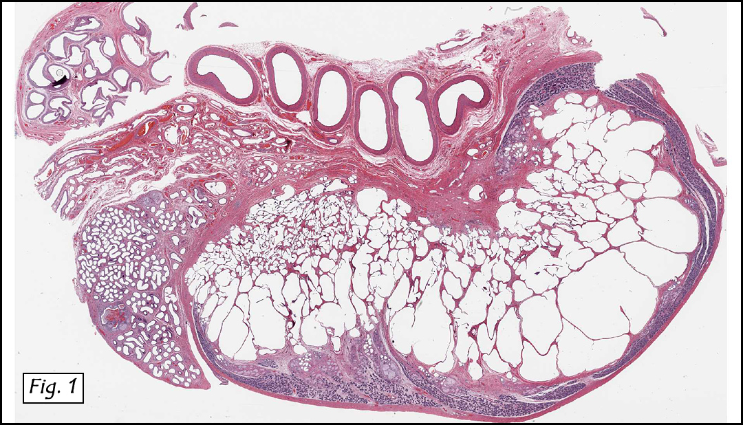
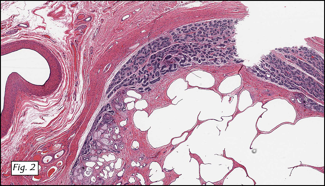
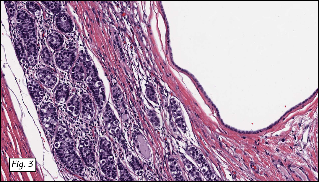
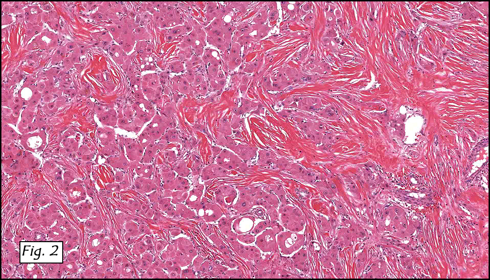
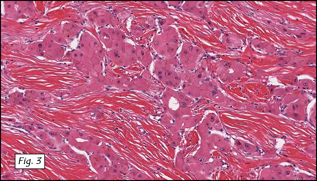
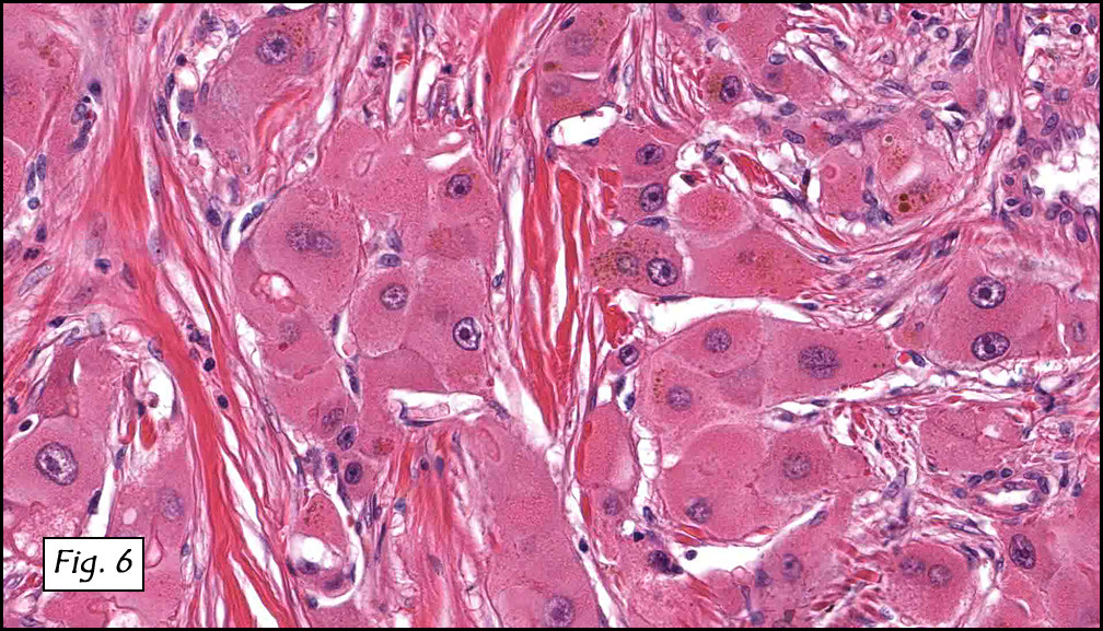
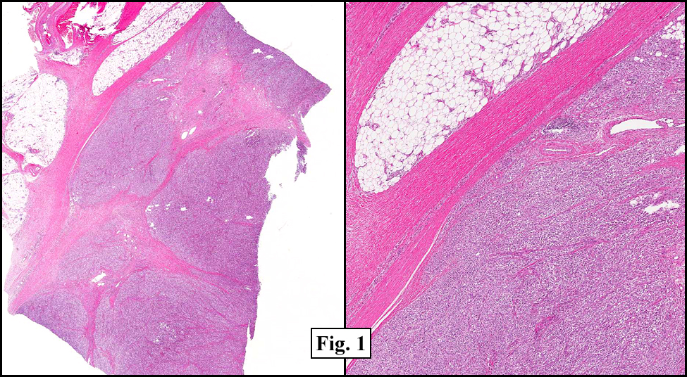
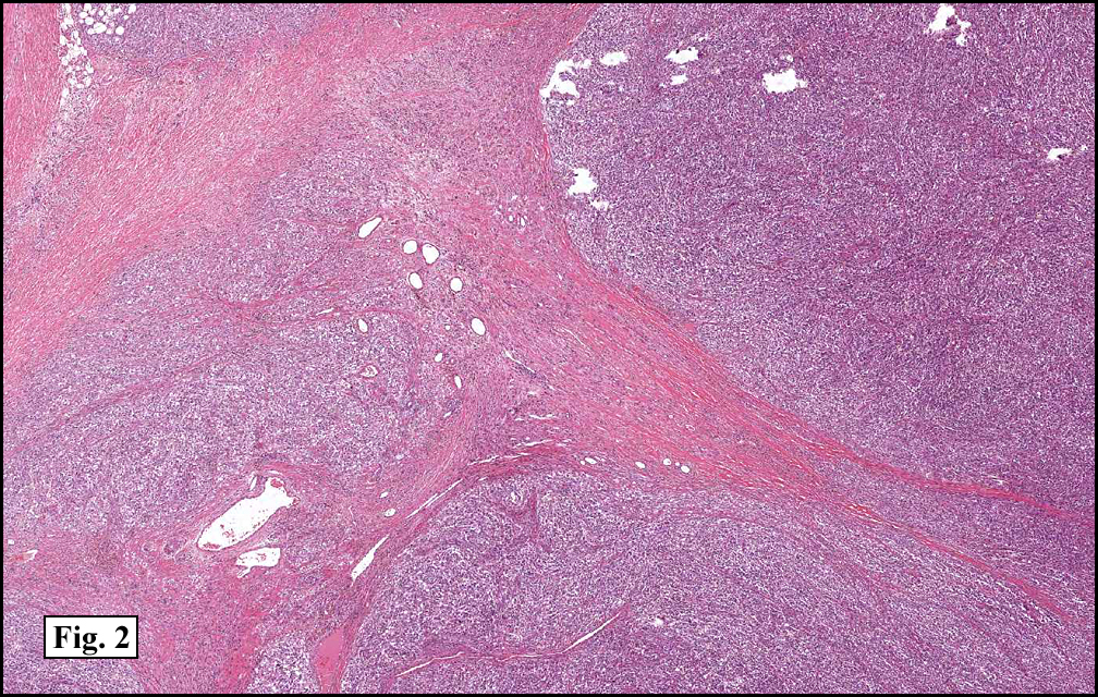
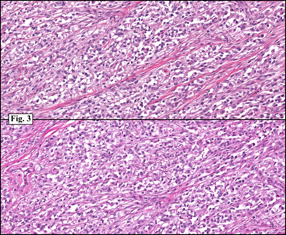
History: A three-year-old boy underwent orchiectomy for a cystic scrotal mass. Microscopically, the rete testis was found to be markedly distorted by cystic dilatation which compressed the adjacent seminiferous tubules (Figs. 1, 2). The cystic rete lining was of low cuboidal epithelium (Fig. 3).

Diagnosis: Cystic dysplasia of the testis
Ellsworth BD, Zuppan C, Chase DR
Department of Pathology & California Tumor Tissue Registry
Loma Linda University Medical Center, Loma Linda, California

Discussion: Cystic dysplasia of the rete testis is a rare anomaly that usually presents in young boys as a unilateral painless scrotal mass. Approximately 50 cases have been documented. The cystic changes are well-appreciated by ultrasound and may be suspicious for malignancy. On ultrasound, variably-sized cysts are seen within the mediastinum testis with no changes in the testicular tissue and epididymis. Histologically, the rete testis is markedly dilated and cystic but lined by flat cuboidal epithelium. This process compresses and often obliterates the nearby testicular parenchyma which can render the testis infertile. Ipsilateral urinary tract anomalies are almost invariably present, most commonly renal agenesis or dysplastic kidney. For this reason, diagnosis of cystic dysplasia of the rete testis warrants further imaging of the urinary tract to look for additional anomalies.

The dysplastic changes are thought to occur as a result of improper development of the mesonephric duct. During normal embryogenesis, the mesonephric duct gives rise to the ureteral bud at its cephalic end which stimulates nephrogenesis of the metanephric blastema. From its caudal end, the extra testicular ducts (efferent ducts, epididymis, and vas deferens) derive. Defects in the mesonephric duct can result in anomalies in any of these structures. Normally, the rete testis and testicular cords (derived from the germinal epithelium) anastomose with the mesonephric-derived extra testicular ducts at the efferent ductules. Failure of this process causes anomalies and results in cystic distortion of the rete testis. With this relationship in mind, Nistal et al have theorized that inappropriate (pre-pubertal) secretory activity by the rete testis and fluid entrapment are responsible forecyst development, a process that occurs until puberty when the seminiferous tubules canalize and develop lumens allowing the fluid to relocate. This process seems to have occurred in two reported cases of resolved cystic dysplasia. In both cases, resolution was relatively sudden and occurred near the time of (reference – Thomas 2003).

Treatment commonly consists of unilateral orchiectomy. Testis-sparing enucleation is occasionally performed to spare hormonally active tissue, but yields a chance of recurrence. As no malignant behavior has been observed and two reported cases have regressed, conservative therapy or watchful waiting has been suggested.

Suggested Reading:

Keetch DW, McAlister WH, Manley CB, Dehner LP. Cystic dysplasia of the testis. Pediatr Radiol. 1991;21:501-503.

Nistal M, Regadera J, Paniagua R. Cystic dysplasia of the testis. Light and electron microscopic study of three cases. Arch Pathol Lab Med.1984;108:579-583.

Thomas AD, Wu H, Canning DA, et al. Spontaneous regression of cystic dysplasia of the testis. J Urol. 2003;169:645.

Wojcik LJ, Hansen K, Diamond DA, et al. Cystic dysplasia of the rete testis: a benign congenital lesion associated with ipsilateral urological anomalies. J Urol. 1997;158:600-604.

Zaragoza M, Buckler L, Parikh M. Cystic dysplasia of the testis: An unusual cause of a pediatric scrotal mass. Urology. 1996;47:244-247.

Robson W, Thomason M, Minette L. Cystic dysplasia of the testis associated with multicystic dysplasia of the kidney. Urology. 1998;51:477-479.

Camassei F, Francalanci P, Ferro F, et al. Cystic dysplasia of the rete testis: Report of two cases and review of the literature. Pediatr Dev Pathol. 2002;5:206-210.

Erberli D, Gretener H, Dommann-Scherrer C, et al. Cystic dysplasia of the testis: A very rare pediatric tumor of the testis. Urol Int.2002;69:1-6.

Fisher JE, Jewett Jr TC, Nelson SJ, et al. Ectasia of the rete testis with ipsilateral renal agenesis. J Urol 1982;128:1040-3.

January 2012: A 30 year old woman with a mass in a transplanted liver

History: A 30 year-old Caucasian woman, status-post hepatectomy for a liver malignancy, presented four years later with an enlarging 4.5 cm right-sided mass in her “new” transplanted liver. The tumor was 4.2 x 3.1 x 2.2 cm and was well-circumscribed, yellow-green, and had a central scar.

Microscopically, the tumor was solid, with prominent ramifying bands of fibrous tissue (Figs. 1, 2) which encased atypical hepatocytes arranged in medium to large-sized nests (Figs. 3, 4). The cells had large vesicular nuclei with prominent nucleoli and abundant granular eosinophilic cytoplasm (Figs. 5, 6). Bile pigment was focally present (Fig. 6). The tumor extended into perihepatic fat (Figs. 7). Neither hemorrhage nor necrosis were seen.

Diagnosis: Hepatocellular carcinoma, fibrolamellar variant

Kate Grogan, MD, and Donald R. Chase, MD
Department of Pathology & California Tumor Tissue Registry
Loma Linda University and Medical Center, Loma Linda, California

Discussion: First described by Edmondson in 1956, fibrolamellar hepatocellular carcinoma (also known as polygonal cell type HCC) is an uncommon variant of hepatocellular carcinoma which typically occurs in young patients mean age of 26 years) who usually lack the common risk factors associated with conventional HCC. Although it was previously thought that there was a slight female predominance, current studies suggest that it occurs in equal frequency among men and women. Almost half of liver cell carcinomas in the United States occur in patients under the age of 35, and most are of this type. It is associated with a better prognosis overall than patients with HCC and cirrhosis, and the prognosis appears to be similar to that in patients with typical HCC without cirrhosis. Since it was first described, more than 180 cases have been described in the literature. Grossly, the tumors are solitary, well-circumscribed, yellow-brown and vaguely nodular. Frequently, they may have a fibrous central scar (similar to the central scar seen in focal nodular hyperplasia). Serum AFP levels are typically within normal limits.

Microscopically, the tumor consists of well-differentiated polygonal cells with abundant granular eosinophilic cytoplasm, which are arranged in nests, sheets or cords. The cytoplasm often contains pale bodies or PAS positive hyaline globules. In the background are dense acellular collagen bundles. As in other forms of HCC, tumors may also have focal acinar structures, bile, multinucleated tumor cells, copper, or fat.

Fibrolamellar carcinomas typically express both hepatic (CK 8 and 18) and biliary (CK 7, 19) cytokeratins. Recent studies have shown that expression of Hep-Par-1 and CD99 may be supportive of the diagnosis. Neuroendocrine markers may be focally positive, but they are of uncertain significance.

Cytogenetic findings: These tumors are often diploid, and overall show fewer chromosomal abnormalities than do classic HCC. Tumors with no cytogenetic abnormalities appear to behave less aggressively.

Differential Diagnosis:

• Hepatocellular carcinoma, sclerosing variant may be histologically similar, however, the tumor cells are typically smaller and pseudoglandular formation is common. Also diffuse lamellar fibrosis combined with oncocytic cellular features is not typical of usual hepatocellular carcinoma
• Cholangiocarcinoma may show glandular formation, and are usually mucin positive.
• Metastatic tumors with extensive fibrosis may mimic the collagen seen in fibrolamellar carcinoma, but is usually arranged in a more haphazard manner and lacks the directional growth characteristic of fibrolamellar carcinoma.

Complete resection of the involved lobe is the therapy of choice. When tumor is deemed unresectable, liver transplantation is an option (as in this case). The tumor metastasizes primarily to regional lymph nodes, peritoneum and lung.

Suggested Reading:

Odze R, Goldblum J. Surgical Pathology of the GI Tract, Liver, Biliary Tract, and Pancreas (2nd edition). Philadelphia: Saunders/Elsevier Inc. 1306-7, 2009.

Liu s, Chan KW, Wang B, Qiao L. Fibrolamellar hepatocellular carcinoma. Am J Gastroenterol 2009; 104: 2617-24.

Kakar S, Burgart LJ, Batts KP et al. Clinicopathologic features and survival in fibrolamellar carcinoma: comparison with conventional hepatocellular carcinoma with and without cirrhosis. Mod Pathol 2005; 18: 1417-23.

MacSween R, Burt A, Portmann B, Ishak K, Scheuer P, Anthony P. Pathology of the Liver (4th edition). London: Churchill Livingstone/Harcourt Publishers Limited. 740-1, 2002.

Torbenson M. Review of the clinicopathologic features of fibrolamellar carcinoma. Adv Anat Pathol 2007; 14: 217-23.

Moreno-Luna LE, Arrieta O, Garcia-Leiva J et al. Clinical and pathologic factors associated with survival in young adult patients with fibrolamellar hepatocarcinoma. BMC Cancer 2005; 5: 142.

December 2011: A 36 year old man with an anterior leg mass

History: A 36 year-old man underwent surgical resection of a 10.0 x 5.5 x 4.0 cm ill-defined left anterior leg mass The excised specimen had a tan, vaguely lobulated cut surface with central hemorrhage It grossly appeared to abut the deep fascial resection margin.

Microscopically, the mass was well-circumbscribed (Fig. 1). It had lobules of nested spindled cells separated by fibrous septae (Fig. 2, 3). The cytoplasm was generally clear and the nuclei were vesicular, many times with prominent nucleoli (Fig. 4). Rare mitotic figures were seen (Fig. 5, right). Immunohistochemistry revealed that the tumor cells were positive for melanoma cocktail, S100, and HMB45, and were negative for cytokeratin and desmin (Fig. 6).

Diagnosis: Clear cell sarcoma

Kate Grogan, MD, and Donald R. Chase, MD
Department of Pathology & California Tumor Tissue Registry
Loma Linda University and Medical Center, Loma Linda, California

Discussion: Initially described by Franz Enzinger in 1965, clear cell sarcoma (CCS or “Malignant melanoma of soft parts”) is a rare aggressive neoplasm of adolescents and young adults (with a mean age of 31 years). It typically presents as a mass in the deep tissues of the lower extremities, usually adjacent to tendons, fascia or aponeuroses.

Histologically, the CCS consists of nests or fascicles of spindled cells with a vesicular chromatin pattern, prominent nucleoli and abundant clear to weakly eosinophilic cytoplasm. In general, CCS tends not to be highly pleomorphic. Mitotic figures are scarce, but multinucleated tumor giant cells are frequently identified. Melanin is present in approximately 50% of cases.

The great majority of cases show diffuse staining for S-100 and frequently also express HMB-45, MiTF and Melan-A.

Cytogenetic findings: One of the most defining diagnostic features of clear cell sarcoma is a reciprocal translocation of chromosomes 12 and 22, more specifically t(12;22)(q13;q12). This translocation results in the fusion of EWS with ATF-1, with the resultant protein mimicking the action of melanocyte stimulating hormone (MSH).

Differential Diagnosis:

  • Malignant melanomas typically involve the dermis, whereas clear cell sarcomas traditionally originate in deep structures, with rare dermal involvement. In ambiguous cases, FISH analysis and RT-PCR can be performed to evaluate for the hallmark EWS-ATF1 gene fusion, which is not seen in malignant melanoma.
  • Cellular Blue Nevi often occur in similar age groups and locations. The blue nevi cells tend to be smaller and nuclei have a less vesicular pattern. Often recurrent cases have pronounced cytologic atypia, and molecular genetic analysis may be indicated to differentiate atypical blue nevus from clear cell sarcoma.
  • Fibrosarcoma, synovial sarcoma and peripheral nerve sheath tumors also have a fascicular growth pattern, however are usually easily distinguished from CCS with immunohistochemistry.
  • Poorly preserved or degenerating specimens may be confused with a round cell sarcoma, particularly alveolar rhabdomyosarcoma, but immunohistochemistry should help delineate them.

In general, complete surgical excision with generous tumor-free margins is the treatment of choice. Chemotherapy is typically ineffective in these patients. Recurrences, which reflect the adequacy of initial excision, range from 14% to 39%. Up to one half of these patients will develop lung or lymph node metastases within 2-8 years. Late metastases (up to 20 years post surgical intervention) have been reported. Prognostic factors include size, necrosis, and nodal metastases.

Suggested Reading:

Chase DR, Rosai J, Argani, P. Clear Cell Sarcoma of Tendon Sheath. CTTR 129th Semi-Annual Slide Seminar. 37-9, 2010.

Antonescu CR, Dal Cin P, Nafa K, Teot LA, Surti U. Fletcher CD, Ladanyi M. EWSR1-CREB1 Is the Predominant Gene Fusion in Angiomatoid Fibrous Histiocytoma. Genes, Chromosomes & Cancer 2001; 46: 1051-1060.

Antonescu CR, Tschernyavsky SJ, Woodruff JM, Jungbluth AA, Brennan MF, Ladanyi M. Molecular Diagnosis of Clear Cell Sarcoma. Detection of EWS-ATF1 and MITF-M Transcripts and Histopathological and Ultrastructural Analysis of 12 cases. Journal of Molecular Diagnostics 2002 4: 44-52.

Enzinger FM, Clear Cell Sarcoma of Tendons and Aponeuroses. An Analysis of 21 cases. Cancer 1965; 18: 1163-74.

Chung EB, Enzinger FM. Malignant melanoma of soft parts. A reassessment of clear cell sarcoma. Am J Surg Pathol 1983; 7: 405-13.

Meis-Kindblom JM. Clear Cell Sarcoma of Tendons and Aponeuroses: A historical Perspective and Tribute to the Man Behind the Entity. Adv Anat Pathol 2006; 13: 286-292.

Weiss S, Goldblum J. Enzinger and Weiss’s Soft Tissue Tumors (5th edition). Philadelphia: Mosby/Elsevier Inc. 926-34, 2008.

Zucman J, Delattre O, Desmaze C, et al. EWS and ATF-1 gene fusion induced by t(12:22) translocation in malignant melanoma of soft parts. Nature Genetics 1993; 4: 341-345.

December 2012

Adventures in Molecular Pathology
December 2, 2012
San Francisco, CA
Jennifer L. Hunt, M.D.


Seminar Objectives: At the conclusion of this seminar, attendees will be able to:

  1. Explain the basic steps in fundamental molecular testings.
  2. Select molecular assays for specific tumor types.
  3. Discuss molecular testing pros and cons with clinical colleagues.
  4. Utilize the correct tissue type for different molecular procedures.

Materials: 1. Glass slides representative of tumors in which molecular studies may be helpful in diagnosis, therapy, and counseling. 2.Correlating clinical histories, 3. Six hour lecture, incorporating projected photographs of the study cases and other illustrative materials, 4. Projected and written material (charts, graphs, tables) to illustrate the basics in molecular diagnoses, issues in PCR and RT-PCR, and others, 5.Comprehensive printed color syllabus, including diagnoses, discussion, and appropriate references from pertinent medical literature. CME applies only to the day of the seminar.

Scroll to top