February 2013: A 36 year old woman with pelvic pain and an abdominal mass

History: A 36 year old female presented with pelvic pain. An abdominal ultrasound was performed and identified a mass on the round ligament.

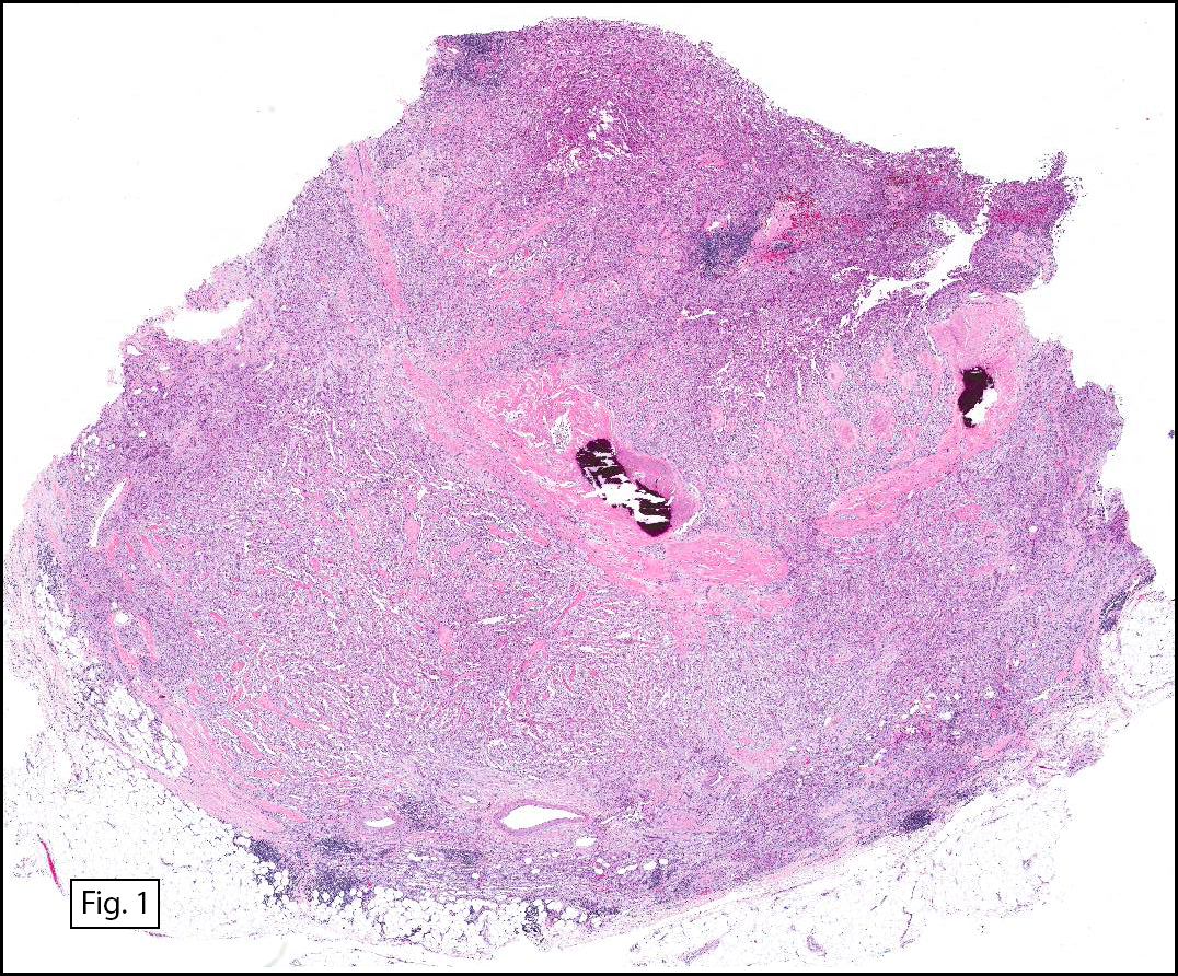
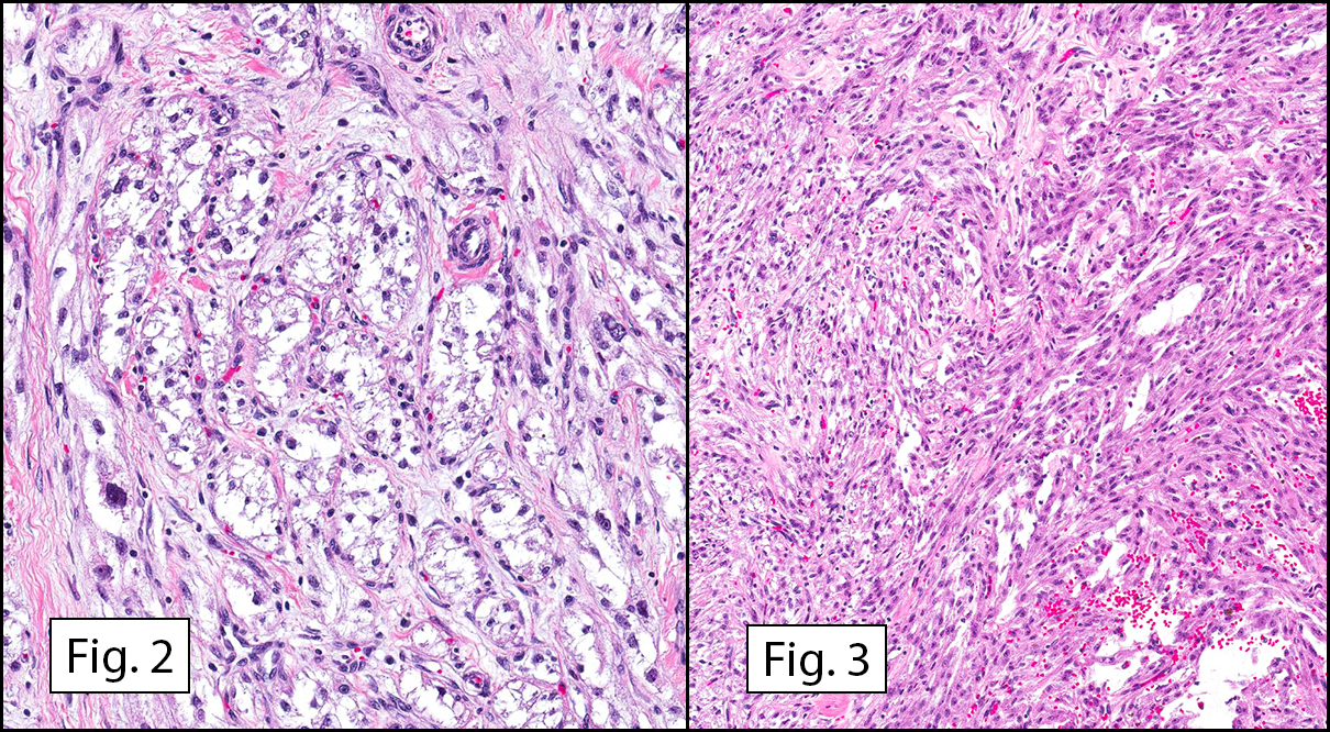
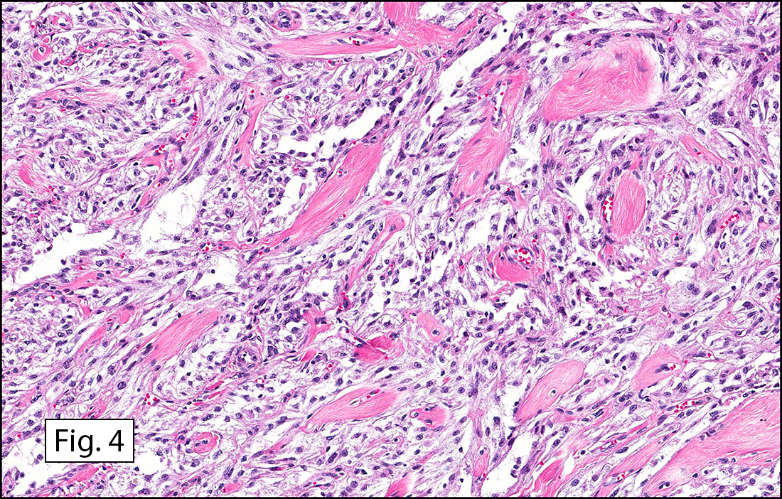
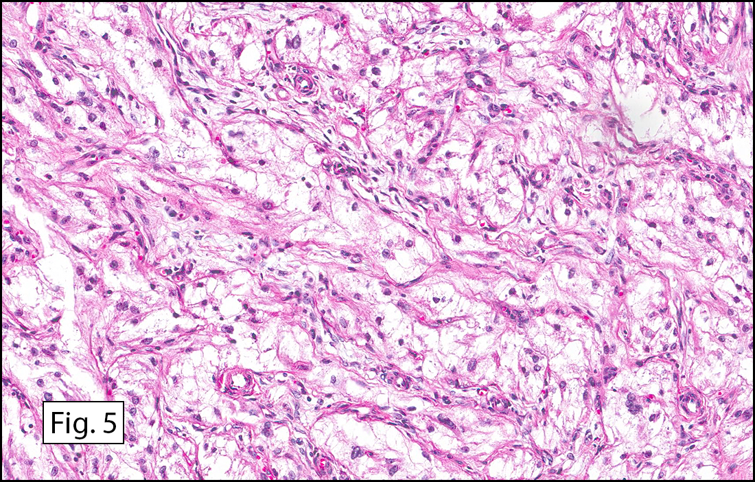
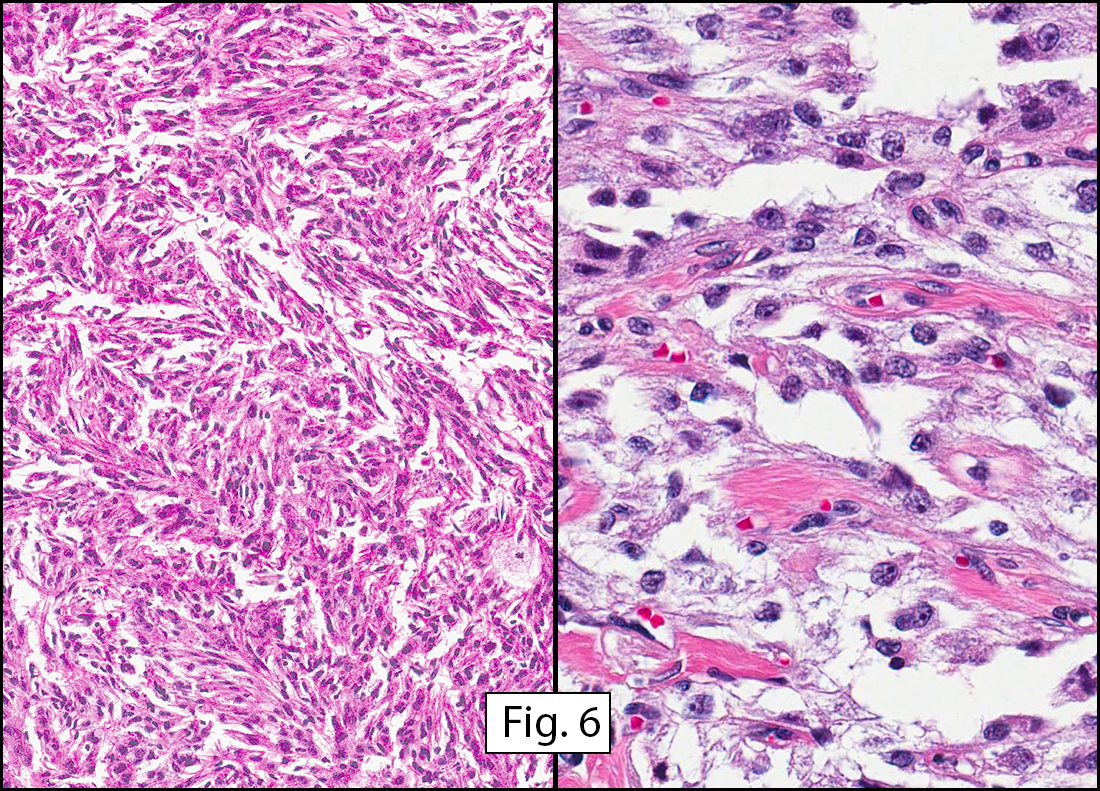
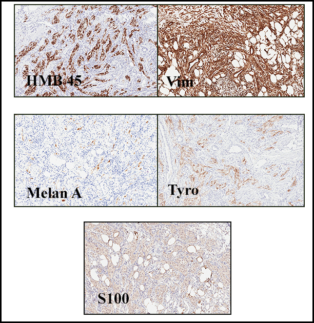
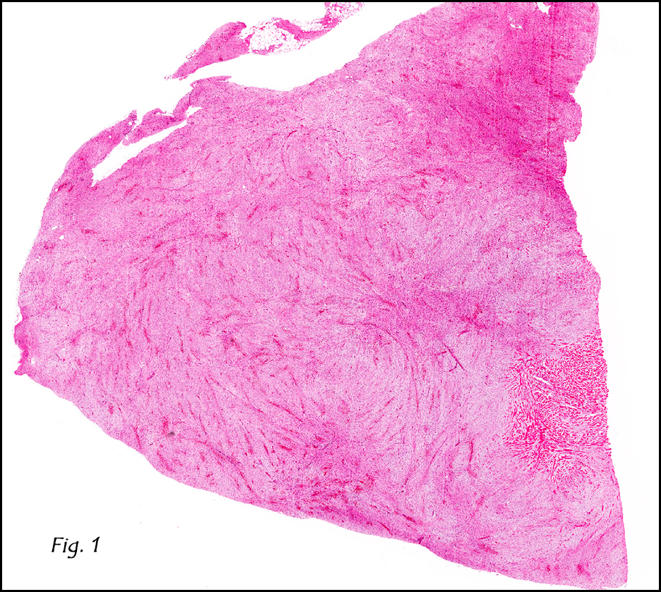
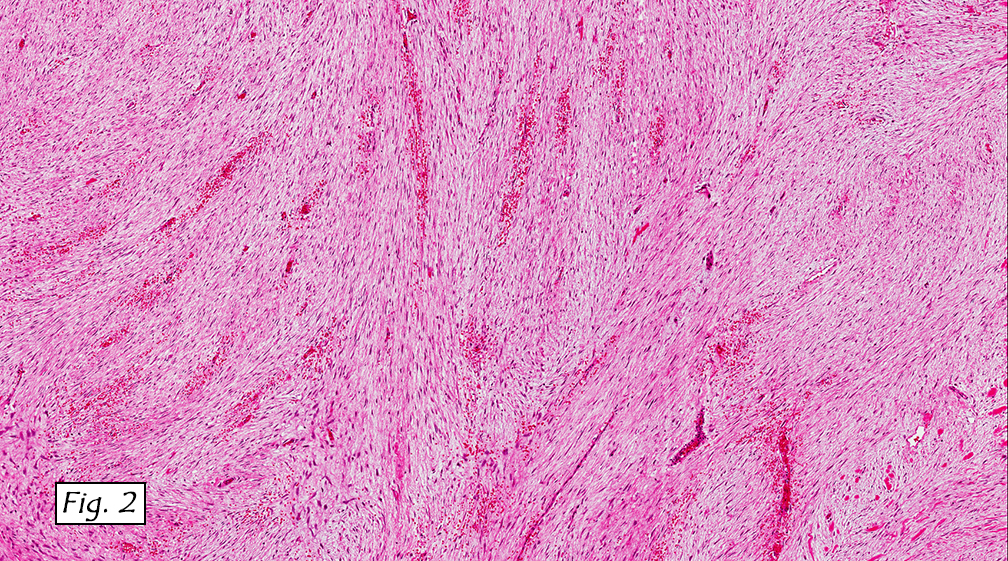
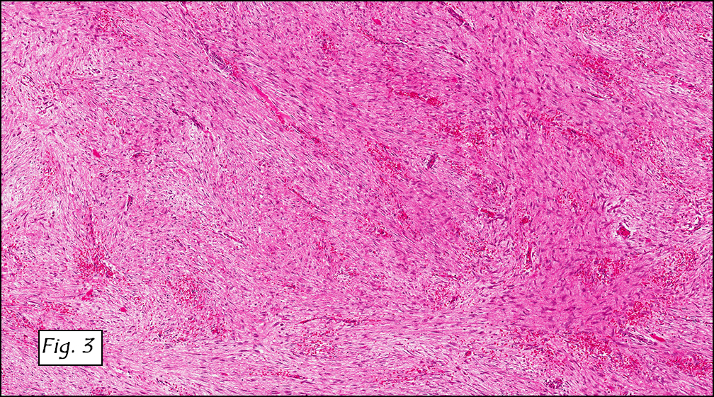
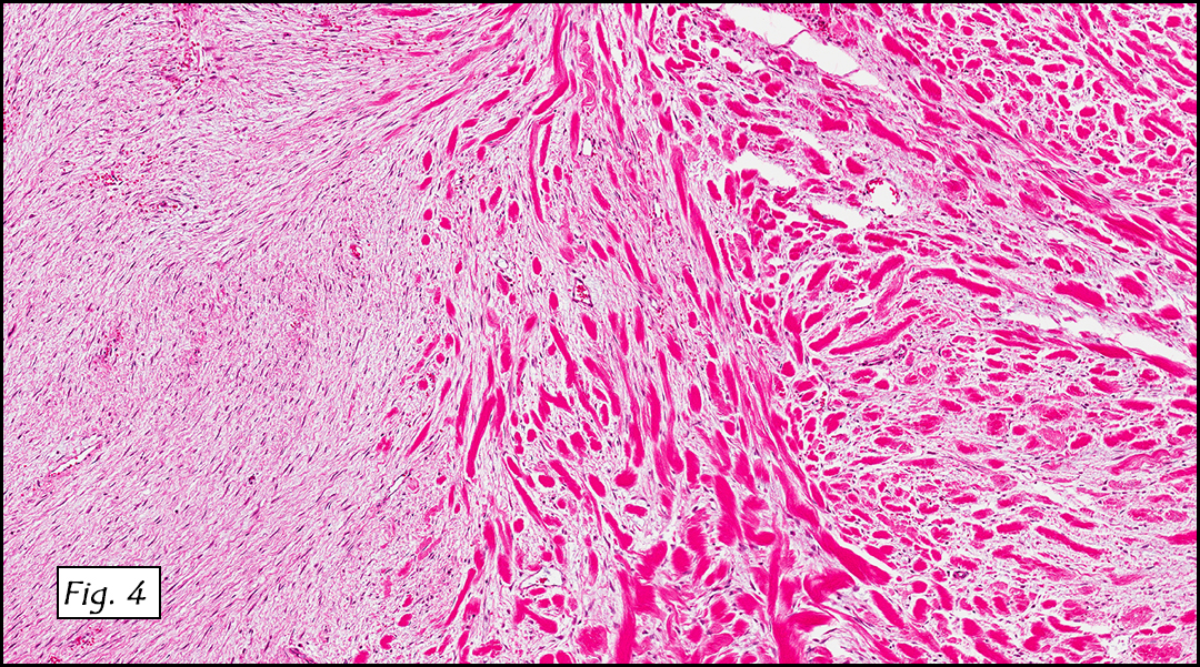
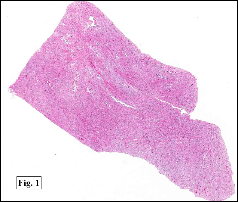
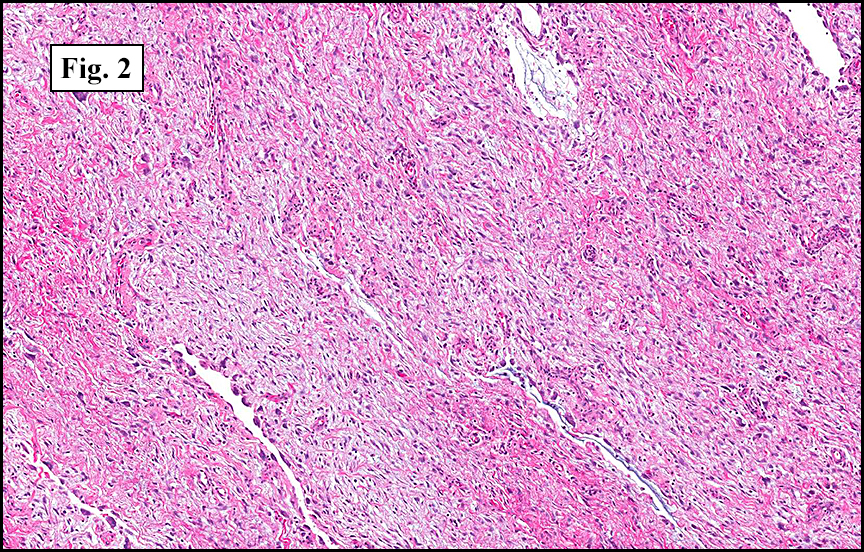
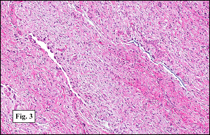
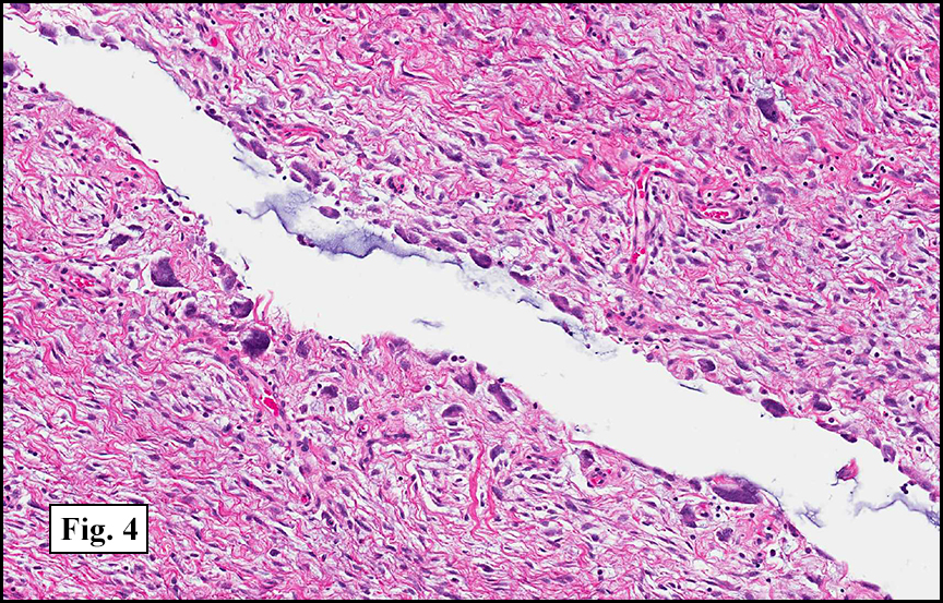
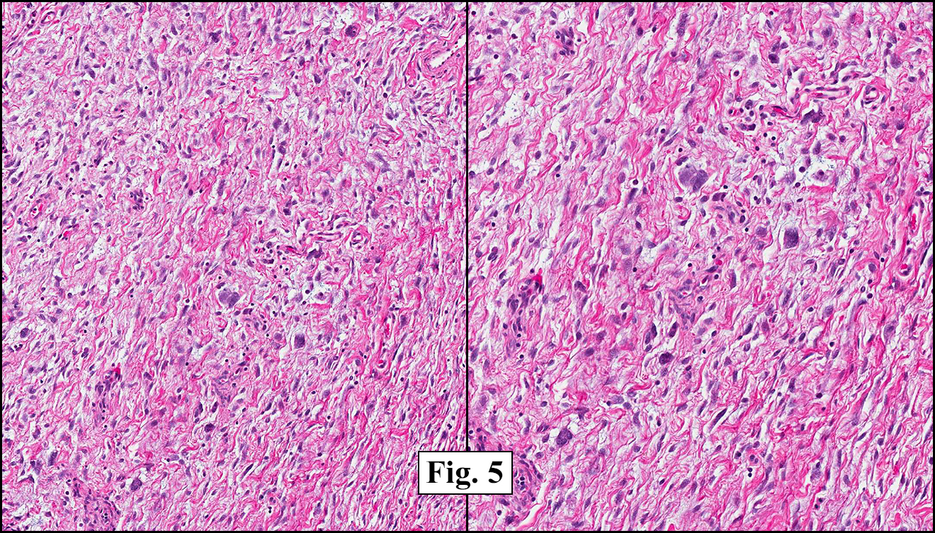
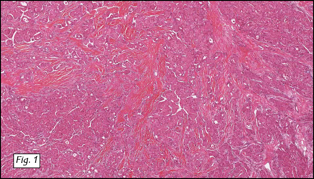
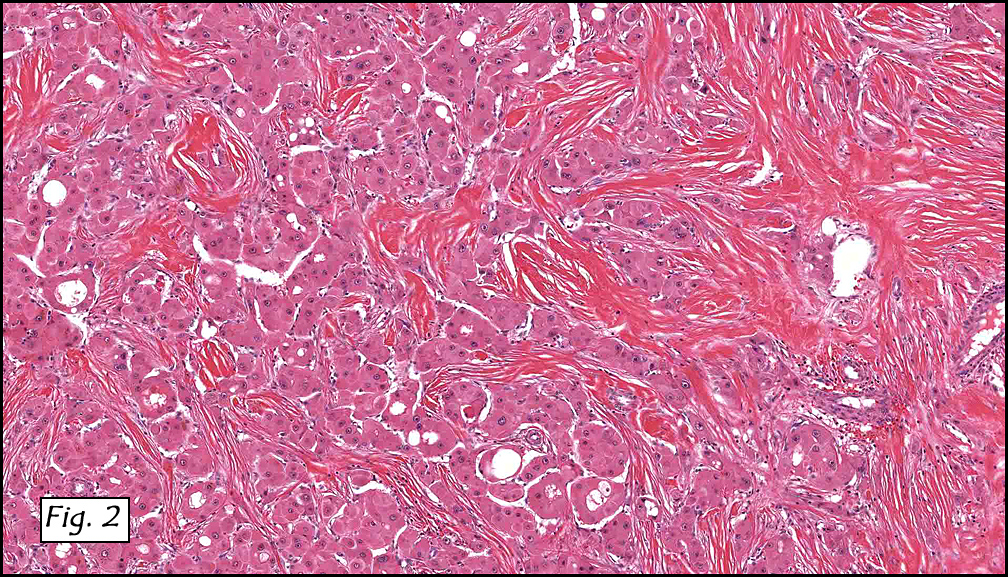
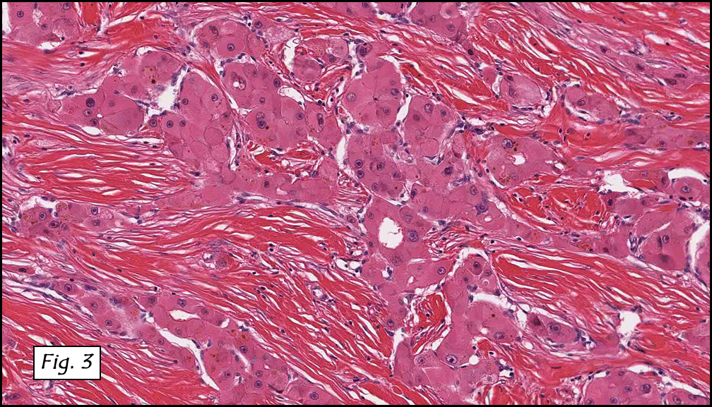
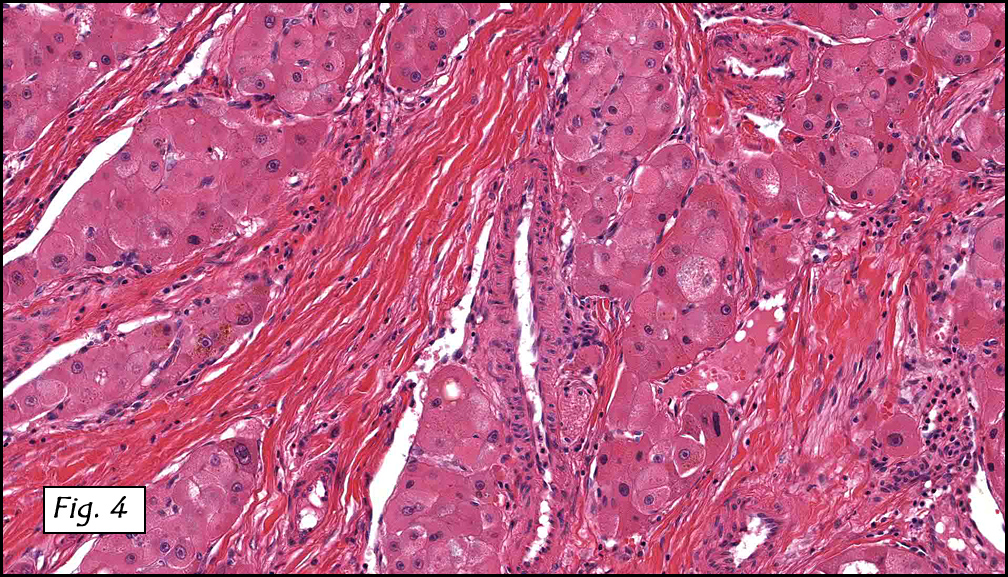
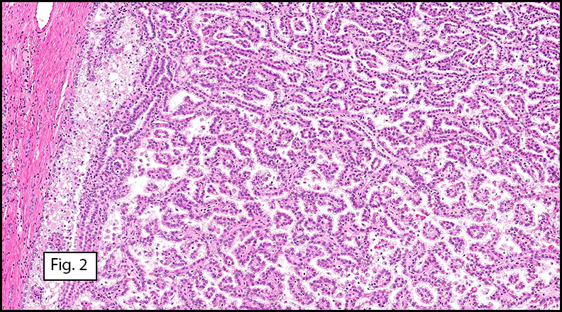
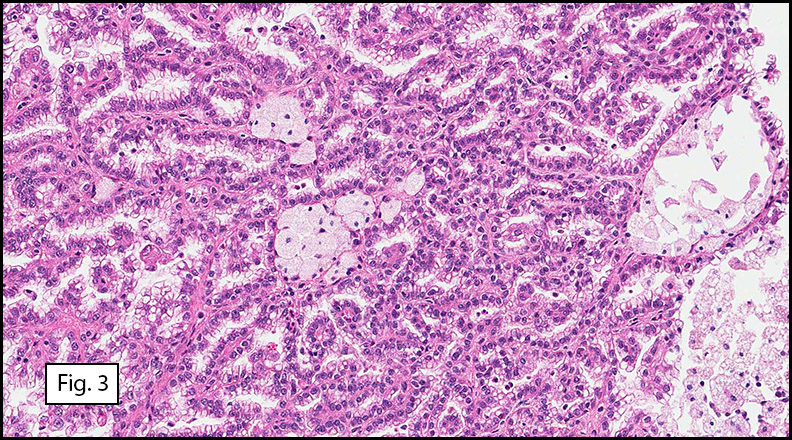
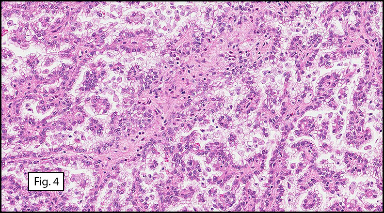
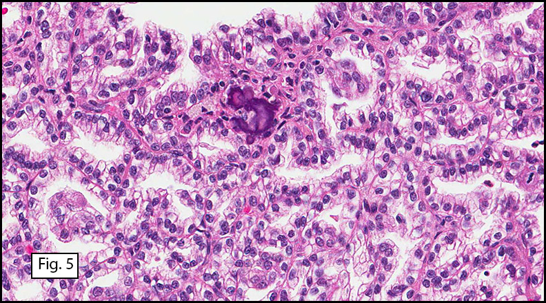
Grossly the tumor was 3 cm in diameter, well-circumscribed and encased within surrounding fat. The cut surface was gritty with focal cystic changes and peripheral fibrosis. Microscopically, the tumor had well defined margins with a thin fibrous capsule and focal calcifications (Fig. 1). The tumor consisted of spindled and polygonal cells arranged in alveolar and fascicular patterns (Fig. 2,3) with intervening dense, hyalinized collagen (Fig. 4). The cells displayed clear to eosinophilic cytoplasm with indistinct borders (Fig. 5) and round to oval nuclei demonstrating large, prominent nucleoli and coarse chromatin (Fig. 6). Mitotic figures were not identified and there was no evidence of necrosis. Immunohistochemistry revealed diffuse positivity for S100, HMB45, and vimentin with focal positivity for melanA and tyrosinase (Fig. 7).

Diagnosis: “Clear Cell Sarcoma (Malignant Melanoma of Soft Parts)”

Drew G. Davis, MSIV, and Donald R. Chase, M.D.
Department of Pathology and Human Anatomy, Loma Linda University and Medical Center, Loma Linda, California
California Tumor Tissue Registry, Loma Linda, California

Discussion: Clear cell sarcoma was first described by Dr. Franz M. Enzinger in 1965. Subsequently, several histologic and ultrastructural similarities with melanoma were noted including melanin pigment, presence of melanosomes, and immunohistochemical staining for S-100 and HMB-45. These findings resulted in the moniker malignant melanoma of soft parts. Despite these similarities clear cell sarcoma and melanoma appear to be two different entities. Recent chromosomal analyses have demonstrated a characteristic translocation in clear cell sarcoma, t(12;22), resulting in a novel fusion gene product which is not found in melanoma.

A majority of cases are localized in the extremities, predominantly the lower legs, with the ankle being the most commonly affected site. Tumors have been noted on the trunk and neck, but intra-abdominal tumors are exceedingly rare. The tumors are intimately involved with adjacent tendon sheaths and aponeuroses and rarely involve the epidermis although occasional dermal extension has been noted.
Most clear cell sarcomas arise between the ages of twenty and forty but they have been noted in a wider range from seven to eighty three. There is a slight female predominance. Clinically they are recognized by gradually enlarging, occasionally painful masses present for months to years. Grossly, the rubbery, firm tumors are lobulated or multinodular and are often encapsulated or partially circumscribed. The cut surface is gray-white and homogeneous with occasional foci of necrosis or hemorrhage. Pigmentation may be observed in some cases.

Histologically, clear cell sarcoma is distinguished by alveolar and fascicular patterns of spindled to polygonal cells with clear to eosinophilic cytoplasm separated by characteristic collagenous septa. The nuclei display vesicular chromatin and one or more prominent nucleoli. Melanin pigment may be seen in up to 50% of cases. Occasional multinucleated giant cells are identified and rare mitoses are seen as well. Immunohistochemical stains are a useful adjunct in the diagnosis and show a similar staining pattern to that of melanoma with varying degrees of S-100, HMB-45, Melan-A and vimentin positivity. Metastatic tumor deposits typically show a similar histologic pattern but increased nuclear pleomorphism, disorderly growth and multiple foci of hemorrhage and necrosis have been observed.

The differential diagnosis includes sarcomas with predominantly fascicular growth patterns, including synovial sarcoma and fibrosarcoma, as well as melanin producing tumors such as cellular blue nevi and paraganglioma-like dermal melanocytic tumor (PDMT). The cytologic features of clear cell sarcoma including clear cytoplasm and prominent melanoma-like nucleoli, along with its immunophenotypic profile, differentiate it from other sarcomas. More important is the distinction between it and other melanocytic lesions. Although the survival rates of both melanoma and clear cell sarcoma are poor, the clinical management differs drastically. The location of the tumor, especially its investiture in adjacent tendons and aponeuroses, and its general lack of epidermal and dermal involvement are helpful clues. The uniform, spindled appearance of clear cell sarcoma also contrasts with the epidermoid appearance of nodular melanomas. Cytologic features are useful in distinguishing clear cell sarcoma from cellular blue nevi, which typically have smaller, pinpoint nucleoli and lack atypia. As a last resort, molecular genetic analysis for the t(12;22) translocation will establish the diagnosis of clear cell sarcoma. Paraganglioma-like dermal melanocytic tumors may also show cells with clear to eosinophilic cytoplasm but the presence of zellballen-like nests, low grade nuclei and their traditional location in the dermis set them apart from clear cell sarcomas.

The established treatment of choice is surgical excision with wide margins. Chemotherapy and radiation for both local and metastatic control have been used with little success. Metastases are common and affect up to 67% of patients, often years after the primary excision. Prognostic factors include necrosis, increased tumor size and the presence of metastases.

Suggested Reading:

Enzinger FM. Clear cell sarcoma of tendons and aponeuroses: An analysis of 21 cases. Cancer. 1965; 18:1163-1174.
Chung EB, Enzinger FM. Malignant melanoma of soft parts: A reassessment of clear cell sarcoma. Am J Surg Pathol. 1983; 7(5):405-13.
Lucas DR, Nascimento AG and Sim FH. Clear cell sarcoma of soft tissues: Mayo Clinic experience with 35 cases. Am J Surg Pathol. 1992; 16(12):1197-1204.
Montgomery EA, Meis JM, Ramos AG, Frisman DM and Martz KL. Clear cell sarcoma of tendons and aponeuroses: A clinicopathologic study of 58 cases with analysis of prognostic factors. Int J Surg Pathol. 1993; 1(2):89-100.
Langezaal SM, Graadt von Roggen JF, Cleton-Jansen AM, Baelde JJ and Hogendoorn PCW. Malignant melanoma is genetically distinct from clear cell sarcoma of tendons and aponeuroses (malignant melanoma of soft parts). Br J Cancer. 2001; 84(4):535-8.
Weiss S, Goldblum J. Enzinger and Weiss’s Soft Tissue Tumors (5th edition). Philadelphia: Mosby/Elsevier Inc. 926-34, 2008.

January 2013: A 66 year old man with an abdominal mass

History: A 66-year-old male dwarf underwent surgical excision of a 6.0 cm mesenteric mass. The cut surface resembled white scar tissue.

Microscopically, the mass was of fibrous tissue with prominent blood vessels (Fig. 1). Spindle-shaped cells were evenly spaced among mature collagen arranged in long sweeping fascicles (Fig. 2). The stroma showed variable density (Fig. 3) and there was an area which consisted of thick bundles of collagen (keloidal fibrosis), (Fig. 4).

Diagnosis: “Mesenteric Fibromatosis”

Matthew S. Johnson, PSF, and Donald R. Chase, MD
Department of Pathology & California Tumor Tissue Registry, Loma Linda University and Medical Center, Loma Linda, California

Discussion: Mesenteric fibromatosis (MF) is classified as an intra-abdominal fibromatosis, and one of a part of a larger group of fibromatoses that are microscopically similar. Fibromatoses in general are benign fibroproliferative processes that may demonstrate infiltrative growth but do not metastasize. Deep fibromatoses have about a 50% recurrence rate, and the superficial variants about 10%. MF accounts for a mere 8% of all fibromatosis cases. These tumors typically arise in the mesentery of the small bowel and the ileocecal region, but occasionally occur in the omentum and retroperitoneum. They usually present as asymptomatic abdominal masses or are found incidentally in patients of various ages (mean age of 41 years) and are more common in males. It is common for them to measure 10 cm or more. Grossly, they well-circumscribed, white masses that may be either soft or firm.

Histologically, MF is comprised of a dense collagenous stroma with evenly dispersed spindle or stellate cells and prominent blood vessels. The cellularity may be variable with stroma ranging from densely fibrous to markedly myxoid. Foci of keloidal fibrosis (thick collagen bundles) are a hallmark feature, present in approximately 50% of cases.

Immunohistochemical studies can help distinguish MF from other tumors. MF displays strong nuclear staining for β-catenin but is negative for CD117, CD34, and S-100.

Differential Diagnosis:

• Gastrointestinal stromal tumor (GIST) is the most common misdiagnosis. They usually have greater cellularity and atypia then do MFs. Also helping to distinguish are the keloidal fibrosis, infiltrative growth pattern, and prominent thin-walled blood vessels which are commonly found in MF but not in GIST. The tumors are also separated by their immunostaining patterns. GIST tends to be positive for CD117, CD34, and β-catenin, and MF is not.

• Sclerosing mesenteritis may also present as a large mass in the mesentery of small bowel, but the histological findings of fat necrosis, chronic inflammation, less fibrosis, and lack of β-catenin expression distinguish it, as does the negative CD 117.

• Inflammatory myofibroblastic tumor of the mesentery or retroperitoneum is a consideration, but usually shows increased cellularity, cytologic atypia, and a greater degree of inflammation. These may also be positive for ALK-1 protein.

• Idiopathic retroperitoneal fibrosis typically displays lymphoplasmacytic inflammation dispersed among bands of dense hyalinized collagen.

MF is associated with familial adenomatous polyposis (FAP), in fact fibromatosis is found in approximately 10 to 15% of patients with FAP and often develops within two years after surgical excision of the affected intestinal tract. Diligent post-operative observation in FAP patients and endoscopic examination of the bowel in MF patients without known FAP is warranted.

Local recurrence is common. Greater recurrence rates have been observed in patients with FAP (90%) versus those without (12%). Recurrent tumors in FAP patients also tend to behave more aggressively and respond worse to therapy. Treatment involves complete excision of the tumor, which is often complicated by intestinal attachment. Alternative therapeutic options including postoperative radiation, chemotherapy, and anti-estrogenic agents have shown mixed results.

Suggested Reading:

Rodriguez JA; Guarda LA; Rosai J. Mesenteric fibromatosis with involvement of the gastrointestinal tract. A GIST simulator: a study of 25 cases. Am J Clin Pathol 2004 Jan;121(1): p93-8.

Pai SA; Zaveri SS. Intra-abdominal fibromatosis of the jejunum and mesentery. J Clin Pathol 2004 Oct;57(10): p1119.

Montgomery E; Torbenson MS; Kaushal M; Fisher C; Abraham SC. Beta-catenin immunohistochemistry separates mesenteric fibromatosis from gastrointestinal stromal tumor and sclerosing mesenteritis. Am J Surg Pathol 2002 Oct;26(10): p1296-301.

Church J; Berk T; Boman BM; Guillem J; Lynch C; Lynch P; Rodriguez-Bigas M; Rusin L; Weber T. Staging intra-abdominal desmoid tumors in familial adenomatous polyposis: a search for a uniform approach to a troubling disease. Dis Colon Rectum 2005 Aug;48(8): p1528-34.

Holubar S; Dwivedi AJ; O’Connor J. Giant mesenteric fibromatosis presenting as small bowel obstruction. Am Surg 2006 May;72(5): p427-9.

December 2012: A 13 month old boy with a chest wall mass

History: A 13-month-old male infant underwent surgical resection of a 6.0 x 5.6 x 2.0 cm subxiphoid body wall mass. The cut surface was soft and homogenously gray-tan.

The stroma was loosely arranged and consisted of undulating spindle cells (Fig. 1, 2) intermixed with irregularly shaped “pseudovascular” spaces (Fig. 3). Discontinuous rows of hyperchromatic giant cells lined the peripheries of these spaces (Fig. 4). Giant cells and multinucleated-appearing cells were scattered throughout the stroma (Fig. 5).

Diagnosis: “Giant Cell Fibroblastoma, Chest Wall”

Matthew S. Johnson, PSF, and Donald R. Chase, MD
Department of Pathology and Human Anatomy
California Tumor Tissue Registry
Loma Linda University and Medical Center, Loma Linda, California

Discussion: Initially described by Shmookler and Enzinger in 1982, giant cell fibroblastoma (GCF) is classified as a fibrohistiocytic tumor commonly felt to be the juvenile form of dermatofibrosarcoma protuberans (DFSP). It is usually diagnosed in infancy or childhood, with two-thirds being discovered by 5 years of age and about 90% by 10 years (median diagnostic age is 3 years). Rare cases have been reported in adults. GCF usually affects males and typically presents as a painless mass in the abdominal wall, groin, or back. Grossly, they are poorly circumscribed and gelatinous. The cut surface is usually gray to yellow.

Histologically, GCFs are variably cellular and composed of spindle and stellate cells within a collagenous, sometimes myxoid stroma. The nuclei are small and uniform without significant numbers of nucleoli. Scattered throughout the stroma are hyperchromatic giant cells with large vesicular nuclei, often with prominent nucleoli. Occasionally the giant cells appear multinucleated; however, ultrastructural examination suggests that most actually have a single, multilobated nucleus. The defining feature of this tumor is the intervening, large, irregular pseudovascular spaces that are partially lined by the giant cells. No endothelial lining is present.

Immunohistochemical studies show the tumor stroma and pseudovascular lining to be negative for S-100, cytokeratin, desmin, and CD31. CD34 is typically weakly positive in the stromal elements.

GCF appears to be closely related to DFSP. Cases of GCF recurring as DFSP (and vice versa) have been documented, and some tumors show both neoplasms. Cytogenetic analysis has revealed the presence of a (17:22) translocation in both phenotypes. Evidence suggests that a linear arrangement of this abnormality occurs in GCF while a ring form takes place in DFSP.

Differential Diagnosis:

• Myxoid liposarcoma may resemble GCF with myxoid stroma; however, the tendency to occur in deep tissues, the presence of lipoblasts, a delicate vasculature, and lack of giant cells separates it from GCF.
• Myxoid malignant fibrous histiocytoma (myxofibrosarcoma) may have histologically similar stroma and can have large cells demonstrating pleomorphism and multinucleation. A vascular network and mitotic activity differentiates it from GCF, as does a lack of space-lining pleomorphic cells.
• Angiosarcomas demonstrate irregular branching vascular channels that are lined by multiple layers of cells with large hyperchromatic nuclei. These lesions are distinguished from GCF by their high mitotic activity, atypical nuclei of the vascular lining cells, and expression of CD31.
• Lymphangiomas contain angiectatic spaces, but these spaces are often surrounded by a layer of smooth muscle, and not by pleomorphic cells. Lymphoid aggregates may also be present in the surrounding stroma.

About 50% of GCF cases recur. Recurrences are not aggressive and metastatic disease has not been reported. Treatment of choice is wide local excision; however, less invasive therapy with routine follow-up is also a consideration.

Suggested Reading:

Terrier-Lacombe MJ; Guillou L; Maire G; Terrier P; Vince DR; de Saint Aubain Somerhausen N; Collin F; Pedeutour F; Coindre JM. Dermatofibrosarcoma protuberans, giant cell fibroblastoma, and hybrid lesions in children: clinicopathologic comparative analysis of 28 cases with molecular data – a study from the French Federation of Cancer Centers Sarcoma Group. Am J Surg Pathol 2003 Jan;27(1): p27-39.

Sandberg AA; Bridge JA. Updates on the cytogenetics and molecular genetics of bone and soft tissue tumors. Dermatofibrosarcoma protuberans and giant cell fibroblastoma. Cancer Genet Cytogenet 2003 Jan 1;140(1): p1-12.

Pascual A; Sanchez-Martinez C; Moreno C; Burdaspal-Moratilla A; Lopez-Rodriguez MJ; Rios L. Dermatofibrosarcoma protuberans with areas of giant cell fibroblastoma in the vulva: a case report. Eur J Gynaecol Oncol 2010;31(6): p685-9.

Najarian DJ; Morrison C; Sait SN; Meguerditchian AN; Kane J; Cheney R; Zeitouni NC. Recurrent giant cell fibroblastoma treated with Mohs micrographic surgery. Dermatol Surg 2010 Mar;36(3): p417-21.

Layfield LJ; Gopez EV. Fine-needle aspiration cytology of giant cell fibroblastoma: case report and review of the literature. Diagn Cytopathol 2002 Jun;26(6): p398-403.

Aliagaoglu C; Bakan V; Atasoy M; Sahin O; Toker S; Albayrak M. A very large, rapidly developing, congenital giant cell fibroblastoma in a 5-month old infant. J Dermatol 2006 Mar;33(3): p182-6.

November 2012: A five month old girl with a right cheek mass

History: A healthy Caucasian girl was delivered vaginally at full term, with no complications. At two weeks, a soft, non-discrete, non-tender right cheek mass was noted. The mass was non-pulsatile, did not bleed or interfere with feeding or respiration, nor did the size of the mass fluctuate with crying or movement. There were no abnormalities of the overlying skin or mucosa. The mass was closely observed for the next few months. Ultrasound demonstrated a 4 x 3 x 1.4 cm solid whorled mass anterior to the parotid; MRI revealed a well-circumscribed mass involving the right masseter muscle, adjacent to the mandible with increased T2-signal within the bone marrow of the mandible. At five months of age, she underwent intraoral excision of the mass under anesthesia. The patient, now 16 months old, recovered from surgery without complications. Post-operative MRI demonstrated a residual and/or recurrent 3 cm right masseter mass with no other nodules or masses have been observed. No family history of similar lesions or syndromic conditions was elicited.

Resection of the lesion yielded an aggregate of approximately 2.0 x 1.5 x 0.4 cm homogenous brown soft tissue. Microscopically, the lesion appeared to be well-circumscribed but extended to the inked margins (Fig. 1). Skeletal muscle differentiation and maturation were observed with cross striations and haphazard arrangement of irregular fascicular bundles of muscle cells with scant to moderate eosinophilic cytoplasm (2,3,4). No definite immature cells were identified, and no myxoid stroma was noted. No areas of hypercellularity, increased mitotic activity, pleomorphism or necrosis were observed.

Diagnosis: “Fetal rhabdomyoma, intermediate (cellular) type, cheek”

Evelyn Choo MD1, Rachel Conrad MD1, Anwar Raza, MD1, Donald Chase MD1,2

1. Department of Pathology and Human Anatomy, Loma Linda University and
Loma Linda Medical Center, Loma Linda, California
2. California Tumor Tissue Registry, Loma Linda, California

Discussion: Fetal rhabdomyoma is a rare but benign skeletal muscle neoplasm, first described by Dehner in 1972. It typically occurs in the head and neck region of young children and demonstrates varying degrees of skeletal muscle differentiation.

The term rhabdomyoma encompasses a complex classification system of several distinct benign skeletal muscle neoplasms. It is organized by location (cardiac versus extracardiac), and the extracardiac type is divided into adult, genital and fetal categories based on tissue differentiation and clinical presentation. The adult form tends to occur in head and neck region of 40-60 year old males and displays prominent eosinophilic polygonal cells with vacuolated cytoplasm. The genital form typically presents as a polypoid lesion in vulva and vagina of middle-aged women; microscopically, long strap-like muscle fibers with prominent cross-striations are seen in a collagenous and myxoid matrix. The fetal form shows immature skeletal muscle differentiation and is more common in the head and neck region of children below age four.

The fetal form is further subdivided into a myxoid type and an intermediate type. The myxoid subtype (also known as “classic”) is comprised almost entirely of immature primitive spindle cells in a myxoid stroma and tends to appear in postauricular soft tissue. In contrast, a wider spectrum of myocyte maturation is seen in the intermediate subtype (also known as “cellular” or “juvenile”), initially described by Di Sant’Agnese and Knowles in 1980. This subtype of rhabdomyoma appears more often in soft tissue of face or mucosal tissue and represents a more differentiated lesion than classic fetal subtype.

Clinically, fetal rhabdomyoma presents in children less than four years old, may often be congenital and is more common in males than females (2.4:1). The lesion consists of a solitary, well-defined, non-tender mass involving soft tissue or mucosa. It grows very slowly and seldom ulcerates the overlying skin or mucosa. Multiple extracardiac rhabdomyomata have been associated with nevoid basal cell carcinoma syndrome (also known as Gorlin-Goltz syndrome) and may show a mutation in PTCH on chromosome 9q22. However, no correlation is seen with tuberous sclerosis as in cardiac rhabdomyoma.

The gross appearance of fetal rhabdomyoma consists of a well-circumscribed, smooth, polypoid mucosal lesion, typically two to six centimeters in diameter. Lesions are typically solitary. Cross-sectioning reveals a grey to pink surface, often with a myxoid appearance.

Microscopically, these lesions display either a myxoid or an intermediate pattern. The myxoid version is comprised of a myxoid matrix surrounding scattered or short bundles of immature spindle-shaped or oval cells with immature skeletal muscle fibers. The small uniform nuclei contain delicate chromatin and are enveloped by tapered eosinophilic cytoplasmic processes. Rare cross-striations are present. The intermediate type closely resembles classic adult head and neck rhabdomyoma, and contains a wider spectrum of myocyte differentiation with few immature spindle-shaped muscle precursors and numerous prominent strap-shaped muscle cells. The myocytes have central vesicular nuclei, scant to moderate eosinophilic cytoplasm, and frequent cross-striations with occasional vacuolation and glycogen. They are haphazardly arranged in irregular fascicular bundles. Ganglion-like rhabdomyoblasts with prominent nucleoli may be present. In both myxoid and intermediate types, the absence of significant atypia, necrosis, or mitotic activity is noted. Invasion and destruction of surrounding tissues should not be seen.

Immunohistochemical staining shows positivity for muscle-specific actin (MSA), myoglobin, and desmin; focal positivity may be seen for smooth muscle actin (SMA), S100, glial fibrillary acidic protein (GFAP), and vimentin.

Electron microscopy will show thick and thin myofilaments in the more mature myocytes; z-bands and glycogen may also be demonstrated in the cytoplasm. The immature myocytes may lack specific ultrastructural features of myocyte differentiation.

The recommended therapy for fetal rhabdomyoma is complete excision. Prognosis is excellent. Local recurrence is rare and is typically associated with incomplete resection. Metastases have never been described in the literature. Only two controversial cases of possible malignant transformation have been reported but may have been misdiagnosed as benign initially, and recurrent cases should be carefully evaluated for a misdiagnosis of rhabdomyosarcoma.

The differential diagnosis of fetal rhabdomyoma includes rhabdomyosarcoma (both embryonal and spindle cell types), adult rhabdomyoma, genital rhabdomyoma, Triton tumor (neuromuscular hamartoma), rhabdomyomatous mesenchymal hamartoma of the skin, and infantile fibromatosis.

1. Rhabdomyosarcoma consists of pronounced diffuse cellular immaturity, necrosis, mitotic figures, and infiltration of adjacent tissues with frank destruction. The presence of nuclear atypia in rhabdomyosarcoma is the most important criteria to distinguish it from fetal rhabdomyoma.
2. Adult rhabdomyoma has distinctive polygonal “spider” cells with vesicular nuclei, central nucleoli, and eosinophilic cytoplasm. It tends to occur in the head and neck region of adult males.
3. Genital rhabdomyoma demonstrates predominately mature long, strap-like striated muscle cells and occasional immature myocytes in a background of myxoid material and collagen. It typically presents as an asymptomatic, slow-growing, small polypoid lesion in the vulvo-vaginal region of middle-aged women. Rare occurrences in the male urogenital tract have also been described.
4. Triton tumor is composed of nerve fascicles mixed with mature striated skeletal muscle. It is associated with neurofibromatosis in younger patients and typically affects the axial skeleton.
5. Rhabdomyomatous mesenchymal hamartoma consists of normal dermal elements with mature striated skeletal muscle.
6. Infantile fibromatosis is not as well-circumscribed as fetal rhabdomyoma. It tends to involve regions deeper than the subcutis, has a fasciculated spindle cell pattern, lacks cytoplasmic striations, and contains interspersed fat cells.

Fetal rhabdomyoma is a rare neoplasm of immature skeletal muscle. Distinguishing it from the more common, more aggressive rhabdomyosarcoma is important. Clinical characteristics (location, behavior) and microscopic traits (mitoses, necrosis, and especially nuclear atypia) may be helpful. Prognosis is excellent after surgical excision. Recognition of this benign neoplasm is important in communicating accurate prognostic information and in preventing over-aggressive treatment.

Suggested Reading:

1. Dehner LP, Enzinger FM, Font RL. Fetal rhabdomyoma: an analysis of nine cases. Cancer. 1972;30:160-166.
2. Di Sant’Agnese PA, Knowles DN. Extracardiac rhabdomyoma: a clinicopathologic study and review of the literature. Cancer. 1980;56:780-789.
3. Kapadia SB, Barr FG. Rhabdomyoma. In: Fletcher CDM, Unni K, Mertens F, eds. World Health Organization Classification of Tumours. Pathology and Genetics of Tumors of Soft Tissue and Bone. Lyon, France: IARC Press, 2002:142-145.
4. Premalata CS, Kumar RV, Saleem KM, Fathima LJ, Das K. Fetal rhabdomyoma of the lower extremity. Pediatr Blood Cancer. 2009;52(7):881-883.
5. Walsh SN, Hurt MA. Cutaneous fetal rhabdomyoma: a case report and historical review of the literature. Am J Surg Pathol. 2008;32(3):485-491.
6. Weiss SW, Goldblum JR. Enzinger & Weiss’s Soft Tissue Tumors. 5th ed. Philadelphia: Mosby-Elsevier, 2008:583-592.
7. Yang S, Zhao C, Zhang Y, Liao S. Mediastinal fetal rhabdomyoma in nevoid basal cell carcinoma syndrome: a case report and review of the literature. Virchows Arch. 2011; 459:235-238.

October 2012: A 61 year old woman with a left kidney mass

History: A sixty-one year old woman underwent a hand-assisted laparoscopic nephrectomy. The 256.0 gram, 12.0 x 6.5 x 2.5 cm left kidney contained a single 4.3 x 4.0 x 3.2 cm homogeneous tan-white, pseudo-encapsulated mass in the mid ante-hilar pole. The mass was adjacent to but did not penetrate the renal capsule. No tumor involvement of the perirenal adipose, hilar vessels, renal pelvis or ureter was noted. The residual renal parenchyma had a normal red-brown appearance and showed a distinct cortico-medullary junction. No other lesions were identified.

Microscopically, the well-circumscribed tumor approached but did not extend through the renal capsule (Fig. 1). A distinctive tubulo-papillary architecture was observed (Fig. 2), with fibrovascular cores lined by a single layer of tumor cells. The tumor cells had moderate to focally abundant basophilic cytoplasm. The low-grade nuclei displayed open chromatin and rare nucleoli, consistent with Fuhrman Nuclear Grade 2. Occasional foamy histiocytes (Fig. 3), clear cells (Fig. 4), and psammoma bodies were seen (Fig. 5). Sarcomatoid features, tumor necrosis, and lymphovascular invasion were absent. The adjacent non-neoplastic kidney demonstrated chronic inflammation and hyalinized glomeruli, consistent with mild benign nephrosclerosis.

Diagnosis: “Papillary Renal Cell Carcinoma, Type 1 (Basophilic)”

Rachel Conrad, MD1; Pamela Boswell, DO2; Manju Aron, MD3; Mahul Amin, MD3; Donald R. Chase, MD1,4.

1. Department of Pathology and Human Anatomy, Loma Linda University and
Loma Linda Medical Center, Loma Linda, California
2. Scripps Clinic Medical Laboratories, La Jolla, California
3. Cedars Sinai Medical Center, Los Angeles, California
4. California Tumor Tissue Registry, Loma Linda, California

Discussion: Papillary renal cell carcinoma is a relatively rare type of renal cell carcinoma with unique morphologic, immunohistochemical, and genetic features. It tends to occur in the sixth to seventh decades with a male predominance (3:1).

Papillary renal cell carcinoma has been associated with several different clinical syndromes. Hereditary/familial papillary renal cell carcinoma (7q31 c-MET mutation) results in multiple bilateral type 1 papillary renal tumors. Hereditary leiomyomatosis and renal cell cancer (1q42-43 mutation) leads to a solitary unilateral type 2 papillary renal cell carcinoma, usually around age thirty, along with cutaneous and uterine leiomyomas and leiomyosarcomas. Birt-Hogg-Dube syndrome (17p11.2 mutation) has multiple chromophobe and oncocytoma renal cell tumors with rare clear cell or papillary renal cell carcinomas. Fibrofolliculomas, lung cysts, and a history of spontaneous pneumothorax are frequently noted. Familial paraganglioma syndrome displays multiple renal tumors with benign and malignant paragangliomas and papillary thyroid carcinoma. PTEN hamartoma tumor syndrome results in unifocal papillary RCC and chromophobe RCC with gastrointestinal polyposis, tumors of the breast, thyroid and uterus, and frequently, lipomas and trichilemmomas.

The gross appearance of papillary renal cell carcinoma consists of a well-circumscribed nodule with a fibrous pseudocapsule. Lesions are often bilateral and multifocal, and hypovascularity may be seen on radiographic studies. Cross-sectioning reveals a yellow-brown surface with regions of cystic degeneration, hemorrhage, and necrosis.

Microscopically, the tumors contain fibrovascular cores lined by neoplastic cells containing foamy histiocytes and occasional neutrophils. Solid-papillary and tubulopapillary growth patterns may also be observed. Sarcomatoid dedifferentiation is seen in five percent of papillary renal cell carcinomas and confers a poor prognosis. Additional findings may include stromal and intracellular hemosiderin deposition, occasional psammoma bodies, necrosis, and clear cell change in areas of hemorrhagic degeneration.

Papillary renal cell carcinoma has been classified by Delahunt and Eble (1997) into two subtypes. Type 1 (also known as “basophilic”) shows a single layer of cells with scant basophilic-to-amphophilic finely granular cytoplasm and low-grade nuclei arranged along the papillary basement membrane. Type 2 shows pseudostratified cells with abundant eosinophilic granular cytoplasm and large nuclei displaying a higher Fuhrman grade. As expected, the higher grade tumors have a worse prognosis.

Immunohistochemical staining shows membranous positivity for CK7, with stronger expression in Type 1 papillary renal cell carcinoma. A granular cytoplasmic pattern is seen for alpha methylacyl CoA-racemase (AMACR). Other positive stains include AE1/AE3, CAM5.2, EMA, CD10, PAX2, PAX8, RCC, and PN15/gp200. Negative staining is seen for CAIX, Cathepsin-K, TFE3, and 34-beta-E12.

Cytogenetic studies often show trisomy 7, trisomy 17, and loss of Y without abnormalities of 3p. Additional genetic anomalies may be observed.

The recommended therapy for papillary renal cell carcinoma is complete extirpation. The overall prognosis is better than for conventional renal cell carcinoma, with a 5 year survival rate of 45-70%. Survival drops to 15-20% if extension to the renal vein or perinephric fat is observed. Metastases are frequently seen in tumors larger than 3 cm and tend to involve the lungs, lymph nodes, bones, and liver.

The differential diagnosis of papillary renal cell carcinoma includes metanephric adenoma, conventional clear cell renal cell carcinoma with papillary foci, clear cell papillary renal cell carcinoma, oncocytic papillary renal cell carcinoma, Xp11 translocation renal cell carcinoma, collecting duct carcinoma, and mucinous tubular and spindle cell carcinoma of the kidney.

1. Metanephric adenoma tends to occur in young to middle aged females. The microscopic appearance is consistent with developing metanephric tubular epithelium and displays tiny tubules and papillae with bland nuclei and scant stroma. This lesion is usually small in size and benign in nature. Immunostaining shows rare focal CK 7 positivity and negativity for EMA and AE1/AE3.
2. Conventional clear cell renal cell carcinoma with papillary foci is fairly common. It may be distinguished from papillary renal cell carcinoma by its hypervascularity on radiographic imaging, golden yellow color on sectioning, and solid or acinar architecture with clear-to-eosinophilic cells separated by thin vascular septae. True papillae are rarely seen; instead, small papillations protrude into necrotic or cystic spaces. Chromosome studies frequently show deletion of the von Hippel-Lindau gene (3p), and inactivation of this gene can be detected by diffuse strong membranous staining for CAIX. Negative immunohistochemical staining is observed with CK7 and AMACR.
3. Clear cell papillary renal cell carcinoma is a small indolent tumor associated with end-stage renal disease. Microscopically, the papillary fronds are lined by clear cells with low Fuhrman nuclear grade and characteristic subnuclear vacuoles. Smooth muscle metaplasia and cystic changes are common. No hemosiderin deposition, foamy histiocytes, or psammoma bodies are seen. Immunohistochemistry shows a cup-like pattern for CAIX and CK7, with patchy positivity for 34-beta-E12; CD10 and AMACR are negative.
4. Oncocytic renal cell carcinoma with papillary features has a distinctive mahogany-brown color with a central scar on sectioning. Microscopic features include abundant granular eosinophilic cytoplasm and a linear arrangement of low-grade apical nuclei. Pseudostratification is absent, and papillary architecture is focal, not diffuse. IHC staining is positive for parvalbumin and negative for RCC and CK7.
5. Xp11 translocation renal cell carcinoma is the most common pediatric renal cell carcinoma subtype and may be associated with previous chemotherapy. Extensive psammomatous calcification may be seen radiographically and microscopically. Papillary architecture with clear-to-eosinophilic cells and vesicular high-grade nuclei is common, but cystic, solid, or nested architecture can also be seen. Immunohistochemical staining is positive for Cathepsin-K, TFE3, and PAX8, with focal staining for melanocytic markers. Weak expression of cytokeratins and epithelial markers may be seen. Cytogenetic studies often show translocation of ASPL and TFE3, t(X;17)(p11.2q25).
6. Collecting duct carcinoma tends to display prominent desmoplasia, frequent invasion, and high grade nuclear features. IHC stains are negative for CD10, AMACR, and PN15/gp200.
7. Mucinous tubular and spindle cell carcinoma of the kidney can be distinguished due to its mucinous stroma and overall architectural pattern. It may share focal papillary growth, mucin production, foam cells, and AMACR positivity with papillary renal cell carcinoma, but it will lack the classic diffuse papillary pattern.

Due to its better prognosis, associated familial syndromes, and tendency toward multiple bilateral hypovascular tumors, papillary renal cell carcinoma is important to separate from other lesions with papillary architecture. Characteristic microscopic traits include true fibrovascular cores containing foamy histiocytes and hemosiderin. Immunohistochemical stains (CK7, AMACR) and cytogenetic studies (trisomy 7/17, loss of Y) can be useful in challenging cases. Treatment consists of surgical excision and careful assessment for metastases. The astute observer will be alert to the unique aspects of this rare tumor.

Suggested Reading:

1. Delahunt B, Eble JN. Papillary renal cell carcinoma. In: Eble JN, Sauter G, Epstein JI, Sesterhenn IA, eds. World Health Organization Classification of Tumours. Pathology and Genetics of Tumors of the Urinary System and Male Genital Organs. Lyon, France: IARC Press, 2004:15-22.
2. Merino MJ, Eccles DM, Linehan WM, et al. Familial renal cell carcinoma. In: Eble JN, Sauter G, Epstein JI, Sesterhenn IA, eds. World Health Organization Classification of Tumours. Pathology and Genetics of Tumors of the Urinary System and Male Genital Organs. Lyon, France: IARC Press, 2004:15-22.
3. Mester JL, Zhou M, Prescott N, Eng C. Papillary renal cell carcinoma is associated with PTEN hamartoma tumor syndrome. J Urol. 2012; 79(5):1187.e1-7
4. Sukov WR, Lohse CM, Leibovich BC, Thompson RH, Cheville JC. Clinical and pathological features associated with prognosis in patients with papillary renal cell carcinoma. J Urol, 2012; 187(1):54-59.
5. Ross H, Martignoni G, Argani P. Renal cell carcinoma with clear cell and papillary features. Arch Pathol Lab Med. 2012;136(4):391-9.
6. Rosai J. Rosai and Ackerman’s Surgical Pathology. China: Mosby, 2004:1251-1264.
7. Delahunt B, Eble JN. Papillary renal cell carcinoma: a clinicopathologic and immunohistochemical study of 105 tumors. Mod Pathol. 1997;10:537.
8. Murphy WM, Grignon DJ, Perlman EJ. AFIP Atlas of Tumor Pathology Series 4: Tumors of the Kidney, Bladder, and Related Urinary Structures. Washington, D.C.: American Registry of Pathology, 2004.

Scroll to top