September 2012: A 70 year old sandblaster with pulmonary nodules

History: A 70-year-old retired sandblaster underwent a left lower lobectomy for lung cancer. He had stopped smoking one year earlier, after smoking 1.5 packs per day for approximately 50 years. Four years later, two new nodules were discovered, resulting in a right lower lobectomy.

Gross examination revealed a dominant, ill-defined, marbled, gray-and-white tan 6 cm tumor which involved the pleural surface and showed central cystic degeneration, as well as numerous smaller satellite lesions of similar appearance. Three hilar lymph nodes were also identified.

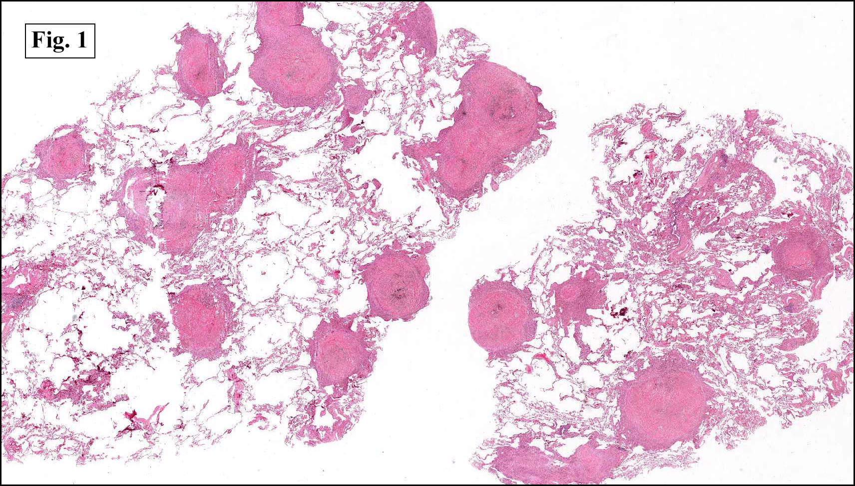
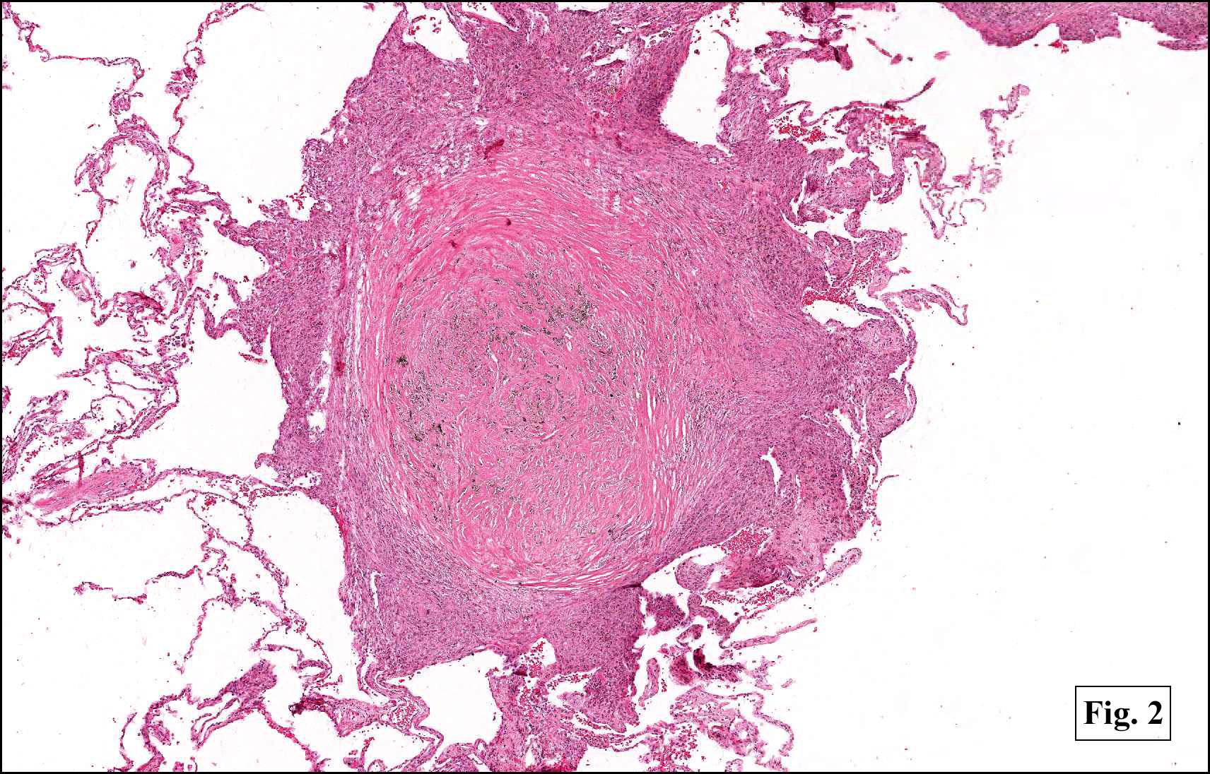
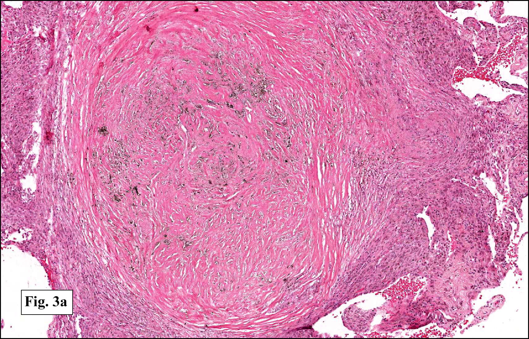
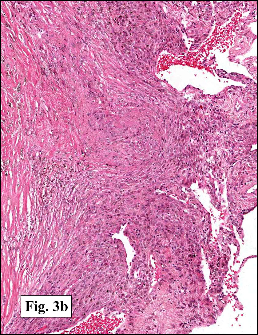
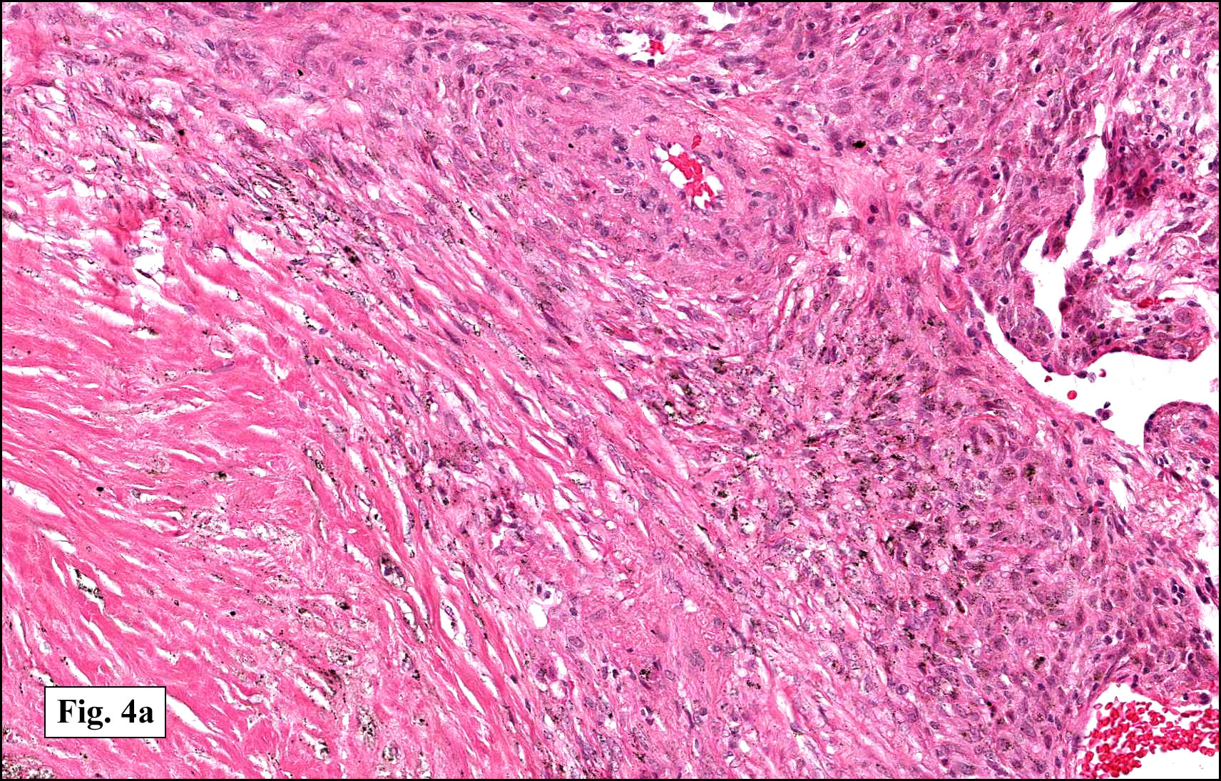
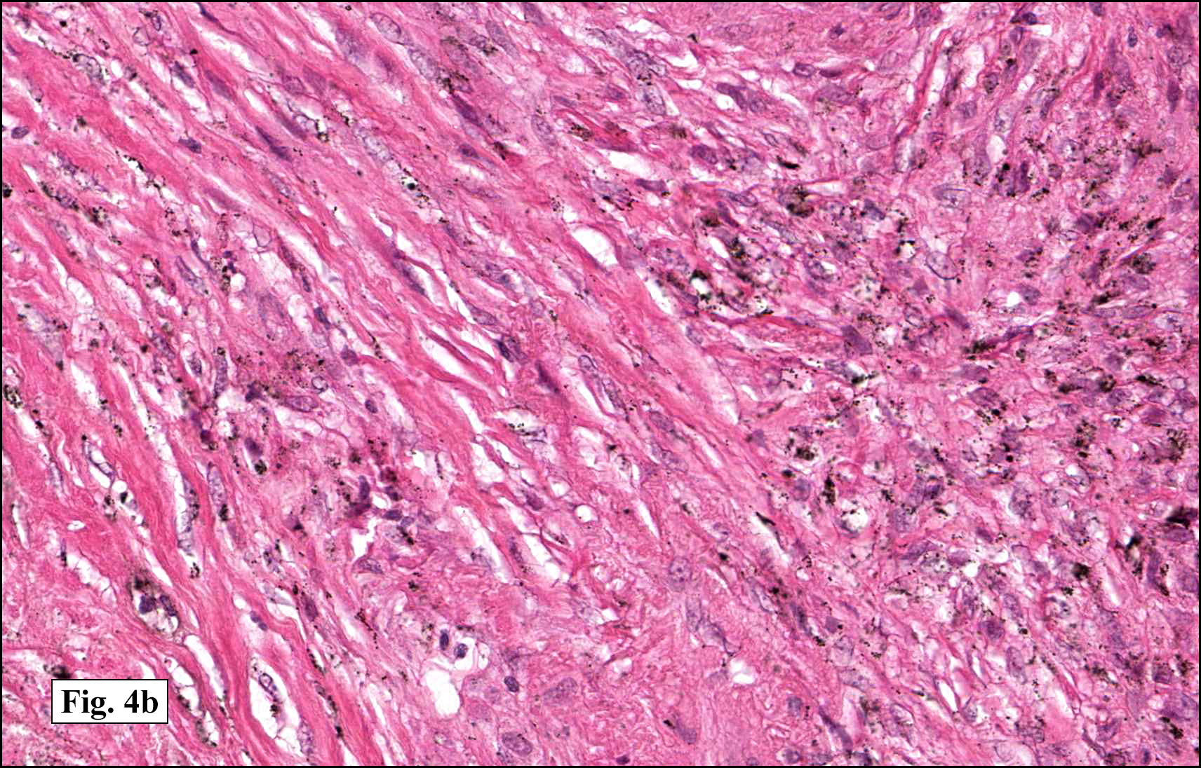
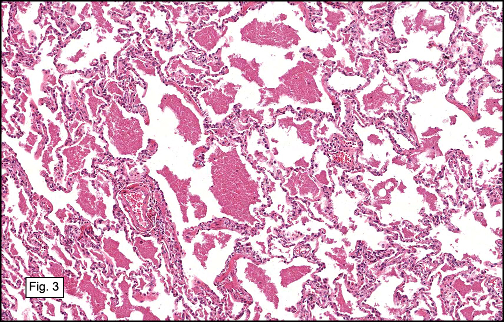
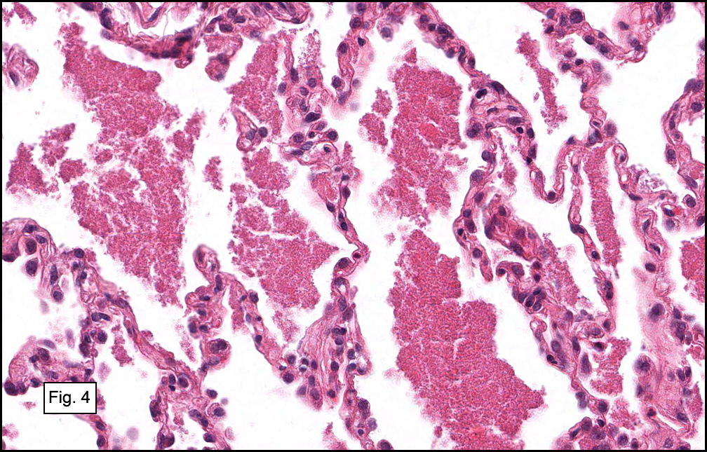
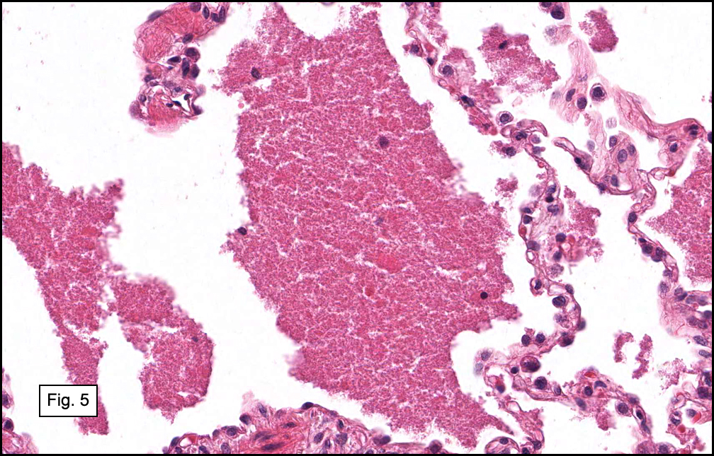
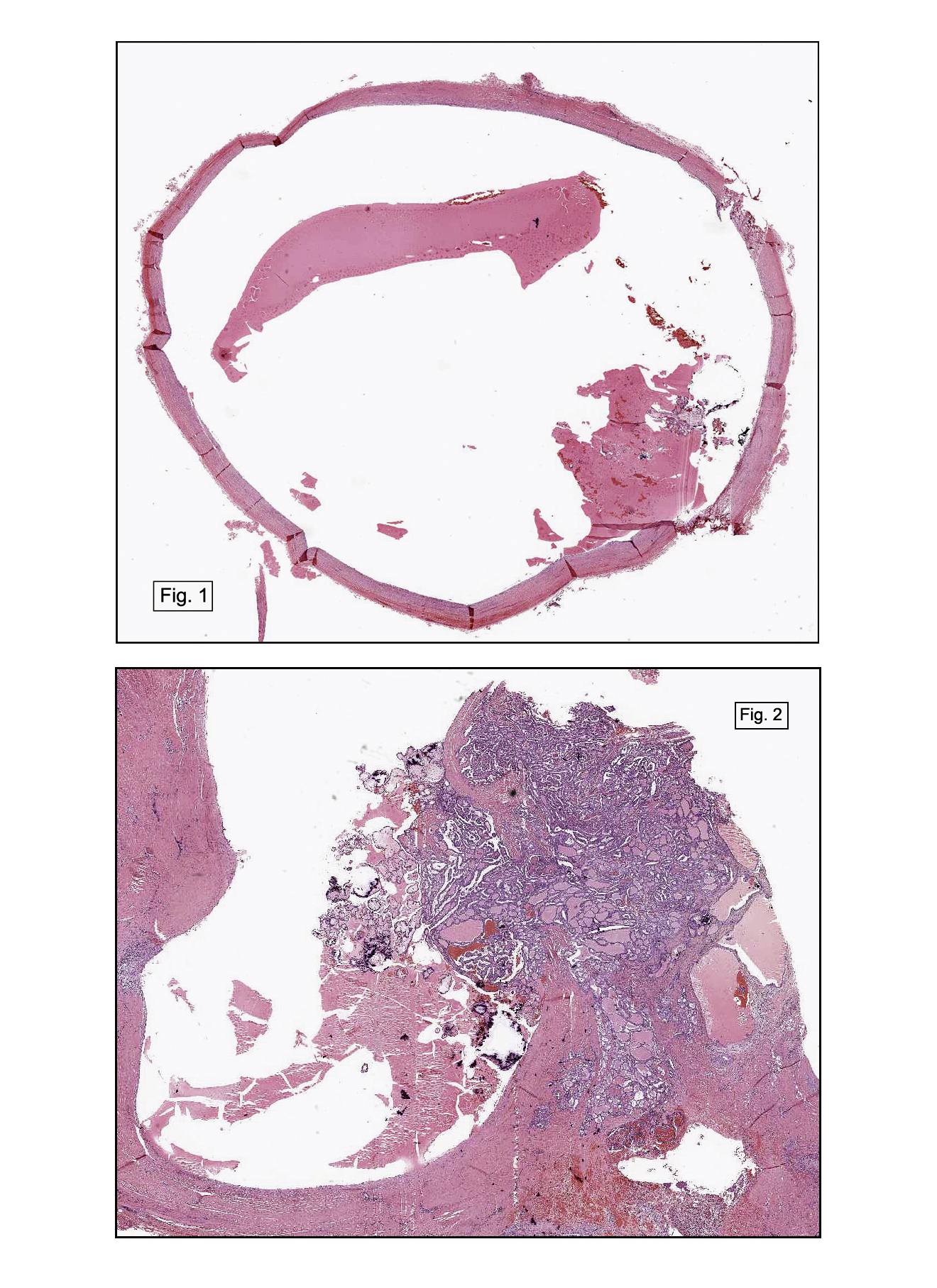
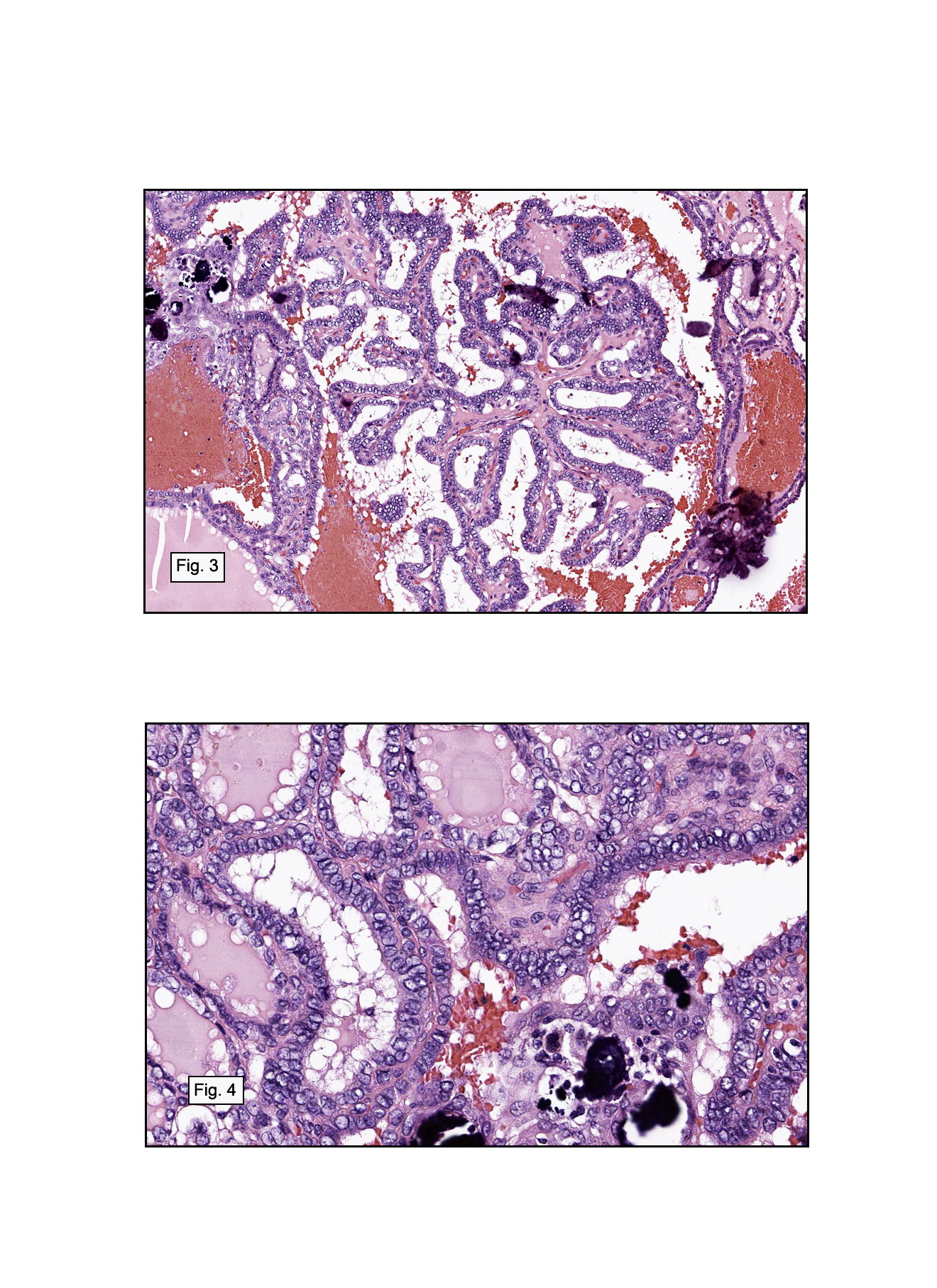
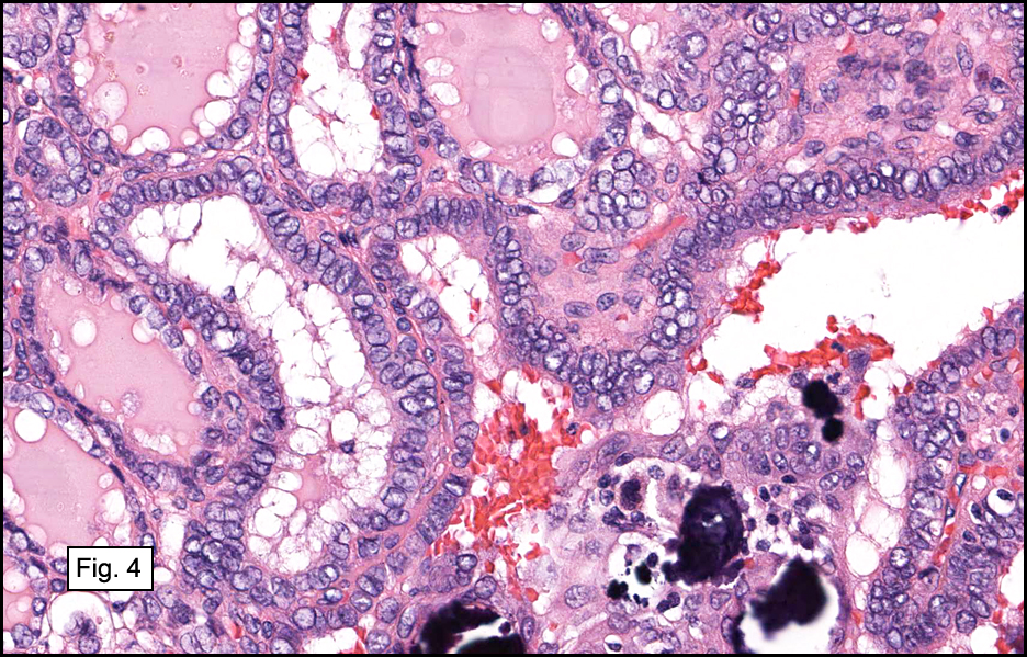
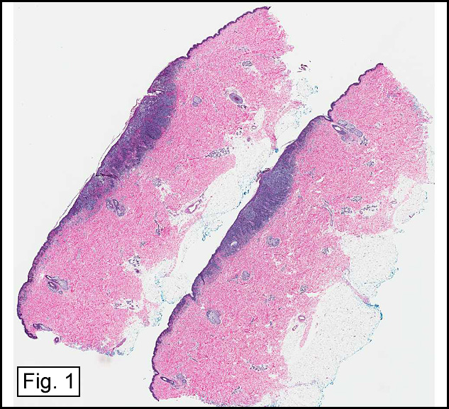
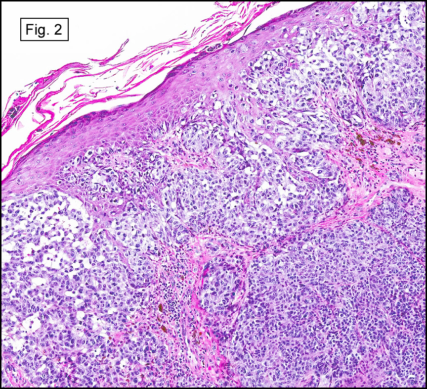
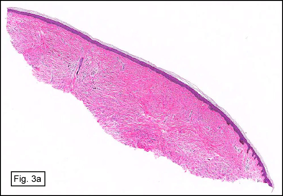
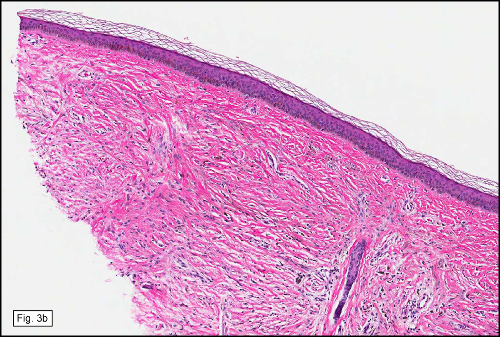
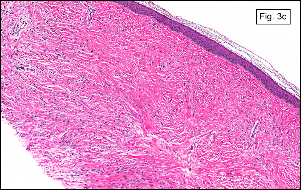
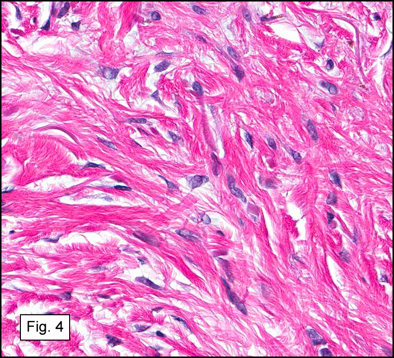
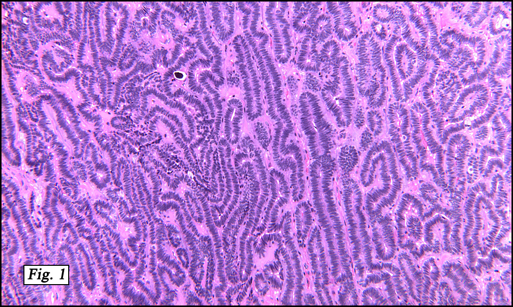
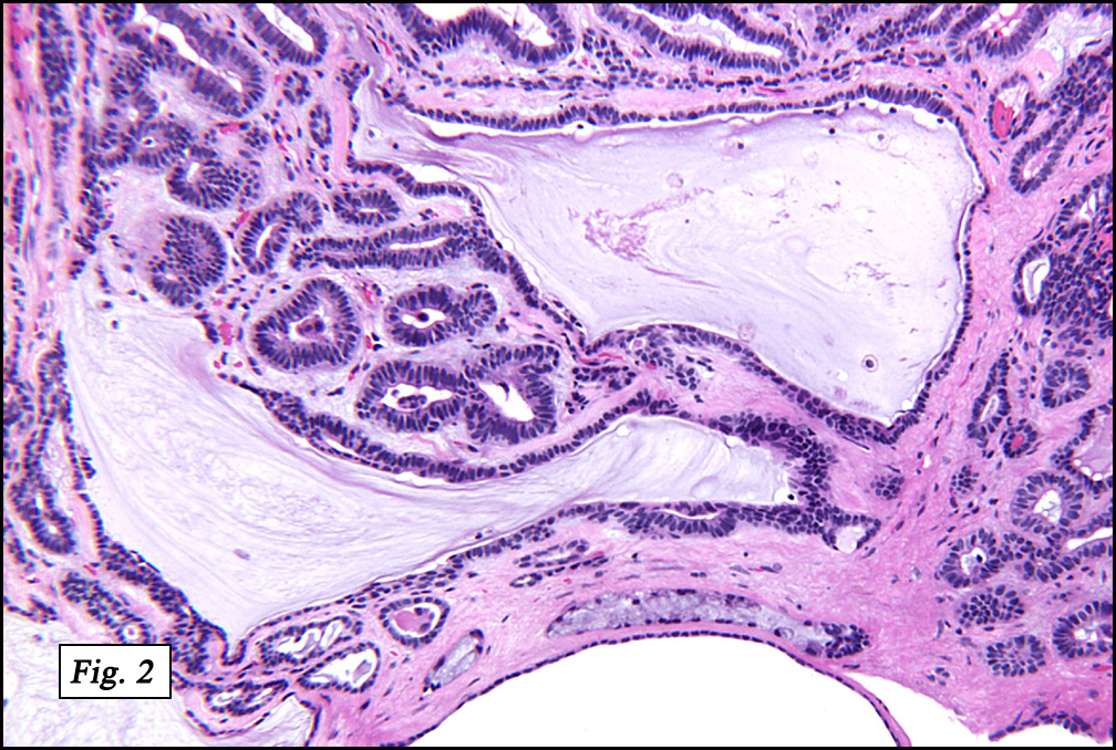
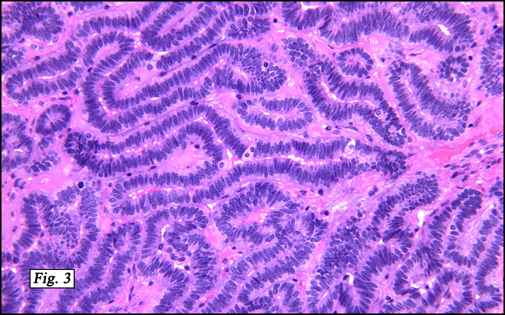
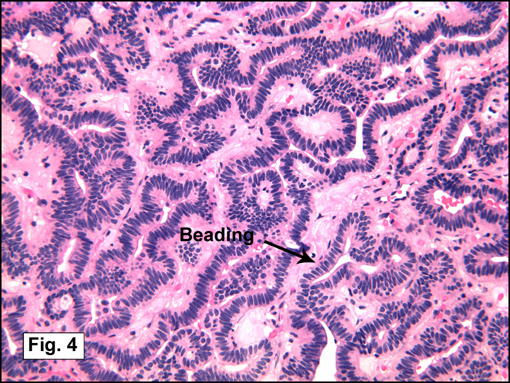
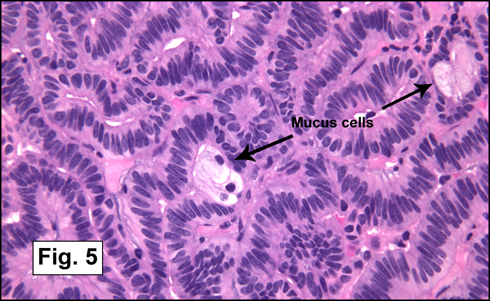
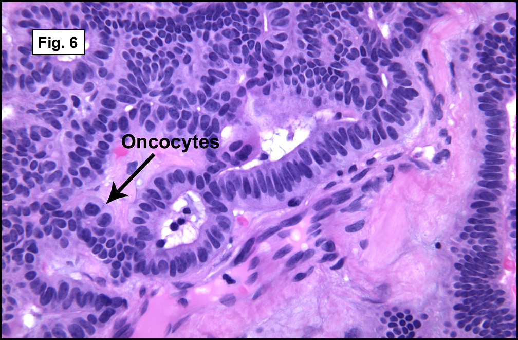
In some sections (not illustrated), squamous cell carcinoma was identified. The point of interest for us, however, was the background lung. Scattered throughout the parenchyma were numerous dense, homogenous nodules composed of brightly-eosinophilic, concentrically-layered material (Fig. 1, 2, 3a, 3b). Dust-filled macrophages were observed, as was emphysematous change (Fig. 4a, 4b). The eosinophilic nodules also involved the accompanying lymph nodes.

Diagnosis: “Silicosis Co-Existent With Pulmonary Adenocarcinoma”

Christina M. Birsan M.D., and Donald R. Chase, M.D.
Department of Pathology and Human Anatomy, Loma Linda University and
Medical Center, Loma Linda, California
California Tumor Tissue Registry, Loma Linda, California

Discussion: Silicosis, a chronic lung disorder, is characterized by progressive development of parenchymal nodules and pulmonary fibrosis as a result of inhalation of crystalline silica. Most cases are related to exposure to quartz, the most abundant mineral in the earth’s crust and also the most abundant form of crystalline silica. Identified in Egyptian mummies, silicosis is the oldest recognized occupational lung disease, with exposure occurring in various rock-cutting, sand-blasting, and ceramics industries.

Clinically, silica-induced lung disease may be acute, accelerated, or chronic. Acute silicoproteinosis is similar to pulmonary alveolar proteinosis, and occurs in cases of heavy exposure. Patients are generally symptomatic within 3 years of exposure, and most cases are fatal. When the disease manifests between 3 to 10 years after exposure, accelerated silicosis is the suggested term. Chronic (classic or nodular) silicosis is the most common form, usually occurring decades after exposure to relatively low levels of silica. Classically, it requires at least 20 years after onset of exposure. It may manifest as simple silicosis (nodules 10 mm or less) or complicated silicosis (nodules greater than 1 cm). Simple silicosis is generally asymptomatic until advanced stages when dyspnea develops. Complicated silicosis results from coalescing smaller nodules over time (also called conglomerate silicosis). Cough and sputum production may occur, but are often the result of chronic bronchitis or concurrent infections. Crackles and digital clubbing are rare in silicosis, and suggest alternate diagnoses. Infections may complicate the process (mycobacterial infections in particular) and can cause fever and weight loss. Severe, longstanding silicosis and fibrosis may cause signs of cor pulmonale. Rheumatoid factor, antinuclear antibodies, serum immune complexes, and polyclonal increase in immunoglobulins may be detected in some cases. In fact, connective tissue disorders (most commonly scleroderma, rheumatoid arthritis, and systemic lupus erythematosus) have been associated with silica exposure. Signs and symptoms in those cases are more specific to the individual entities, and pulmonary silicosis is not always radiologically detectable. Significant associations between silicosis and lung cancer have been reported as well.

Radiologically, classic silicosis manifests as multiple nodules which mostly involve upper lobes. Best seen on CT scans, these nodules are well-circumscribed, uniform in size, and usually less than 5 mm in diameter. They may progress to form conglomerate masses, resulting in larger size. Complicated silicosis is usually bilateral and causes marked loss of lung volumes in the upper lobes, expansion and hyperinflation of the lower lobes, and superior retraction of the hila. Hilar lymph nodes are usually involved and may show a peripheral “eggshell” type calcification. Nodules have also been described in liver, spleen, bone marrow, and abdominal lymph nodes.

Grossly, chronic silicosis demonstrates firm, rounded, well-demarcated nodules, usually about 3 to 6 mm and slate gray to black. A cuff of pigmentation may be seen surrounding nodules on the pleural surface.

Microscopically, the same discrete nodules are apparent. Their formation begins with the accumulation of dust-filled macrophages along lymphatic routes, bronchovascular bundles, and in the pleura and septa. Early stages of disease may demonstrate only these accumulated macrophages. Increased reticulin and fibrosis in the sheets of macrophages represent developing nodules, which then enlarge and become discrete and round, replaced by dense lamellar collagen. They may become calcified, hyalinized, or may undergo central degeneration including cavitation and necrosis, in which case infection (especially tuberculosis) should be carefully ruled out. Polarized light reveals weakly birefringent silica and strongly birefringent round or oval silicate particles within nodules and in surrounding macrophages. If exposure stops, nodules may become fibrous with a few surrounding histiocytes. Emphysematous change is common in surrounding lung. Acute silicosis (silicoproteinosis) resembles pulmonary alveolar proteinosis (PAP), with an eosinophilic, granular, PAS-positive material filling the alveolar spaces. Unlike PAP, however, interstitial inflammation and fibrosis and irregular hyaline scars may be present. Silicotic nodules are usually poorly-formed or absent in such cases.

The pathogenesis of silicosis involves the inhalation of particles less than 10 micrometers in diameter, with the most pathogenic particles being approximately 1 micrometer. The intensity and duration of exposure are the most important factors, as the damage occurs once the silica particles are deposited in the lung. Direct cytotoxicity or the production of oxidants and other mediators may be involved, however the exact sequence of events is unknown. It appears that alveolar macrophages ingest the silica particles and die, perhaps liberating fibroblast-stimulating factors which promote fibrosis. Evidence also suggests that silica interferes with the ability of macrophages to inhibit the growth of mycobacteria, hence the association with tuberculosis. Toxic effects of silica on alveolar type 2 cells has been implicated in silicoproteinosis (acute silicosis).

The diagnosis of silicosis rests upon an appropriate exposure history, consistent radiologic findings, and the absence of other diseases which could explain the radiologic evidence. Observing silica, silicates, silicotic nodules, or increased dust in lymph nodes alone is not sufficient for the diagnosis, as virtually all adults have a small amount of silica in their lungs.

Unfortunately, no effective therapy exists once the inhalation of silica dust has occurred. Most affected patients are asymptomatic, however, and have a normal lifespan, with only a small percentage experiencing progressive respiratory disability.

Suggested Reading:

Travis W, Colby T, Koss M, et al. Non-neoplastic disorders of the lower respiratory tract. In King D, ed. Atlas of Nontumor Pathology. Washington, DC: American Registry of Pathology, Armed Forces Institute of Pathology; 2002.

Katzenstein AL III. Katzenstien’s and Askin’s Surgical Pathology of Non-neoplastic Lung Disease. 3rd edition. Philadelphia, PA: WB Saunders Company; 1997.

Hammar S. Pleural diseases. In: Dail D, Hammar S, eds. Pulmonary Pathology. 2nd ed. New York, NY: Springer-Verlag; 1994.

Erren TC, Morfeld P, Glende CB, et al. Meta-analyses of published epidemiological studies, 1979-2006, point to open causal questions in silica-silicosis-lung cancer research”. Med Lav 2011 Jul-Aug; 102(4):321-35.

August 2012: A 34 year old man with fever and a lung infiltration.

History: A 34-year-old Caucasian male accountant for a powder factory and brass foundry experienced low-grade fever, debility (fatigue?), and weakness. He was found to have a slight infiltration in the right lower and mid lung fields. Cultures were negative, however he continued to have progressive dyspnea for the next two years. During that time he also developed a chronic cough productive of a copious amount of white sputum which was occasionally greenish or streaked with blood. He was admitted with severe dyspnea on exertion.

A physical examination revealed no significant findings, however the chest x-ray showed diffuse bilateral mottling in all lung areas, mostly in the lower lobes. Hemoglobin levels were slightly high at 18.2, and sputum cultures grew alpha streptococcus and staphylococcus epidermis. A left thoracotomy was performed. No pleural adhesions were noted, however the surgeons observed that the lung did not expand normally. Biopsies were taken from the left lingular segment and the left upper lobe.

Two tissue samples were examined. One was soft, fluffy, red-pink, and well aerated. The other contained firm, gray nodules up to 0.8 cm in greatest diameter.

Histologic sections showed focal, nodular areas (Fig. 1) comprised of alveoli which were filled with an intensely eosinophilic material made up of tiny granules and amorphous debris (Figs. 2,3, 4, 5). These areas were immediately adjacent to essentially normal lung tissue, without a surrounding inflammatory response. No other significant abnormalities were identified.

Diagnosis: “Pulmonary Alveolar Proteinosis”

Christine M. Birsan M.D., and Donald R. Chase, M.D.
Department of Pathology and Human Anatomy, Loma Linda University and Medical Center, Loma Linda, California
California Tumor Tissue Registry, Loma Linda, California

Discussion: Pulmonary alveolar proteinosis (PAP) is a rare condition in which a lipid-rich, granular proteinaceous eosinophilic material fills the alveoli. “Primary” cases are found in isolation, while “secondary” PAP may occur in the setting of infection (tuberculosis, Pneumocystis jirovecii), malignancy (especially leukemia and lymphoma), immune deficiency (chemotherapy, congenital alymphoplasia, hypogammaglobulinemia, juvenile dermatomyositis), environmental dust exposure (wood, aluminum, silica, kaolin), and lysinuric protein intolerance. A history of smoking is present in many, but not all cases. Most patients present between the ages of 20-50, however 18% of cases occur in infants and children. A congenital form is described by Leslie and Wick as a lethal disease caused by defects in surfactant production and metabolism, some of which are in association with mutations of the surfactant protein B gene and genes related to secretion of surfactant proteins. “Secondary” PAP has also been observed in infants, most commonly seen with viral infections (respiratory syncytial virus, cytomegalovirus, and parainfluenza virus). “Primary” PAP is unusual in infants and children, but may be seen in adolescents. Males are affected more often than females at a ratio of approximately 2:1-4:1, and the disease may be more common in Caucasians.

The pathogenesis of PAP is unknown. Studies have suggested that macrophage dysfunction may play a role, in which there is a reduced ability to process surfactant (a significant component of the accumulated intra-alveolar material). Mutant mice lacking the gene for granulocyte-macrophage-colony-stimulating factor (GM-CSF) develop a similar disease process which is reversed when GM-CSF is replaced. In addition, antibodies against GM-CSF have been discovered in human cases of PAP (especially “primary” PAP), further supporting this hypothesis. Alternatively, defective production of surfactant may be a consideration, as the pathologically accumulated surfactant lacks its usual surface-active properties, while surfactant extracted from uninvolved areas of the patient’s lung shows normal activity. It has been shown experimentally that the material from PAP produces macrophage dysfunction in normal human blood monocytes and reduces activity of lymphocytes, raising the possibility that the macrophage dysfunction may be a secondary result rather than a primary cause. The wide variety of clinical situations in which this process is observed suggests that PAP represents a common tissue reaction to a broad range of insults.

Clinically, the onset of PAP is often insidious. About one-third of patients are asymptomatic at presentation despite having extensive radiologic abnormalities. Patients who are symptomatic may have a nonproductive cough, a cough productive of chunky, gelatinous material, and/or streaky hemoptysis. Dyspnea on exertion, fatigue, weight loss, chest pain, and low-grade fever may also be present. Occasionally clubbing and cyanosis are observed. Crackles are sometimes heard on auscultation however they are often absent.

Radiographic findings usually include bilateral and symmetric areas of vaguely nodular airspace consolidation or hazy ground-glass opacity. Peri-hilar regions and lower lobes are most severely affected. Interlobular septal thickening and ground-glass opacities seen on CT scans produce a characteristic “crazy paving” appearance. This pattern, although suggestive of the diagnosis, is also seen in other conditions.

Pulmonary function testing most commonly demonstrates a restrictive process, and a decrease in the diffusion capacity for carbon monoxide (DLCO) out of proportion to the reduced lung volume is sometimes seen. Hypoxemia and compensated respiratory alkalosis are frequent and are made worse by exercise. An elevated shunt fraction is usually present.

Lab values may reveal polycythemia, hypergammaglobulinemia, and increased lactate dehydrogenase. Serum levels of lung surfactant proteins A and D (SP-A, SP-D) have been found to be markedly high which can help narrow the diagnosis, however, other lung processes can have similar findings. Elevated levels of several tumor markers have been identified in the BAL fluid from some patients, including carcinoembryonic antigen (CEA), carbohydrate antigens sialyl Lewis (CA19-9), and sialyl SSEA-1 (SLX). KL-6, a mucin-like glycoprotein may also be useful for diagnosis when found in serum or BAL specimens. Cultures may reveal Nocardia or other opportunistic mycobacterial, fungal, and viral agents thought to represent secondary infection.

Cytologic preparations may suggest the diagnosis of PAP. In BAL specimens, abundant lipoproteinaceous material causes an opaque appearance, and large, acellular eosinophilic bodies may be seen in a background of eosinophilic granules. PAS-positive proteinaceous material as well as macrophages engorged with PAS-positive material point to the diagnosis.

Tissue examination is frequently necessary for definitive diagnosis. Grossly, lung tissue is heavy and viscid, containing yellow fluid which leaks from cut surfaces. Firm, yellowish-white nodules scattered throughout the parenchyma range from a few millimeters to 2 cm. Microscopically, eosinophilic proteinaceous granular material fills the alveoli and occasionally involves bronchioles and alveolar ducts, leaving the interstitial architecture of the lung generally intact. A diffuse pattern is usually seen, however focal or patchy involvement also exist, as seen in this case. Hyperplastic, cuboidal, type 2 pneumocytes are often line the alveolar septae. Sharply demarcated round, empty spaces, cholesterol clefts, and small, dense, globular eosinophilic clumps are distinctive findings within the accumulated material which may also contain cellular debris, foamy macrophages, ghosts of degenerated cells, and detached type 2 pneumocytes. It is usually PAS-positive and diastase-resistant, while staining for antibody to surfactant apoprotein may be positive especially in “primary” disease. Alcian and mucicarmine stains are negative. Frozen sections shows abundant lipid, highlighted with Oil Red O staining. Examination by electron microscopy demonstrates concentrically laminated myelin figures and lamellar bodies within the proteinaceous material which look identical to the cytoplasmic inclusions of type 2 pneumocytes, suggestive of surfactant. Polarization microscopy may reveal birefringent needle-like particles if there has been an exposure to dust. Interstitial fibrosis or inflammation are typically not prominent and if present may indicate an associated infection, but may also indicate long-standing or recurrent PAP.

The differential diagnosis for PAP includes pulmonary edema, alveolar mucinosis, and Pneumocystis jirovecii pneumonia. Pulmonary edema and alveolar mucinosis lack the granularity of PAP, as well as the intense eosinophilic staining, cholesterol clefts, and engorged macrophages containing PAS-positive debris. The intra-alveolar exudate seen in Pneumocystis jirovecii may be distinguished from PAP by the cysts or organisms which appear as “bubbles” within the eosinophilic material which stain positive for Gomori methenamine silver (GMS).

Treatment for PAP should be initiated when the patient becomes sufficiently symptomatic, as many patients have little or no impairment and spontaneous remission may occur. Severe dyspnea and hypoxemia at rest or with exercise warrant therapeutic whole lung lavage via a double-lumen endotracheal tube, the most widely accepted and effective treatment. Patients often feel dramatically better after this treatment, although potential complications include malpositioning of the endotracheal tube, saline spillover into unlavaged and ventilated lung, and hydropneumothorax. Thirty to forty percent of patients require lavage only once, while some patients require repeat lavages at intervals of 6 to 12 months. Corticosteroids or other immunosuppressives should not be used, as there is concern that they may increase mortality by aggravating or inducing secondary opportunistic infections. Further study is necessary to determine the potential use of GM-CSF as a therapeutic option.

Suggested Reading:

Travis W, Colby T, Koss M, et al. Non-neoplastic disorders of the lower respiratory tract. In King D, ed. Atlas of Nontumor Pathology. Washington, DC: American Registry of Pathology, Armed Forces Institute of Pathology; 2002.

Katzenstein AL III. Katzenstien’s and Askin’s Surgical pathology of non-neoplastic Lung disease. 3rd edition. Philadelphia, PA: WB Saunders Company; 1997.

Hammar S. Pleural diseases. In: Dail D, Hammar S, eds. Pulmonary Pathology. 2nd ed. New York, NY: Springer-Verlag; 1994.

Leslie K, Wick M. Practical pulmonary pathology. A diagnostic approach. 1st edn. Philadelphia: Churchill-Livingstone; 2005.

July 2012: A 50 year old man with an anterior neck mass

History: A 50 year old man presented with a 2 cm fluctuant midline anterior neck mass. The removed specimen was a unilocular cyst which exuded serous fluid. A papillary nodule was noted on its luminal surface.

Microscopically, the cyst had a denuded epithelial lining supported by fibrous tissue with varying numbers of chronic inflammatory cells (Fig. 1). It also showed a papillary excrescence protruding into the cyst wall (Fig. 2). Tumor cells were crowded and arranged in papillary fronds and follicular patterns (Fig. 3). They had enlarged nuclei and occasional calcifications. Some showed longitudinal clefts. Nucleoli were inconspicuous and peripherally located. Many of the cells showed central cytoplasmic clearing giving the cells an “Orphan Annie” appearance (Fig. 4).

Diagnosis: “Classic Papillary Thyroid Carcinoma” arising in thyroglossal duct cyst”

David Panther, MSIV, and Donald R. Chase, M.D.
Department of Pathology and Human Anatomy, Loma Linda University and Medical
Center, Loma Linda, California
California Tumor Tissue Registry, Loma Linda, California

Discussion: Thyroid tissue has its embryologic origin at the foramen cecum at the base of the tongue from which it migrates into the lower anterior neck. The tract then degenerates. However, incomplete regression of this tract, can accumulate fluid and form a cyst (thyroglossal duct cyst [TDC]) usually lined with columnar epithelium. Occasionally the epithelium can undergo squamous metaplasia, or become denuded and/or inflamed. During its descent, the thyroid may leave ectopic tissue anywhere along the path and up to 60% of TDCs are associated with residual thyroid tissue. These elements are physiologically similar to those in the lower neck, however they lack parafollicular C cells. The remnant tissue can be responsible for any traditional thyroid disease, except medullary carcinoma, a tumor of C cell origin.

Rarely (<1%) residual thyroid tissue give rise to a primary malignancy, mostly papillary thyroid carcinoma (80%), (PTC). Other malignancies include follicular carcinoma and anaplastic carcinoma

Questions have been raised over the origin of PTC in a TDC, namely whether this entity represents a TDC-related primary versus a thyroid primary with local metastasis to a TDC. However, a substantial number of cases have been reported where the thyroid was removed and carefully examined. Roughly 20-50% of the lower neck thyroids have had concurrent tumor, implying that at least half of the upper neck tumors are most likely to be primary TDC tumors. In addition, occult thyroid malignancies are often found at autopsy in asymptomatic individuals, indicating that a significant portion of concurrent TDC and thyroid gland tumors are coincidental. It is generally accepted that the thyroid should not be automatically excised unless clinical evidence of a mass or abnormal scintiscan is/are present in the lower neck thyroid gland.

The differential diagnosis includes other thyroid epithelial neoplasms, as well as squamous cell carcinoma. Although metastatic disease has not yet been reported in TDCs, possible look-alikes could include metastases from clear-cell variants of lung and renal carcinoma, melanoma, and mucinous gynecologic malignancies.

– Clear Cell (“Sugar”) Tumor of the Lung: cells have a large cytoplasmic glycogen droplet, displacing the nucleus peripherally. In addition, it stains positive for HMB-45.
– Clear Cell Renal Cell Carcinoma: central nuclei are surrounded by clear cytoplasm, with cells often arranged in nests surrounded by branching fibrovascular tissue.
– Melanoma: features include haphazard arrangement of cells, nuclei that are pleomorphic and readily seen nucleoli and mitoses. Key immunohistochemical markers include HMB-45, MART-1, Melan A, and S-100.
– Mucinous Cystadenocarcinoma: the cyst may be multiloculated. The epithelial component may be several cells thick, with atypical nuclei containing prominent nucleoli, pale cytoplasm, and extracellular mucin. CEA is the most important immunohistochemical marker.

Suggested Reading:

DeLellis RA, Nikiforov YE. Thyroid and Parathyroid. In: Gnepp, DR. Diagnostic Surgical Pathology of the Head and Neck. Saunders Elsevier, Philadelphia, PA. 2009.

Luna MA, Pfaltz M. Cysts of the Neck, Unknown Primary Tumor, and Neck Dissection. In: Gnepp, DR. Diagnostic Surgical Pathology of the Head and Neck. Saunders Elsevier, Philadelphia, PA. 2009.

LiVolsi, VA. Surgical Pathology of the Thyroid. Volume 22 in the series: Bennington, JL, ed. Major Problems in Pathology. WB Saunders Co., Philadelphia, PA. 1990.

Hartl DM, Al Ghuzlan A, Chami L, et al. High rate of multifocality and occult lymph node metastases in papillary thyroid carcinoma arising in thyroglossal duct cysts. Ann Surg Oncol. 2009;16:2595-601.

Peretz A, Leiberman E, Kapelushnik J, Hershkovitz E. Thyroglossal duct carcinoma in children: case presentation and review of the literature. Thyroid. 2004;14:777-85.

Motamed M, McGlashan JA. Thyroglossal duct carcinoma. Curr Opin Otolaryngol Head Neck Surg. 2004;12:106-9.

June 2012: A 59 year old man with an enlarging abdominal scar

History: A 59-year-old man had a cutaneous melanoma of his right abdomen excised three years earlier. He presented with an ulcerated, enlarging scar at the site of previous excision.

The original melanoma was classic, composed mostly of polygonal melanocytes, without spindled elements (Figs. 1, 2). The peripheral margins were microscopically free of tumor (Fig. 1). The more recent excision had a cicatrix appearance (Figs. 3a,3b,3c). Upon closer examination, the tumor had an infiltrative pattern consisting most of fibrosing spindle cells (Fig. 4). The cytology of these spindle cells was rather bland, resembling fibroblasts. Interspersed between the spindle cells were (rarely) macrophages filled with dark pigment (Fig. 5). Polygonal cells, which dominated in the original tumor, were not present. Immunohistochemical stains showed the spindled cells to be positive for both HMB-45 (Fig. 6 left) and Melan A (Fig. 6 right).

Diagnosis: “Recurrent Melanoma with Desmoplastic Phenotype (“Desmoplastic Melanoma”)

Jonathan Zumwalt, MSIV, Resa L. Chase, MD, and Donald R. Chase, MD
Department of Pathology and Human Anatomy, Loma Linda University and Medical
Center, Loma Linda, California
California Tumor Tissue Registry, Loma Linda, California

Discussion: In 1812, Rene Laennec first described the disease entity currently known as “melanoma”, in a paper entitled “The Melanoses”. Today melanoma is seen in 1/39 Caucasian men and 1/58 Caucasian women in the United States. There are approximately 70,000 cases of invasive melanoma diagnosed every year and almost 9,000 lives are lost every year to this tumor. The incidence of melanoma has markedly increased over the past 10-20 years and may be attributed to three non-biologic factors: 1) increased rates of screening, 2) an increase in the number of biopsies being taken and 3) changes involving histologic interpretation of early evolving lesions. The main risk factors for developing melanoma are: family history, history of atypical nevi, high nevus count, history of increased sun exposure and physical traits (i.e. light colored hair and eyes).

There are a variety of factors to consider when reporting a melanoma:
• Breslow thickness is the most important factor for melanoma staging.
• Ulceration is now the second most powerful predictor of survival.
• Mitotic activity of the tumor shows a correlation between replication and survival.
• Radial and vertical growth phases may help characterize the tumor’s aggressiveness. Tumor infiltrating lymphocytes seem to indicate the presence of vertical growth phase.
• Microscopic satellites cut down the five year survival rate of a patient by more than half.
• Regression is still controversial and experts give different opinions on this phenomenon.
• The anatomic primary tumor site of a melanoma does correlate with patient survival.
• Clark’s levels are no longer used in today’s practice for staging, but many physicians still report them.

Melanomas today are divided into four common variants: superficial spreading, lentigo maligna, acral lentiginous, and nodular.

1. Superficial spreading melanomas make up 75% of melanomas. Histologically, they are asymmetric, poorly circumscribed and lack cellular maturation. Upward invasion of melanocytes into the epidermis is characteristic, as is a prolonged radial growth phase.
2. Lentigo maligna melanomas begin as tan-brown macule on sun damaged skin that evolves into larger, darker areas with asymmetric foci. Histologically, the neoplastic melanocytes are arrayed along the dermal-epidermal junction in a lentiginous pattern against a background of epidermal atrophy and solar elastosis.
3. Acral lentiginous melanomas commonly arise on the palmar, plantar, subungual, and occasionally, mucosal surfaces and typically occur in dark skinned individuals. Histologically, they are characterized by a lentiginous array of atypical melanocytes along the dermal-epidermal junction, with foci of confluent melanocytic growth and progress to large junctional nests that are composed of atypical melanocytes.
4. Nodular melanomas are darkly pigmented polypoid or pedunculated nodules. Histologically, these typically show dermal growth in isolation, but occasionally, in association with an epidermal component.

One rare variant of melanoma is desmoplastic melanoma (DM). It is frequently mistaken for a scar, dermatofibroma and/or sarcoma. Grossly DM generally appears as an amelanotic, pale, and fleshy nodule or papule, usually resembling a scar. A variety of studies have shown that it is highly associated with lentigo maligna (24 percent). Histologically these tumors show a poorly circumscribed vertical growth phase tumor with spindle-shaped malignant cells with a fibrogenic stromal pattern. The nuclei of these cells are typically elongated and hyperchromatic. The morphology of these cells resemble fibroblasts. Immunohistochemical staining usually shows S-100 positivity, while HMB-45, Melan-A, tyrosinase and microphthalmia transcription factor are generally negative.

There has been controversy regarding the prognosis of DM. Original studies found it a “highly malignant, stubbornly recurring and often metastasizing neoplasm.” In the past twenty years this perception has started to change, allowing for an indolent behavior. The confusion of this may be due to the failure of some studies to detect differences between DM and conventional melanoma. A second reason for the conflicting results is the heterogeneity among melanomas declared as desmoplastic.

Differential Diagnosis:

Sclerosing Melanocytic Nevus usually occurs in younger patients, and usually in skin which has not been actinically damaged. Histologically seen from low power, they are sometimes perceived as a circumscribed “silhouette” with little or no cytologic atypia or mitotic activity. In contrast, DM, tends to be asymmetric, infiltrative and poorly circumscribed. Most DMs are associated with a previous or current in-situ melanoma.

Dermal Scar or Dermatofibroma: Without evidence of abnormal melanocytic changes in the epidermis, recognition of a dermal spindle cell proliferation can be most difficult to identify as melanoma. Often DM may be mistaken for a scar. But fibroblasts in scars typically are arranged in parallel to the skin surface, while blood vessels are perpendicular. Dermatofibromas tend to have spindle cells that wrap around the collagen bundles. In contrast, DM has fusiform cells oriented both parallel and perpendicular to the skin surface. Also, DM usually shows some cytologic atypia with elongated hyperchromatic nuclei.

Sarcomas or Sarcomatoid Carcinoma: DM at times can resemble a spindle cell sarcoma, and immunohistochemistry is usually needed to help distinguish these differences. The use of S-100, muscle markers and epithelial markers can help distinguish DM from desmoplastic carcinoma, leiomyosarcoma, pleomorphic sarcoma and malignant peripheral nerve sheath tumor. Sarcomatoid carcinoma usually stains strongly for 34BE12, which does not stain melanomas.

Suggested Reading:

Busam KJ. Desmoplastic Melanoma. Clin Lab Med. 2011 Jun;31(2):321-30.

Divito SJ, Ferris LK. Advances and short comings in the early diagnosis of melanoma. Melanoma Res. 2010 Dec;20(6):450-8.

Garbe C, Eigentler TK, Bauer J, et al. Histopathological diagnostics of malignant melanoma in accordance with the recent AJCC classification 2009: Review of the literature and recommendations for general practice. J Dtsch Dermatol Ges. 2011 Jun 9. doi: 10.1111/j.1610-0387.2011.07714.x.

Kong Y, Kumar SM, Xu X. Molecular pathogenesis of sporadic melanoma and melanoma-initiating cells. Arch Pathol Lab Med. 2010 Dec;134(12):1740-9.

Leong SP, Gershenwald JE, Soong SJ, Schadendorf D, Tarhini AA, Agarwala S, Hauschild A, Soon CW, Daud A, Kashani-Sabet M. Cutaneous melanoma: a model to study cancer metastasis. J Surg Oncol. 2011 May 1;103(6):538-49. doi: 10.1002/jso.21816.

Murali R, Zannino D, Synnott M, McCarthy SW, Thompson JF, Scolyer RA. Clinical and pathological features of metastases of primary cutaneous desmoplastic melanoma. Histopathol. 2011 May;58(6):886-95. doi: 10.1111/j.1365-2559.2011.03808.x. Epub 2011 Mar 25.

Murali R, Shaw HM, Lai K, McCarthy SW, Quinn MJ, Stretch JR, Thompson JF, Scolyer RA. Prognostic factors in cutaneous desmoplastic melanoma: a study of 252 patients. Cancer. 2010 Sep 1;116(17):4130-8.

Piris A, Mihm MC Jr, Duncan LM. AJCC melanoma staging update: impact on dermatopathology practice and patient management. J Cutan Pathol. 2011 May;38(5):394-400. doi: 10.1111/j.1600-0560.2011.01699.x. Epub 2011 Mar 9.

Sade S, Al Habeeb A, Ghazarian D. Spindle cell melanocytic lesions–part I: an approach to compound naevoidal pattern lesions with spindle cell morphology and Spitzoid pattern lesions. J Clin Pathol. 2010 Apr;63(4):296-321.

Sade S, Al Habeeb A, Ghazarian D. Spindle cell melanocytic lesions: part II–an approach to intradermal proliferations and horizontally oriented lesions. J Clin Pathol. 2010 May;63(5):391-409.

Whiteman DC, Pavan WJ, Bastian BC. The Melanomas: A synthesis of epidemiological, clinical, histopathological, genetic, and biological aspects, supporting distinct subtypes, causal pathways, and cells of origin. Pigment Cell Melanoma Res. 2011 Jun 27. doi: 10.1111/j.1755-148X.2011.00880.x.

May 2012: A 79 year old woman with an inner cheek mass

History: A 79 year-old woman presented with a two year history of a slow-growing oral cavity mass between the buccal mucosa and cheek, near the lip commissure. Her past medical history was unremarkable.

Sections showed a well-circumscribed, nodular, thinly encapsulated epithelial neoplasm. The tumor was predominantly composed of a solid proliferation of cytologically bland cuboidal to columnar basaloid cells with dark nuclei and scant eosinophilic cytoplasm (Fig. 1). Microcyst formation was focally present (Fig. 2), sometimes associated with fine papillae. The periphery showed the mucous pattern of minor salivary glands. Lesional cells were usually bilayered in branching/anastomosing cords (i.e. trabecular pattern) (Fig. 3) The cords sometimes separated and rejoined repeatedly, creating a “beaded” appearance (Fig. 4). Scattered foci of mucous cells (Fig. 5), oncocytic cells (Fig. 6), and squamous metaplastic cells were seen. Psammoma bodies were scattered throughout the tumor (Fig. 7).

Diagnosis: “Canalicular Adenoma of Minor Salivary Gland Origin”

Melissa Skaugset MSIV, Mia Perez MD, Donald Chase MD
Department of Pathology and Human Anatomy,
Loma Linda University and Medical Center
California Tumor Tissue Registry, Loma Linda, California

Discussion: Canalicular adenoma (CanA) is a benign epithelial neoplasm of the salivary glands that, by the WHO definition, is composed of columnar epithelial cells arranged in thin, anastomosing cords often with a beaded pattern. The stroma is characteristically paucicellular and highly vascular. They are benign neoplasms, only rarely recurring after local excision. Synonyms include:

• Basal cell adenoma, canalicular type
• Monomorphic adenoma, canalicular type
• Adenomatosis of minor salivary gland

Canalicular adenomas most commonly affect the minor salivary glands. The majority arise in the upper lip (70-90%) where they are the second most common salivary gland tumor after pleomorphic adenoma. Their next most common site is buccal mucosa. Only rarely have they been reported in the parotid gland or palate.

CanAs are rare before the fifth decade of life, and have a peak in the seventh decade. No clear gender prevalence has been shown. They usually present as slow growing, painless small mass(es). They are freely mobile, frequently multinodular, and usually are less than 2 centimeters in greatest diameter (mean 1.7cm). They are generally slow growing, and have been reported to have been present for up to 15 years. The majority present as a solitary mass, but up to 22% are reportedly multifocal and/or multiple (Daley, 1984). Grossly, they are well demarcated and frequently encapsulated. The tumor is usually solid and homogenous, but may have cystic inclusions filled with mucoid material.

CanAs were long thought to be a subclass of basal cell adenoma due to its phenotypic similarity, but is now thought to be a distinct entity. It derives its name from the double rows of cuboidal to columnar epithelium that separate and rejoin to form small duct-like structures or canaliculi. The pattern of separation and rejoining evokes the image of beads on a rosary, leading to descriptions of “beading” or “rosary beads”. The epithelium may be cuboidal or columnar and is usually without significant cellular atypia or pleomorphism. The stroma is loose, highly vascular, and usually of low cellularity. Cyst formation is common.

Unlike other salivary gland tumors which may show multiple growth patterns, canalicular adenomas tend to have a predictable patterns. But they may vary in cellular density and amount of cyst formation, and the degree of encapsulation. The diagnosis may be made without special stains, although recognized positive markers include keratin, vimentin and S100. GFAP may be focally positive. The tumors do not react to anti-SMA.

The differential diagnosis includes sebaceous cyst, lipoma, nasolabial cyst, and more commonly, salivary gland tumors that have a “basaloid” pattern, i.e. basal cell adenoma and adenoid cystic carcinoma. Distinguishing basal cell adenoma from canalicular adenoma has limited clinical significance apart from the potential of canalicular adenomas for multifocality. Distinguishing this entity from adenoid cystic carcinoma is much more important. The double rows of columnar cells and canaliculi that typify canalicular adenomas are not seen in adenoid cystic carcinoma, although the cribriform and tubular patterns of the latter can be confused with this pattern. Nuclear irregularity and atypia are present in adenoid cystic carcinoma, but not in an adenoma. Vascularity of the stroma of canalicular adenomas is absent in adenoid cystic carcinoma. Dense collagen may surround cell nests in adenoid cystic carcinoma but is typically present only in the capsule of canalicular adenoma.

Suggested reading:

Barnes L, Eveson JW, Reichart P, Sidransky D. WHO Classification of Tumours: Pathology and Genetics of Head and Neck Tumours, 2005 Ed. IARC Press, International Agency for Research on Cancer, 2005. p 267.

Peel RL, Barnes L. Surgical Pathology of the Head and Neck. Marcel Dekker, New York, 2001. pp 663-670.

Ellis GL, Auclair PL, Rosai J. AFIP Atlas of Tumor Pathology: Tumors of the Salivary Glands. American Registry of Pathology, Armed Forces Institute of Pathology, Washington D.C., 1995. pp95-103.

Kratochvi F, Ellis GL, Auclair P, Gnepp D. Surgical Pathology of the Salivary Glands. W.B. Saunders Company, Philadelphia, 1991. pp 202-212.

Scroll to top