March 2013: A 33 year old woman with a mass in the thigh

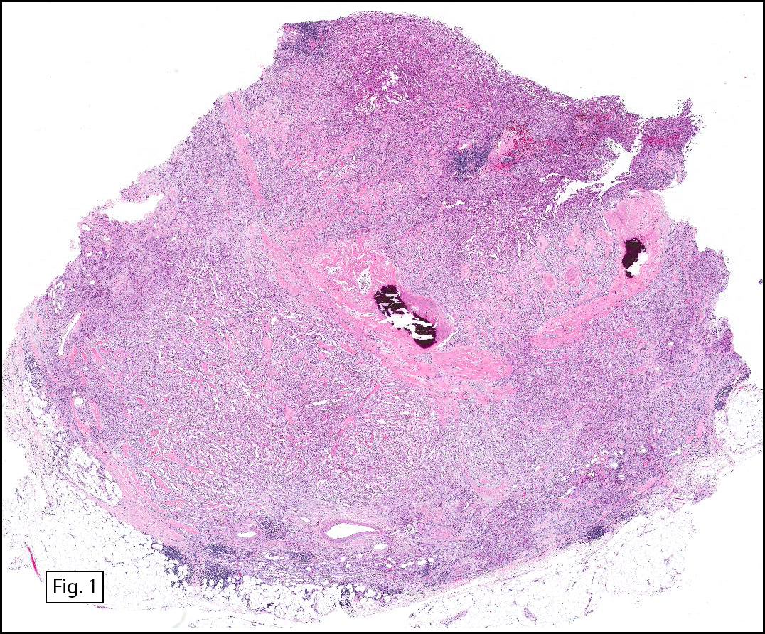
History: A 33 year-old woman presented with a palpable, 6 cm mass in the thigh. At surgery, it was found to deeply reside within the anterior compartment where it involved muscle. Osseous involvement was not seen. The excised specimen was poorly circumscribed, and had a cut surface that was fleshy tan-grey.

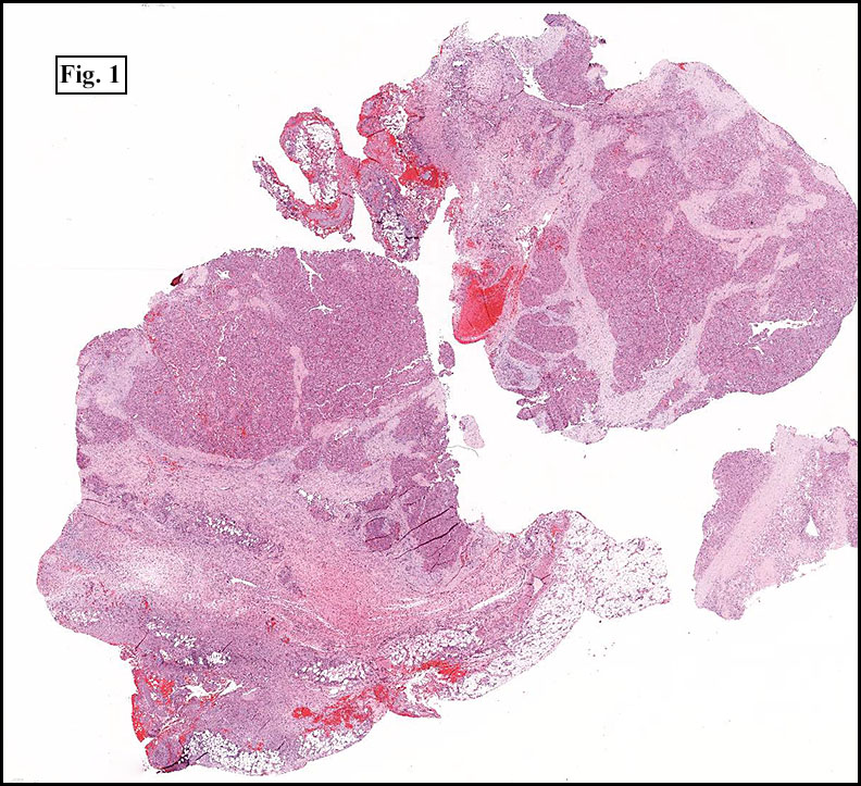
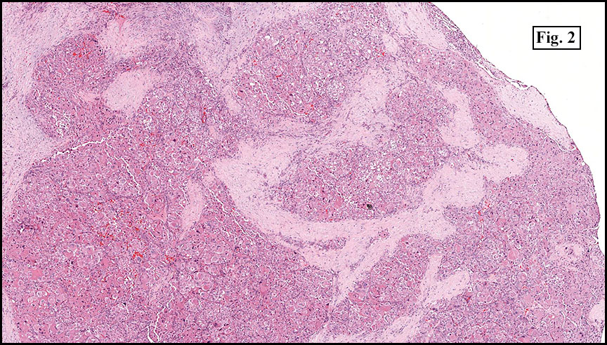
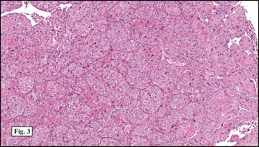
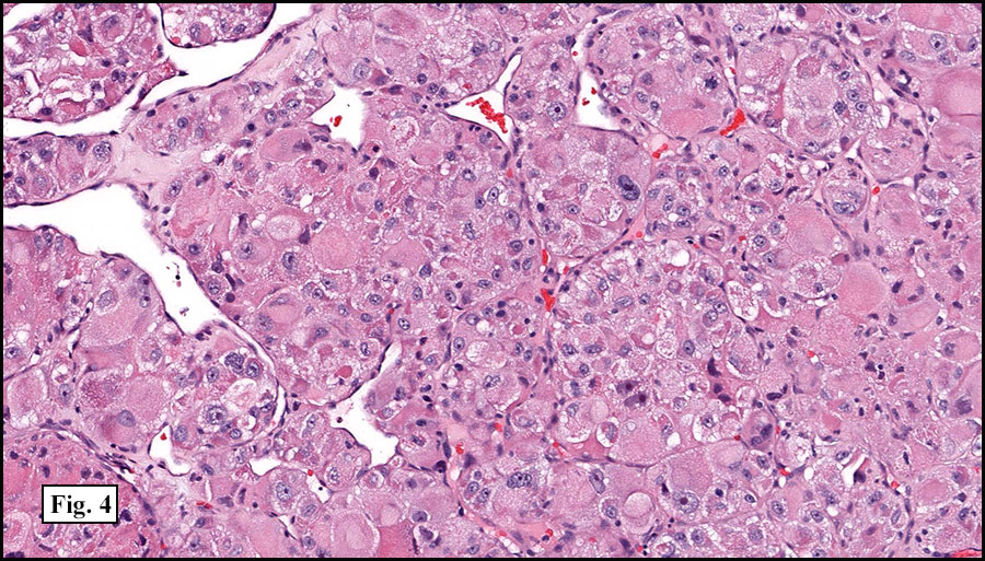
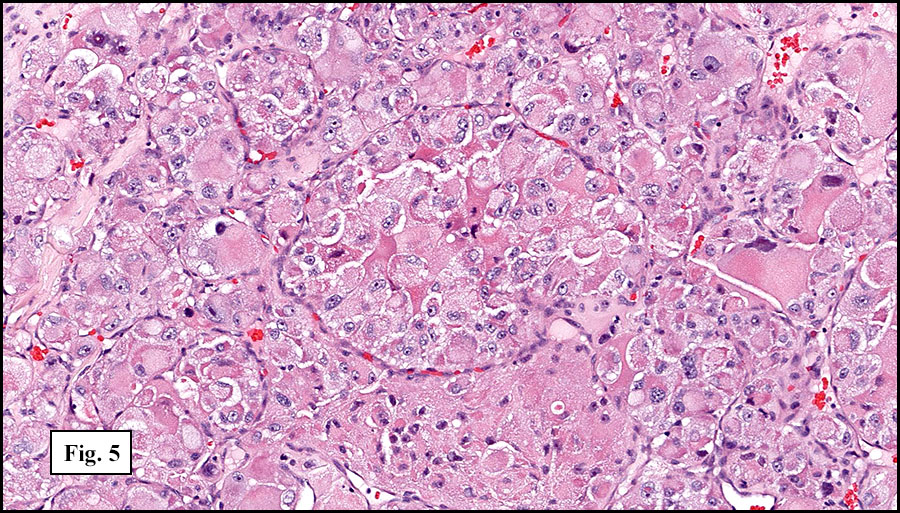
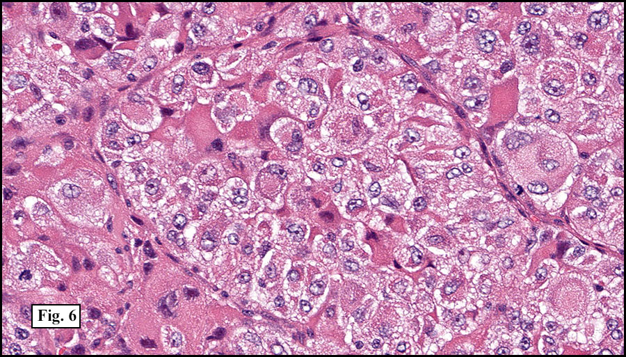
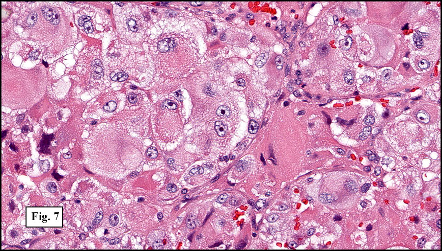
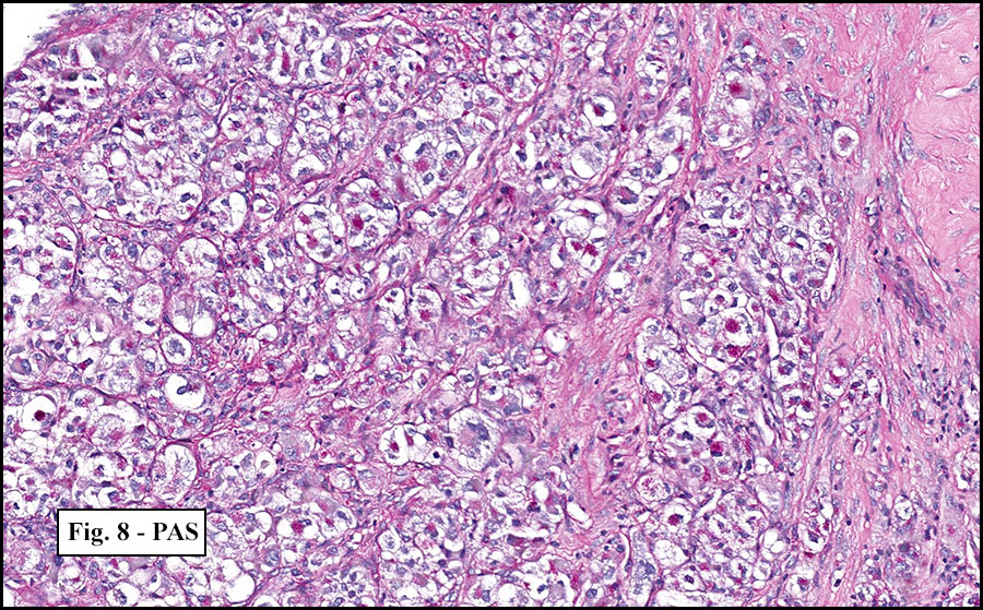
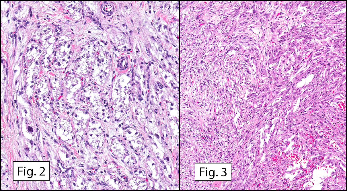
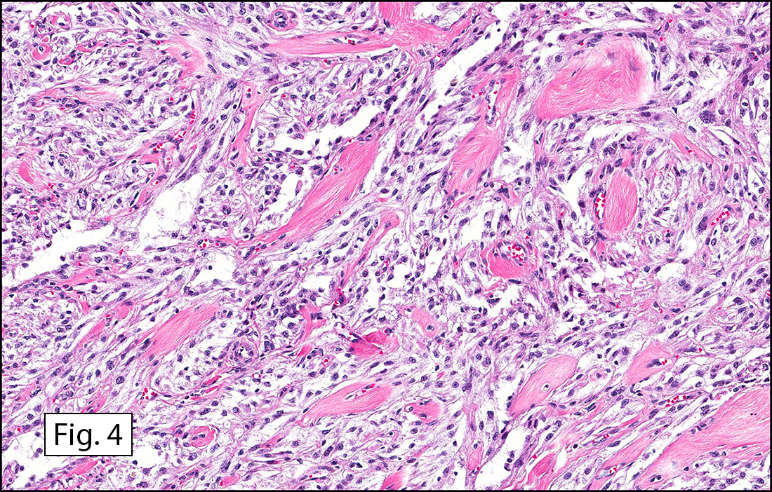
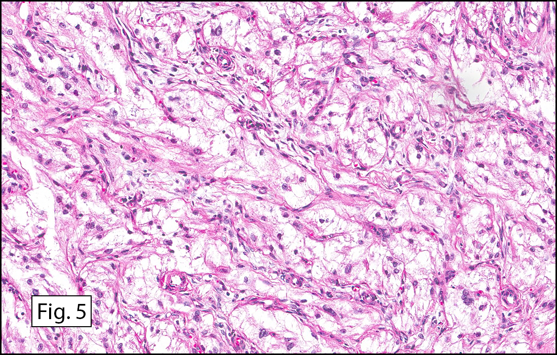
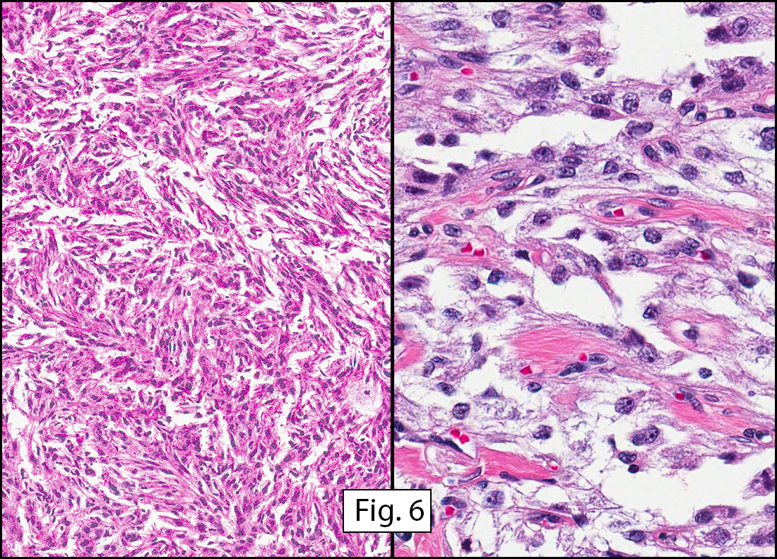
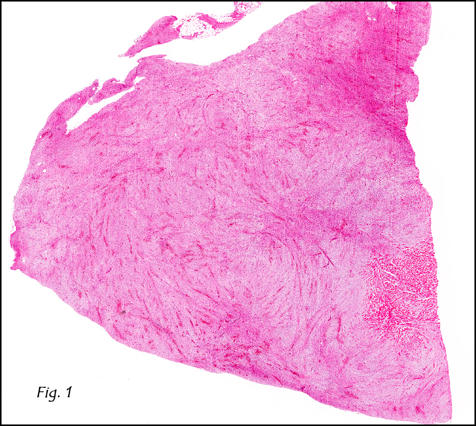
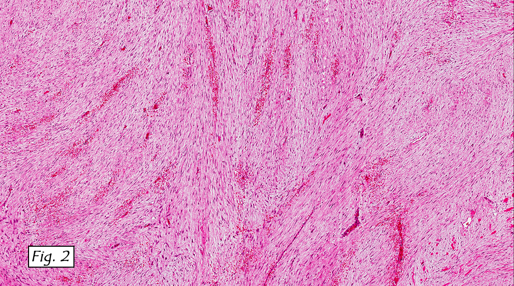
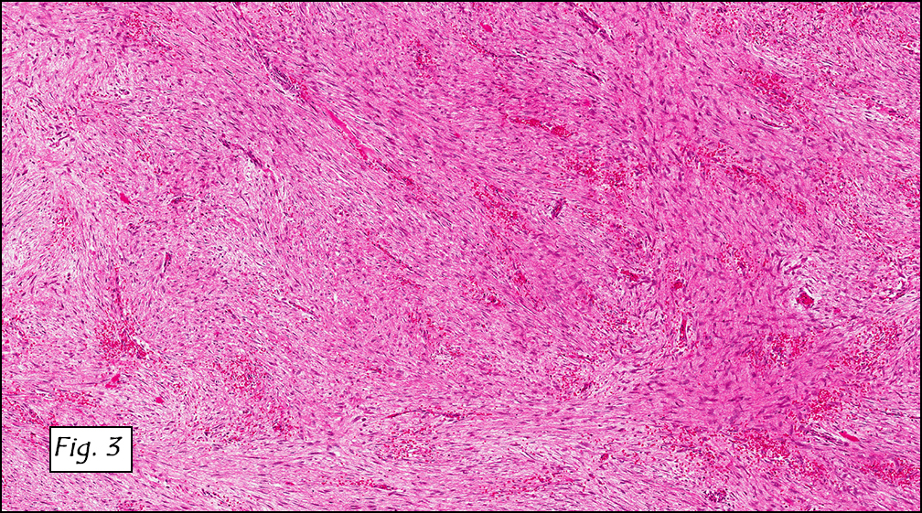
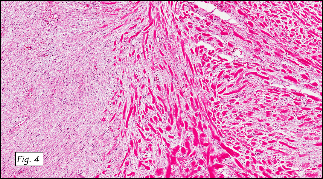
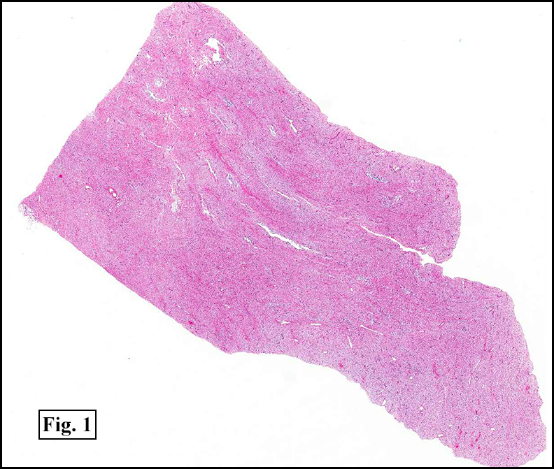
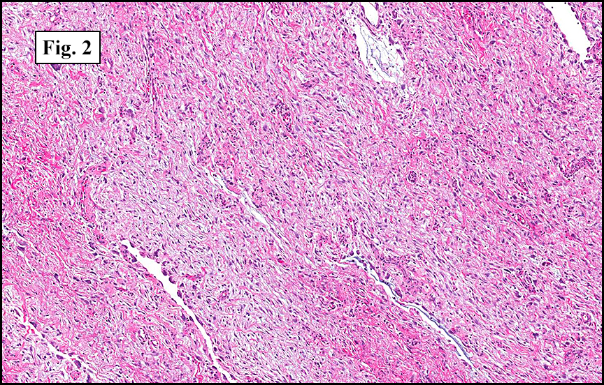
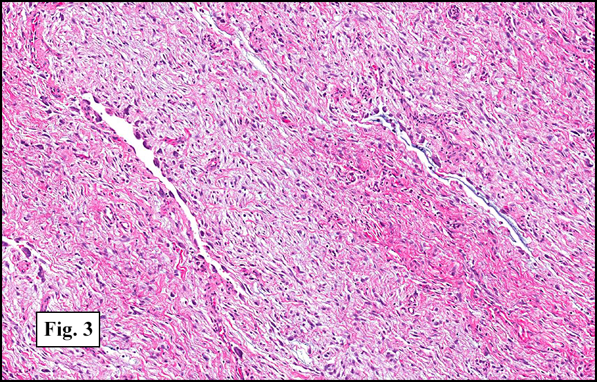
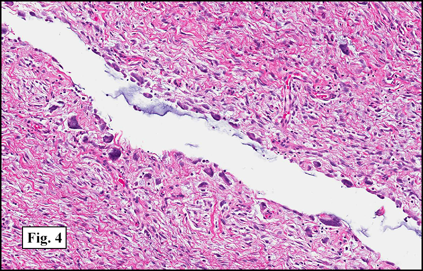
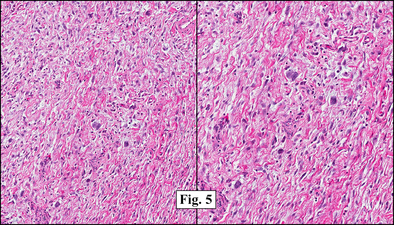
The tumor was compartmentalized, showing islands of cells separated into large and small nodules by fibrous stroma (Figs. 1,2). Smaller nests were bounded by sinusoidal vascular structures, a few of which were dilated (Figs. 3,4). Some of these smaller compartments showed central necrosis and many exhibited clearing around the cells with poor cellular cohesion (Fig. 5). The cells were uniform in both shape and size, and contained abundant pink granular cytoplasm and eccentrically placed, rounded, vesicular nuclei, most having prominent nucleoli (Figs. 6,7). There were scattered binucleated cells (Fig. 7). Mitoses were rare and no significant pleomorphism was present. A PAS stain showed abundant PAS positive granular and crystalloid material within the cytoplasm of many cells (Fig. 8).

Diagnosis: “Alveolar Soft Part Sarcoma”

Cody S. Carter, MSIV, Donald R. Chase, M.D.
Department of Pathology and Human Anatomy, Loma Linda University and Medical Center, Loma Linda, California
California Tumor Tissue Registry, Loma Linda, California

Discussion: Alveolar soft part sarcoma (ASPS) is a rare entity of unknown origin accounting for less than 1% of sarcomas. It usually occurs in ages 15-35, and predominantly affects females, especially in younger patients. In adults, the majority of tumors occur in the lower extremities, especially in the anterior thigh and buttocks. In children, these tumors typically present in the head and neck, especially the orbit and tongue, and tend to be smaller, likely due to earlier detection in these locations. The tumor most commonly presents as a slow-growing painless mass, but can often present with respiratory symptoms or neurologic symptoms like headache, visual changes and nausea due to their striking proclivity for early metastasis to the lung and brain. ASPS are typically very vascular and may be associated with an audible bruit. Radiologically, the diagnosis my be suggested byits hypervascularity with prolonged capillary staining and locally dilated veins on angiography and CT scan.

Grossly, ASPS is usually poorly circumscribed and highly vascular, with increased risk of significant blood loss at the time of surgery. The cut surface is usually yellow-white to gray and can feature areas of necrosis or hemorrhage, making the tumor soft and friable.

Microscopically, ASPS shows little variability. Compartments and nests are divided by fibrous stroma, thin-walled sinusoidal channels, usually lined by flattened endothelial cells. A lack of cellular cohesion and central necrosis is often seen. The dominant pattern of peripherally viable tumor cells with partial central clearing likens this neoplasm to the alveoli of the lung. While these features generally make a histologic diagnosis straightforward, the nest-like pattern can, on rare occasion, be absent, and large sheets of cells are seen together. This variant is primarily seen in infants and young children.

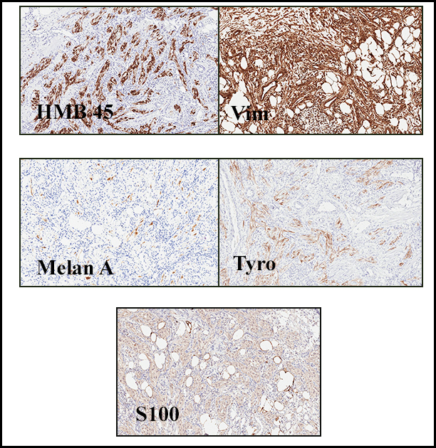
Cells within the nests are large, rounded, and fairly uniform, with eosinophilic granular cytoplasm and vesicular nuclei with distinct nucleoli. Cellular pleomorphism is only rarely present. Vascular invasion is almost invariably present, and reflects the tendency for early blood metastasis. The characteristic histologic pattern can usually be relied on for diagnosis, and the unreliable staining exhibited by ASPS makes immunohistochemistry most important in excluding other neoplasms. Of the histologic stains, PAS will likely show positively staining, diastase-resistant granules or rhomboid and rod-shaped crystals within the cytoplasm. These structures are composed of monocarboxylate tansporter 1 and its chaperone protein CD147. ASPS fails to express cytokeratin, EMA, neurofilament, GFAP, serotonin, synaptophysin and chromogranin. It can sometimes be positive for S-100 and neuron-specific enolase, but these seem to have no significant diagnostic value. Stains for MyoD1 and myogenin have been controversially positive, with one study using positivity in several tumors as support for skeletal muscle differentiation, but several subsequent studies have since failed to reproduce the results.

Cytogenetics may be a useful tool as the unbalanced translocation t(X;17)(p11.2q25) leads to an ASPL-TFE3 fusion gene that is both sensitive and specific for ASPS within the realm of soft tissue sarcomas (a small subset of pediatric renal cell carcinomas have also shown this same fusion gene). This can be more easily confirmed with an immunohistochemical stain for TFE3. This fusion gene has been linked to the overexpression of the promitotic MET receptor tyrosine kinase, which has suggested a model for oncogenesis, as well as a source of targeted therapy that is currently being tested in ongoing clinical trials.

Alveolar soft tissue sarcoma displays indolent growth, but overall prognosis is poor due to early metastasis, which can often be present at initial presentation. ASPS differs from most other adult sarcomas in that local recurrence is less likely after radical excision, but late metastasis can occur years after resection of the primary, and indeed metastases upwards of 30 years later have been recorded. Poorer prognosis is associated with increasing age and increasing tumor size at diagnosis, as well as metastases at initial presentation. Histologic grading is not contributory to prognosis.

Due to slow growth of the tumor, radical surgical excision of both primary and metastatic lesions is the most effective treatment, and conventional radiotherapy and chemotherapy have thus far been shown to be ineffective. Recent advances in targeted therapy are promising, and clinical trials are ongoing.

Differential diagnosis includes:

• Renal cell carcinoma (RCC) can look similar to the histology of ASPS, especially because some variants of the sarcoma can have less eosinophilic cells, and some RCCs can have quite eosinophilic cytoplasm. RCC can be distinguished by its distinct lack of PAS-positive crystalline structures or granules. Also, RCC is immunoreactive to EMA and keratin, but ASPS shows absent staining. TFE3 positivity can also be helpful, but some RCCs can express this antigen in the pediatric population. In this situation, radiologic correlation with renal imaging may be essential.

• Paraganglioma can be differentiated by its positivity for neuroendocrine markers, which are consistently negative in ASPS. Vesicular nuclei and prominent nucleoli are also not characteristics of paraganglioma. Also, paragangliomas typically present in patients over 40 years of age and are not found in the extremities; in contrast, those alveolar soft part sarcomas that arise in the head and neck region are found in pediatric patients.

• Granular cell tumors typically have a solid architecture with cells exhibiting more densely eosinophilic cytoplasm, and can exhibit spindling of the cells, a feature not present in ASPS. Also, cells are uniformly positive for S-100, while ASPS shows variably positivity.

• Alveolar rhabdomyosarcoma (AR) is characterized by smaller cells that are often more pleomorphic and feature dense nuclei. They are also much more likely to mark for desmin, MyoD1, and other muscle markers. The clusters of cells in an AR are surrounded by actual fibrous septa, as opposed to sinusoidal vessels.

Suggested Reading:
Portera CA Jr, Ho V, Patel SR, Hunt KK, Feig BW, Respondek PM, Yasko AW, Benjamin RS, Pollock RE, Pisters PW. Alveolar soft part sarcoma: clinical course and patterns of metastasis in 70 patients treated at a single institution. Cancer. 2001 Feb 1;91(3): 585-91.

Lieberman PH, Brennan MF, Kimmel M, Erlandson RA, Garin-Chesa P, Flehinger BY. Alveolar soft-part sarcoma. A clinico-pathologic study of half a century. Cancer. 1989 Jan 1;63(1):1-13.

Mitton B. Federman N. Alveolar soft part sarcomas: molecular pathogenesis and implications for novel targeted therapies. Sarcoma; 2012:428789. Epub 2012 Apr 8.

Folpe AL, Deyrup AT. Alveolar soft-part sarcoma: a review and update. J Clin Pathol. 2006 Nov;59(11): 1127-32.

Kayton ML, Meyers P, Wexler LH, Gerald WL, LaQuaglia, MP. Clinical presentation, treatment and outcome of alveolar soft part sarcoma in children, adolescents, and young adults. J Ped Surg. 2006 Jan;41(1): 187-93.

M. Ladanyi, M. Y. Lui, C. R. Antonescu et al. The der(17)t(X;17)(p11;q25) of human alveolar soft part sarcoma fuses the TFE3 transcription factor gene to ASPL, a novel gene at 17q25. Oncogene. 2001 Jan 4;20(1):48-57.

Tsuji K, Ishikawa Y, Imamura T. Technique for differentiating alveolar soft part sarcoma from other tumors in paraffin-embedded tissue: comparison of immunohistochemistry for TFE3 and CD147 and of reverse transcription polymerase chain reaction for ASPSCR1-TFE3 fusion transcript. Hum Pathol. 2012 Mar;43(3):356-63.

Weiss S, Goldblum J. Enzinger & Weiss’s Soft Tissue Tumors (5th ed). Philadelphia: Mosby/Esevier Inc. 177-92, 2008.

December 2013

Soft tissue tumors: Morphologic challenges and contemporary approaches to diagnosis
December 8, 2013
San Francisco
Cyril Fisher, MD, DSc, FRCPath

Seminar Objectives: At the conclusion of this seminar, attendees will be able to:

  1. Recognize the morphologic features of a wide variety of soft tissue sarcomas, pseudosarcomas and tumor-like lesions and their differential diagnosis in core biopsy and excision specimens.
  2. Appropriately select and interpret immunohistochemical panels and molecular techniques to discriminate between morphologically similar soft tissue tumor types.
  3. Correlate clinical and pathologic findings for diagnosis, prognosis and management of soft tissue sarcomas.

February 2013: A 36 year old woman with pelvic pain and an abdominal mass

History: A 36 year old female presented with pelvic pain. An abdominal ultrasound was performed and identified a mass on the round ligament.

Grossly the tumor was 3 cm in diameter, well-circumscribed and encased within surrounding fat. The cut surface was gritty with focal cystic changes and peripheral fibrosis. Microscopically, the tumor had well defined margins with a thin fibrous capsule and focal calcifications (Fig. 1). The tumor consisted of spindled and polygonal cells arranged in alveolar and fascicular patterns (Fig. 2,3) with intervening dense, hyalinized collagen (Fig. 4). The cells displayed clear to eosinophilic cytoplasm with indistinct borders (Fig. 5) and round to oval nuclei demonstrating large, prominent nucleoli and coarse chromatin (Fig. 6). Mitotic figures were not identified and there was no evidence of necrosis. Immunohistochemistry revealed diffuse positivity for S100, HMB45, and vimentin with focal positivity for melanA and tyrosinase (Fig. 7).

Diagnosis: “Clear Cell Sarcoma (Malignant Melanoma of Soft Parts)”

Drew G. Davis, MSIV, and Donald R. Chase, M.D.
Department of Pathology and Human Anatomy, Loma Linda University and Medical Center, Loma Linda, California
California Tumor Tissue Registry, Loma Linda, California

Discussion: Clear cell sarcoma was first described by Dr. Franz M. Enzinger in 1965. Subsequently, several histologic and ultrastructural similarities with melanoma were noted including melanin pigment, presence of melanosomes, and immunohistochemical staining for S-100 and HMB-45. These findings resulted in the moniker malignant melanoma of soft parts. Despite these similarities clear cell sarcoma and melanoma appear to be two different entities. Recent chromosomal analyses have demonstrated a characteristic translocation in clear cell sarcoma, t(12;22), resulting in a novel fusion gene product which is not found in melanoma.

A majority of cases are localized in the extremities, predominantly the lower legs, with the ankle being the most commonly affected site. Tumors have been noted on the trunk and neck, but intra-abdominal tumors are exceedingly rare. The tumors are intimately involved with adjacent tendon sheaths and aponeuroses and rarely involve the epidermis although occasional dermal extension has been noted.
Most clear cell sarcomas arise between the ages of twenty and forty but they have been noted in a wider range from seven to eighty three. There is a slight female predominance. Clinically they are recognized by gradually enlarging, occasionally painful masses present for months to years. Grossly, the rubbery, firm tumors are lobulated or multinodular and are often encapsulated or partially circumscribed. The cut surface is gray-white and homogeneous with occasional foci of necrosis or hemorrhage. Pigmentation may be observed in some cases.

Histologically, clear cell sarcoma is distinguished by alveolar and fascicular patterns of spindled to polygonal cells with clear to eosinophilic cytoplasm separated by characteristic collagenous septa. The nuclei display vesicular chromatin and one or more prominent nucleoli. Melanin pigment may be seen in up to 50% of cases. Occasional multinucleated giant cells are identified and rare mitoses are seen as well. Immunohistochemical stains are a useful adjunct in the diagnosis and show a similar staining pattern to that of melanoma with varying degrees of S-100, HMB-45, Melan-A and vimentin positivity. Metastatic tumor deposits typically show a similar histologic pattern but increased nuclear pleomorphism, disorderly growth and multiple foci of hemorrhage and necrosis have been observed.

The differential diagnosis includes sarcomas with predominantly fascicular growth patterns, including synovial sarcoma and fibrosarcoma, as well as melanin producing tumors such as cellular blue nevi and paraganglioma-like dermal melanocytic tumor (PDMT). The cytologic features of clear cell sarcoma including clear cytoplasm and prominent melanoma-like nucleoli, along with its immunophenotypic profile, differentiate it from other sarcomas. More important is the distinction between it and other melanocytic lesions. Although the survival rates of both melanoma and clear cell sarcoma are poor, the clinical management differs drastically. The location of the tumor, especially its investiture in adjacent tendons and aponeuroses, and its general lack of epidermal and dermal involvement are helpful clues. The uniform, spindled appearance of clear cell sarcoma also contrasts with the epidermoid appearance of nodular melanomas. Cytologic features are useful in distinguishing clear cell sarcoma from cellular blue nevi, which typically have smaller, pinpoint nucleoli and lack atypia. As a last resort, molecular genetic analysis for the t(12;22) translocation will establish the diagnosis of clear cell sarcoma. Paraganglioma-like dermal melanocytic tumors may also show cells with clear to eosinophilic cytoplasm but the presence of zellballen-like nests, low grade nuclei and their traditional location in the dermis set them apart from clear cell sarcomas.

The established treatment of choice is surgical excision with wide margins. Chemotherapy and radiation for both local and metastatic control have been used with little success. Metastases are common and affect up to 67% of patients, often years after the primary excision. Prognostic factors include necrosis, increased tumor size and the presence of metastases.

Suggested Reading:

Enzinger FM. Clear cell sarcoma of tendons and aponeuroses: An analysis of 21 cases. Cancer. 1965; 18:1163-1174.
Chung EB, Enzinger FM. Malignant melanoma of soft parts: A reassessment of clear cell sarcoma. Am J Surg Pathol. 1983; 7(5):405-13.
Lucas DR, Nascimento AG and Sim FH. Clear cell sarcoma of soft tissues: Mayo Clinic experience with 35 cases. Am J Surg Pathol. 1992; 16(12):1197-1204.
Montgomery EA, Meis JM, Ramos AG, Frisman DM and Martz KL. Clear cell sarcoma of tendons and aponeuroses: A clinicopathologic study of 58 cases with analysis of prognostic factors. Int J Surg Pathol. 1993; 1(2):89-100.
Langezaal SM, Graadt von Roggen JF, Cleton-Jansen AM, Baelde JJ and Hogendoorn PCW. Malignant melanoma is genetically distinct from clear cell sarcoma of tendons and aponeuroses (malignant melanoma of soft parts). Br J Cancer. 2001; 84(4):535-8.
Weiss S, Goldblum J. Enzinger and Weiss’s Soft Tissue Tumors (5th edition). Philadelphia: Mosby/Elsevier Inc. 926-34, 2008.

January 2013: A 66 year old man with an abdominal mass

History: A 66-year-old male dwarf underwent surgical excision of a 6.0 cm mesenteric mass. The cut surface resembled white scar tissue.

Microscopically, the mass was of fibrous tissue with prominent blood vessels (Fig. 1). Spindle-shaped cells were evenly spaced among mature collagen arranged in long sweeping fascicles (Fig. 2). The stroma showed variable density (Fig. 3) and there was an area which consisted of thick bundles of collagen (keloidal fibrosis), (Fig. 4).

Diagnosis: “Mesenteric Fibromatosis”

Matthew S. Johnson, PSF, and Donald R. Chase, MD
Department of Pathology & California Tumor Tissue Registry, Loma Linda University and Medical Center, Loma Linda, California

Discussion: Mesenteric fibromatosis (MF) is classified as an intra-abdominal fibromatosis, and one of a part of a larger group of fibromatoses that are microscopically similar. Fibromatoses in general are benign fibroproliferative processes that may demonstrate infiltrative growth but do not metastasize. Deep fibromatoses have about a 50% recurrence rate, and the superficial variants about 10%. MF accounts for a mere 8% of all fibromatosis cases. These tumors typically arise in the mesentery of the small bowel and the ileocecal region, but occasionally occur in the omentum and retroperitoneum. They usually present as asymptomatic abdominal masses or are found incidentally in patients of various ages (mean age of 41 years) and are more common in males. It is common for them to measure 10 cm or more. Grossly, they well-circumscribed, white masses that may be either soft or firm.

Histologically, MF is comprised of a dense collagenous stroma with evenly dispersed spindle or stellate cells and prominent blood vessels. The cellularity may be variable with stroma ranging from densely fibrous to markedly myxoid. Foci of keloidal fibrosis (thick collagen bundles) are a hallmark feature, present in approximately 50% of cases.

Immunohistochemical studies can help distinguish MF from other tumors. MF displays strong nuclear staining for β-catenin but is negative for CD117, CD34, and S-100.

Differential Diagnosis:

• Gastrointestinal stromal tumor (GIST) is the most common misdiagnosis. They usually have greater cellularity and atypia then do MFs. Also helping to distinguish are the keloidal fibrosis, infiltrative growth pattern, and prominent thin-walled blood vessels which are commonly found in MF but not in GIST. The tumors are also separated by their immunostaining patterns. GIST tends to be positive for CD117, CD34, and β-catenin, and MF is not.

• Sclerosing mesenteritis may also present as a large mass in the mesentery of small bowel, but the histological findings of fat necrosis, chronic inflammation, less fibrosis, and lack of β-catenin expression distinguish it, as does the negative CD 117.

• Inflammatory myofibroblastic tumor of the mesentery or retroperitoneum is a consideration, but usually shows increased cellularity, cytologic atypia, and a greater degree of inflammation. These may also be positive for ALK-1 protein.

• Idiopathic retroperitoneal fibrosis typically displays lymphoplasmacytic inflammation dispersed among bands of dense hyalinized collagen.

MF is associated with familial adenomatous polyposis (FAP), in fact fibromatosis is found in approximately 10 to 15% of patients with FAP and often develops within two years after surgical excision of the affected intestinal tract. Diligent post-operative observation in FAP patients and endoscopic examination of the bowel in MF patients without known FAP is warranted.

Local recurrence is common. Greater recurrence rates have been observed in patients with FAP (90%) versus those without (12%). Recurrent tumors in FAP patients also tend to behave more aggressively and respond worse to therapy. Treatment involves complete excision of the tumor, which is often complicated by intestinal attachment. Alternative therapeutic options including postoperative radiation, chemotherapy, and anti-estrogenic agents have shown mixed results.

Suggested Reading:

Rodriguez JA; Guarda LA; Rosai J. Mesenteric fibromatosis with involvement of the gastrointestinal tract. A GIST simulator: a study of 25 cases. Am J Clin Pathol 2004 Jan;121(1): p93-8.

Pai SA; Zaveri SS. Intra-abdominal fibromatosis of the jejunum and mesentery. J Clin Pathol 2004 Oct;57(10): p1119.

Montgomery E; Torbenson MS; Kaushal M; Fisher C; Abraham SC. Beta-catenin immunohistochemistry separates mesenteric fibromatosis from gastrointestinal stromal tumor and sclerosing mesenteritis. Am J Surg Pathol 2002 Oct;26(10): p1296-301.

Church J; Berk T; Boman BM; Guillem J; Lynch C; Lynch P; Rodriguez-Bigas M; Rusin L; Weber T. Staging intra-abdominal desmoid tumors in familial adenomatous polyposis: a search for a uniform approach to a troubling disease. Dis Colon Rectum 2005 Aug;48(8): p1528-34.

Holubar S; Dwivedi AJ; O’Connor J. Giant mesenteric fibromatosis presenting as small bowel obstruction. Am Surg 2006 May;72(5): p427-9.

December 2012: A 13 month old boy with a chest wall mass

History: A 13-month-old male infant underwent surgical resection of a 6.0 x 5.6 x 2.0 cm subxiphoid body wall mass. The cut surface was soft and homogenously gray-tan.

The stroma was loosely arranged and consisted of undulating spindle cells (Fig. 1, 2) intermixed with irregularly shaped “pseudovascular” spaces (Fig. 3). Discontinuous rows of hyperchromatic giant cells lined the peripheries of these spaces (Fig. 4). Giant cells and multinucleated-appearing cells were scattered throughout the stroma (Fig. 5).

Diagnosis: “Giant Cell Fibroblastoma, Chest Wall”

Matthew S. Johnson, PSF, and Donald R. Chase, MD
Department of Pathology and Human Anatomy
California Tumor Tissue Registry
Loma Linda University and Medical Center, Loma Linda, California

Discussion: Initially described by Shmookler and Enzinger in 1982, giant cell fibroblastoma (GCF) is classified as a fibrohistiocytic tumor commonly felt to be the juvenile form of dermatofibrosarcoma protuberans (DFSP). It is usually diagnosed in infancy or childhood, with two-thirds being discovered by 5 years of age and about 90% by 10 years (median diagnostic age is 3 years). Rare cases have been reported in adults. GCF usually affects males and typically presents as a painless mass in the abdominal wall, groin, or back. Grossly, they are poorly circumscribed and gelatinous. The cut surface is usually gray to yellow.

Histologically, GCFs are variably cellular and composed of spindle and stellate cells within a collagenous, sometimes myxoid stroma. The nuclei are small and uniform without significant numbers of nucleoli. Scattered throughout the stroma are hyperchromatic giant cells with large vesicular nuclei, often with prominent nucleoli. Occasionally the giant cells appear multinucleated; however, ultrastructural examination suggests that most actually have a single, multilobated nucleus. The defining feature of this tumor is the intervening, large, irregular pseudovascular spaces that are partially lined by the giant cells. No endothelial lining is present.

Immunohistochemical studies show the tumor stroma and pseudovascular lining to be negative for S-100, cytokeratin, desmin, and CD31. CD34 is typically weakly positive in the stromal elements.

GCF appears to be closely related to DFSP. Cases of GCF recurring as DFSP (and vice versa) have been documented, and some tumors show both neoplasms. Cytogenetic analysis has revealed the presence of a (17:22) translocation in both phenotypes. Evidence suggests that a linear arrangement of this abnormality occurs in GCF while a ring form takes place in DFSP.

Differential Diagnosis:

• Myxoid liposarcoma may resemble GCF with myxoid stroma; however, the tendency to occur in deep tissues, the presence of lipoblasts, a delicate vasculature, and lack of giant cells separates it from GCF.
• Myxoid malignant fibrous histiocytoma (myxofibrosarcoma) may have histologically similar stroma and can have large cells demonstrating pleomorphism and multinucleation. A vascular network and mitotic activity differentiates it from GCF, as does a lack of space-lining pleomorphic cells.
• Angiosarcomas demonstrate irregular branching vascular channels that are lined by multiple layers of cells with large hyperchromatic nuclei. These lesions are distinguished from GCF by their high mitotic activity, atypical nuclei of the vascular lining cells, and expression of CD31.
• Lymphangiomas contain angiectatic spaces, but these spaces are often surrounded by a layer of smooth muscle, and not by pleomorphic cells. Lymphoid aggregates may also be present in the surrounding stroma.

About 50% of GCF cases recur. Recurrences are not aggressive and metastatic disease has not been reported. Treatment of choice is wide local excision; however, less invasive therapy with routine follow-up is also a consideration.

Suggested Reading:

Terrier-Lacombe MJ; Guillou L; Maire G; Terrier P; Vince DR; de Saint Aubain Somerhausen N; Collin F; Pedeutour F; Coindre JM. Dermatofibrosarcoma protuberans, giant cell fibroblastoma, and hybrid lesions in children: clinicopathologic comparative analysis of 28 cases with molecular data – a study from the French Federation of Cancer Centers Sarcoma Group. Am J Surg Pathol 2003 Jan;27(1): p27-39.

Sandberg AA; Bridge JA. Updates on the cytogenetics and molecular genetics of bone and soft tissue tumors. Dermatofibrosarcoma protuberans and giant cell fibroblastoma. Cancer Genet Cytogenet 2003 Jan 1;140(1): p1-12.

Pascual A; Sanchez-Martinez C; Moreno C; Burdaspal-Moratilla A; Lopez-Rodriguez MJ; Rios L. Dermatofibrosarcoma protuberans with areas of giant cell fibroblastoma in the vulva: a case report. Eur J Gynaecol Oncol 2010;31(6): p685-9.

Najarian DJ; Morrison C; Sait SN; Meguerditchian AN; Kane J; Cheney R; Zeitouni NC. Recurrent giant cell fibroblastoma treated with Mohs micrographic surgery. Dermatol Surg 2010 Mar;36(3): p417-21.

Layfield LJ; Gopez EV. Fine-needle aspiration cytology of giant cell fibroblastoma: case report and review of the literature. Diagn Cytopathol 2002 Jun;26(6): p398-403.

Aliagaoglu C; Bakan V; Atasoy M; Sahin O; Toker S; Albayrak M. A very large, rapidly developing, congenital giant cell fibroblastoma in a 5-month old infant. J Dermatol 2006 Mar;33(3): p182-6.

Scroll to top