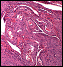
 History: A 48 y/o woman underwent a hysterectomy and right salpingo-oophorectomy ten years previously for an enlarged uterus. A large number of blood vessels were found throughout the endometrium and myometrium that were commented upon as being “unusual/uncertainâ€. Ten years later she presented with microscopic hematuria and severe pain radiating to her back. An IVP identified a pelvic mass which was biopsied and later excised. Following removal of the mass the CA-125, which had been elevated, returned to normal limits.“January 2008: A 48 year old woman with a pelvic mass”Continue reading
December 2007: An arm nodule in a three month old boy
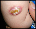 History: A three-month old male presented for evaluation of an enlarging lesion on the right arm that had been present since birth. His mother noted rapid growth over the period of one month with central crusting of the lesion and minimal bleeding. The boy had been born full-term in a normal spontaneous vaginal delivery to a gravida 1, para 1 mother. He had no other medical problems and family history was noncontributory.“December 2007: An arm nodule in a three month old boy”Continue reading
November 2007: A 57 year old man with a mass in the upper back
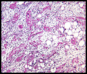 History: A 57 year old male presented with a painless 5 x 3.5 x 2.5 cm mass in the superficial tissues of the right upper back. An excisional biopsy was performed.
Grossly, the mass appeared circumscribed and had a brown-tan lobulated appearance.“November 2007: A 57 year old man with a mass in the upper back”Continue reading
December 2007 Seminar
Advances in Surgical Pathology: New Entities, Emerging Concepts, and New Perspectives on Old Problems
Saul Suster, M.D..
December 9, 2007
8:30 AM – 4:30 PM
Dr. Saul Suster is Professor of Pathology and Vice-Chair for Pathology at Ohio State University. He is also Director of Anatomic Pathology at Ohio State University Medical Center, the Arthur G. James Cancer Center, and the Solove Research Institute. Following medical school in Guayaquil, Ecuador and a rotating internship, he did his initial pathology training at the University of Tel-Aviv Sackler School of Medicine and later did a pathology residency at Mount Sinai Medical Center of Greater Miami. He studied with Juan Rosai at Yale University and developed special expertise in electron microscopy, immunohistochemistry, and molecular biology. His contributions in national and international organizations are numerous. Dr. Suster has published over 100 abstracts, over 200 papers and has written 20 book chapters on topics ranging from thymus and mediastinum, lung and pleura, skin and the endocrine system.
Educational Contents and Media:
- Glass microscopic slides and digital images representative of fifteen to twenty tumors of importance to surgical pathologists.
- Correlating clinical histories
- Six hour lecture, incorporating projected photographs of the cases and other illustrative materials.
- Comprehensive printed color syllabus, including diagnosis, discussion, and appropriate references from pertinent medical literature.
The Palace Hotel
San Francisco, California
2 New Montgomery Street,
San Francisco, CA 94105
(415) 512-1111; FAX (415) 243-8062
October 2007: A 25 year old man with a nodule in the arm
 History: A 25 year old man with a 1 cm nodule in his arm had an excisional biopsy.
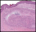
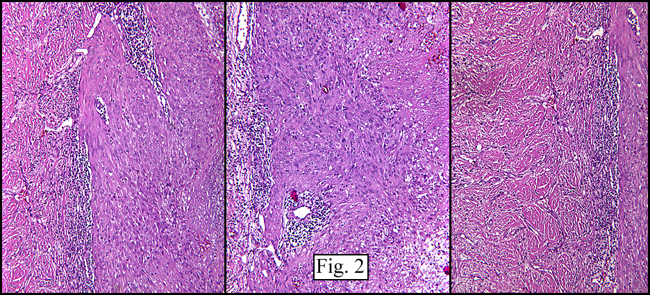
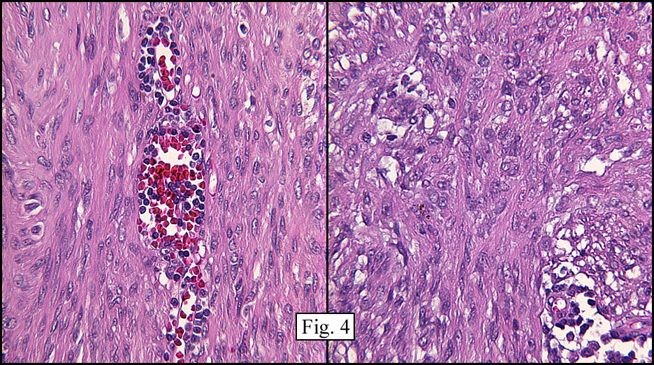
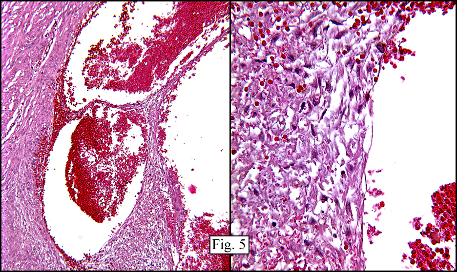
The superficial dermis resembled a simple acrochordon however the deeper dermis had sheets and nodules of spindled cells arranged in short fascicles (Figs. 1,2) These fusiform cells had ovoid nuclei and tapered eosinophilic cytoplasm (Figs. 3,4). At the periphery was a cuff of lymphocytes. There were also cystic spaces filled with erythrocytes (Fig. 5). Hemosiderin pigment was focally evident.“October 2007: A 25 year old man with a nodule in the arm”Continue reading