May 2015: An adult male with a subcutaneous soft tissue mass involving a heel

History: A painful, one cm nodule was found the subcutaneous tissue of the right heel of an adult male. Radiographs showed it to be ill-defined and radio-opaque. The nearest bone was the calcaneus, but it was not involved. There was no history of trauma.

The lesion, at low magnification, consisted of an admixture of adipose tissue, osteoid, trabecular mature bone, and fibrous stroma (Figs. 1, 2). Prominent osteoid, bone and abortive cartilaginous elements were present (Fig. 3). Zones of osteoid were surrounded by cellular or myxoid fibrous stroma and were sometimes rimmed by osteoblasts (Fig. 4).

Diagnosis: Panniculitis Ossificans, heel

Bing Wang, M.D., and Donald R. Chase, M.D.
Department of Pathology and Human Anatomy
Loma Linda University Medical Center, Loma Linda, California
California Tumor Tissue Registry, Loma Linda, California

Discussion: Panniculitis Ossificans (PO) is also referred to as a “fibro-osseous lesion” or “heterotopic ossification”. It has been subclassified based upon its location [1]:

Unlike usual panniculitis, which may show inflammatory infiltrates involving the septa [4], PO commonly lacks inflammation. If inflammation is present it is usually minimal and mostly evident in the tissue surrounding the lesion [2].

PO usually affects young, physically active adolescents and adults. Most cases involve the limbs, and some are seemingly incited by injury. The initial symptoms may include pain or tenderness followed by soft tissue swelling. Later, the swollen tissue becomes circumscribed and indurated. Calcification usually starts three to six weeks following injury [2].

Radiographs show a series of changes, initially with an increase in soft tissue density or opacity. Calcification starts around the end of third week, and as the lesion becomes increasingly calcified, it becomes well-circumscribed with either a solitary region of calcification or scattered regions of calcification [2].

Histologically, PO is usually well-circumscribed and is characterized by the presence of a distinct zonal pattern that reflects different components of this entity, including: normal-appearing adipose tissue, fibrous stroma, osteoid and mature bone. The adipose tissue consists of normal lobules of mature fat. Fat necrosis usually is not seen. Foci of osteoid formation are scattered between the lobules of adipose tissue, and are usually surrounded by a cellular or myxoid fibrous stroma. Trabeculae of mature bone usually begins in the middle of osteoid islands. The content of the mature bone formation depends on the stage of the lesion. Sometimes osteoid and mature bones are rimmed by layers of osteoblasts showing little variation in size and shape. The osteoid and bone are separated from the surrounding tissue by a zone of loose or compressed fibrous tissue. The fibrous tissue consists of fibroblasts and myofibroblasts which may display a mild degree of cellular pleomorphism and rather prominent mitotic activity. In addition, there may be prominent vascular proliferation, focal hemorrhage, or fibrin deposition [2].

Panniculitis ossificans is a benign lesion and its etiology is not fully understood. It is usually caused by some kind of injury. It is proposed that injuries may generate an environment which produces certain proteins such as bone morphogenic protein, which in turn, can induce pluripotential mesenchymal cells to turn into osteogenic cells, which can then form osteoid, which in turn develops into mature heterotopic ossification [3].

There is no convincing evidence of malignant transformation of panniculitis ossificans [2]. The early stage of PO has immature and highly cellular zones that may be confused with extraskeletal osteosarcoma. However, extraskeletal osteosarcoma usually occurs in older patients; in contrast panniculitis ossificans often occurs in younger active people. Unlike panniculitis ossificans, extraskeletal osteosarcoma shows marked cellular atypia and infiltration of neighboring tissues in a destructive manner. Panniculitis ossificans is well-circumscribed with distinct zones of different components. In some cases, PO may contain entrapped atrophic muscle, which may cause confusion with another entity in the same category, myositis ossificans. The difference lies in how much muscle or adipose tissue is present in the lesion [2].

Suggested Reading:
1. Ackerman LV. Extra-osseous localized non-neoplastic bone and cartilage formation (so – called myositis Ossificans): clinical and pathological confusion with malignant neoplasms. J Bone Joint Surg Am 1958; 40-A(2): 279-298.
2. Goldblum JR, Folpe AL and Weiss SW. Soft tissue tumors, sixth edition. Pg 926-932.
3. McCarthy EF, Sundaram M: Heterotopic ossification: a review. Skeletal Radiol.34:609-619 2005.
4. Requena L. Normal subcutaneous fat, necrosis of adipocytes and classification of the panniculitides. Semin Cutan Med Surg 2007; 26:66–70.

April 2015: A 32 year old man with a soft tissue mass adjacent to the right humerus

History: A 32-year-old man presented with a mass in the right arm. It involved the humerus and upon excision was 8.2 x 6.5 x 3.5 cm and weighed 52 grams. It consisted mostly of fatty tissue but had a 3.0 x 2.5 cm, vaguely circumscribed nodule centrally located.

H&E staining of the central lesion revealed marked hyperplasia of lymphoid follicles with expanded germinal centers and well-formed mantle zones (Fig. 1). Some of the follicles had intervening fibrosis (Fig. 2). Germinal centers contained lymphocytes admixed with dendritic cells, polykaryocytes and eosinophilic proteinaceous deposits. Eosinophils were widely interspersed between the follicles with focal micro-abscess formation (Fig. 3), or occasionally infiltrated germinal centers with resultant follicular lysis (Fig. 4). The lesion was well-vascularized. Numerous post-capillary venules lined by flat endothelial cells were present between follicles and within germinal centers (Figs. 5, 6). Epithelioid endothelial cells were not seen.

Diagnosis: Kimura Disease, arm

Li Lei, M.D., Ph.D. and Donald R. Chase, M.D.
Department of Pathology and Human Anatomy
Loma Linda University Medical Center, Loma Linda, California
California Tumor Tissue Registry, Loma Linda, California

Discussion: Kimura disease (KD), named after Kimura for recognizing the vascular component of the entity in 1948, was first described in China in 1937 by Kim and Szeto as eosinophilic hyperplastic lymphogranuloma. It is a chronic inflammatory process characterized by benign reactive lymphoid proliferation with significant eosinophilia, secondary vascular proliferation and fibrosis.

KD is endemic in Asia, particularly in East and Southeast Asia. Other racial groups including Caucasian, African American and Hispanic may also be targeted. There is a striking male predominance with reported male-to-female ratio ranging from 3.5:1 to 19:1. Most of the affected patients are in their twenties to forties.
KD typically presents as a deep subcutaneous mass measuring 2 – 6 cm in size, but giant tumors up to 13 cm have been reported. Commonly it presents as cervical lymphadenopathy with a classic example being a peri-auricular mass involving the parotid gland. Less common sites include the extremities (like our case), axilla, groin, epiglottis, chest wall, spermatic cord and peripheral nerve.

Laboratory tests usually show peripheral blood eosinophilia and elevated serum immunoglobulin (IgE). Imaging is usually nonspecific, but sometimes may be seen as multiple ill-defined, enhancing lesions around the parotid gland with associated lymphadenopathy.
Clinically, up to 16% of cases are associated with proteinuria, nephrotic syndrome, membranous glomerulonephritis, bronchial asthma, ulcerative colitis or necrotizing eosinophilic vasculitis. Patients are otherwise asymptomatic.

Histologically, the tumor consists of dense lymphoid aggregates with prominent germinal centers, which contain IgE-bearing dendritic reticulum cells, polykaryocytes (Warthin-Finkeldey type), nuclear debris and a delicate eosinophilic matrix. Interfollicular dense eosinophilic infiltrate is a common feature and may form microabscesses. Plasma cells, small lymphocytes and mast cells are often increased in the paracortex. There is a moderate proliferation of postcapillary venules which are lined by flat endothelial cells. Long-standing lesions usually develop hyaline fibrosis and become less vascular. Affected lymph nodes demonstrate same histologic features with preserved architecture.
The differential diagnosis prominently includes “Epithelioid hemangioma” (EH), also known as “angiolymphoid hyperplasia with eosinophilia”, a benign vascular neoplasm which shares many features of KD. Both entities affect young to middle-aged adults, presenting in the head and neck. Both have an inflammatory component, a vascular component and increased numbers of eosinophils, but there are differences:

Despite the above differences (table) these two entities appear intertwined. Some lesions have characteristic features of both making specific classification almost impossible. Moreover, KD and EH can coexist in the same patient. Patients with one entity can develop the other during follow-up. These phenomena suggest that EH and KD may be a part of a spectrum of reactive vascular lymphoid proliferations.

In addition to EH, other differentials of KD include:

• Angioimmunoblastic T cell lymphoma at its early phase can have marked follicular hyperplasia with germinal centers, eosinophilia and prominent high endothelial venules. However, patients are often older and present with fever and generalized lymphadenopathy. Lymph node architecture is usually effaced. Perivascular clustering of neoplastic clear cells and increased follicular dendritic cell meshworks are helpful clues. Molecular study shows monoclonal T cell receptor gene rearrangements in the majority of the cases or IgH rearrangements in the minority.
• Langerhans cell histiocytosis can present as indolent lymphadenopathy in young adults. Microscopically, lymph node architecture is effaced by mixed infiltrates of Langerhans cells and eosinophils. CD1a and S100 immunoreactivity help to confirm the diagnosis.
• Follicular lymphoma: Primary cutaneous follicular lymphoma generally affects the elderly. Follicles consist of predominantly centrocytes with admixed centroblasts. In cases of secondary cutaneous involvement by follicular lymphoma, a pertinent medical history, back-to-back follicles with no cell polarization and t(14;18) translocation help reach the correct diagnosis.
• Hodgkin lymphoma has a bimodal age distribution with the first peak at 15-35 years of age. Lymph nodes have a “fish-flesh” cut surface. Despite lymphocytes, eosinophils, and collagen fibers in the background, CD30 and CD15 positive Hodgkin Reed-Sternberg cells are pathognomonic.

The etiology of KD remains unknown. Multiple clues such as elevated serum IgE, peripheral eosinophilia and association with other autoimmune diseases favor immune system hypersensitivity as the underlying pathophysiology. The endemic epidemiology of KD may reflect either inherent genetic susceptibility or an antigenic trigger unique to the geographic area.
Patients with KD normally have a favorable outcome. If untreated, the mass may remain stable or spontaneously regress. The standard care is simple surgical excision. Other options include laser therapy, radiotherapy and systemic corticosteroids. Local recurrence rate vary from 14 – 44%. Characteristics which may predict recurrence include: size ≥ 3.5 cm, peripheral eosinophilia ≥35%, Notch-1 upregulation and Ki-67 proliferation index ≥ 3% outside the germinal center. Metastases are very rare and therefore anecdotal.

Suggested Reading:
Goldblum J, Folpe A, Weiss S. Enzinger & Weiss’ Soft Tissue Tumors, 6th ed: Philadelphia, Elsevier Inc, 2014; 649-654.
Rosai J. Rosai and Ackerman’s Surgical Pathology, 10th ed: Philadelphia, Elsevier Inc, 2011; 1805.
Kung IT, Gibson JB, Bannatyne PM. Kimura’s disease: a clinico-pathological study of 21 cases and its distinction from angiolymphoid hyperplasia with eosinophilia. Pathology. 1984;16:39-44.
Deng WY, Ye SB, Luo RZ, Yan SM, Gao YF, Yang YZ, Guo ZM, Chen YF. Notch-1 and Ki-67 receptor as predictors for the recurrence and prognosis of Kimura’s disease. Int J Clin Exp Pathol. 2014;7: 2402-10.
Buder K, Ruppert S, Trautmann A, Bröcker EB, Goebeler M, Kerstan A. Angiolymphoid hyperplasia with eosinophilia and Kimura’s disease – a clinical and histopathological comparison. J Dtsch Dermatol Ges. 2014;12:224-8.
Liu XK, Ren J, Wang XH, Li XS, Zhang HP, Zeng K. Angiolymphoid hyperplasia with eosinophilia and Kimura’s disease coexisting in the same patient: evidence for a spectrum of disease. Australas J Dermatol. 2012;53:e47-50

March 2015: A 69 year old man with a slowly growing knee mass

History: A 69-year-old man presented with a slowly growing mass in the right knee. Twenty years prior he had trauma to the area and subsequently developed “arthritis” at the site. Physical exam demonstrated the mass but noted that the knee still had full range of motion without pain. MRI showed a 9.8 x 3.8 x 4.4 cm mass with a fat signal at the lateral knee that involved soft tissue and spared the bone. A previous biopsy had been called “lipoma”.

The excised specimen weighed 93 grams and was 10.0 x 5.0 x 3.5 cm. It was multi-nodular with a thin capsule. Villous projections of white-yellow adipose tissue were present consisting of white-yellow lobulated adipose tissue. A central portion showed a 2.0 x 1.8 cm region of calcification.“March 2015: A 69 year old man with a slowly growing knee mass”Continue reading

February 2015: 67 year old woman with a perineal mass

History: A 67-year-old woman presented with a 5 year history of non-tender swelling in the perineal area. There had been no drainage or infection. Physical examination showed a 3 x 2 cm, firm mass attached to the skin just below and lateral to the labia. It was superficial and did not involve underlying tissues.

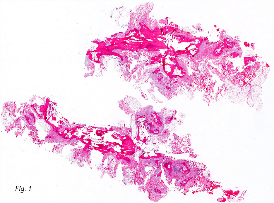
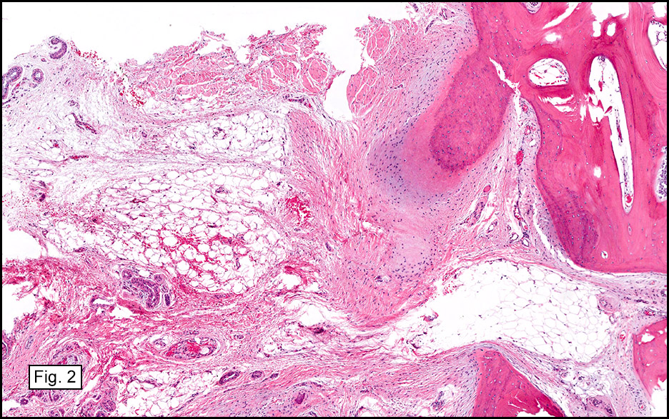
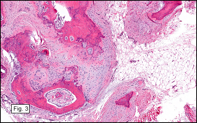
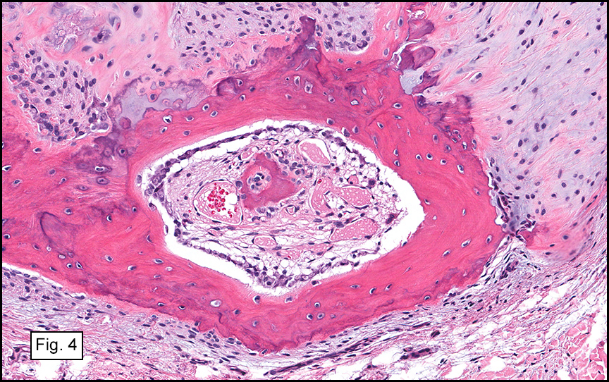
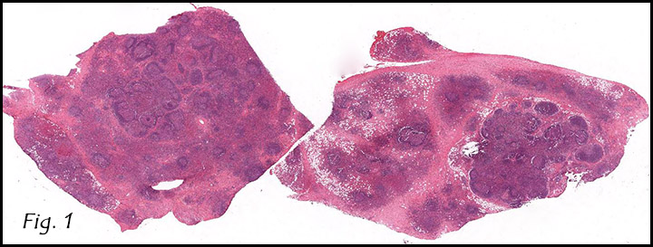
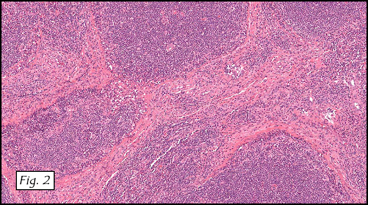
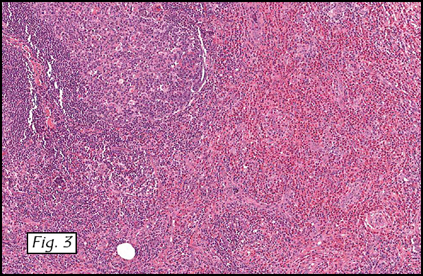
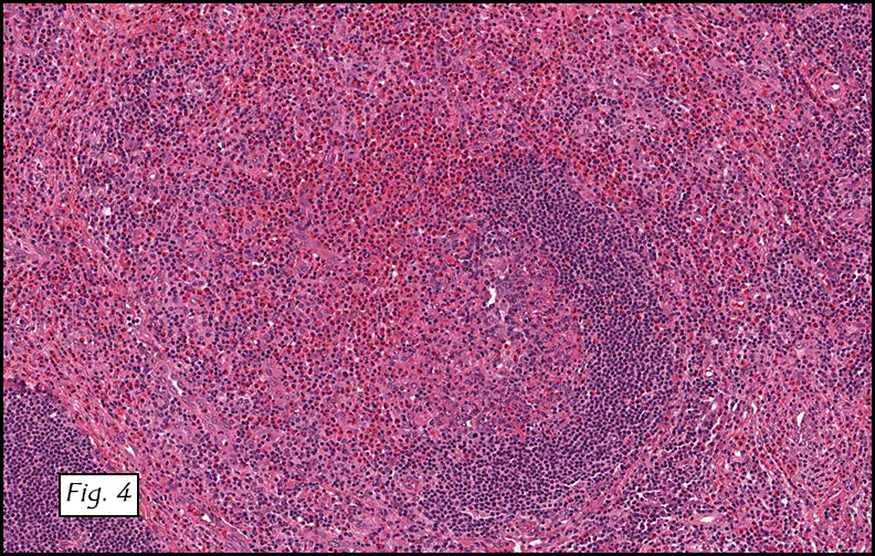
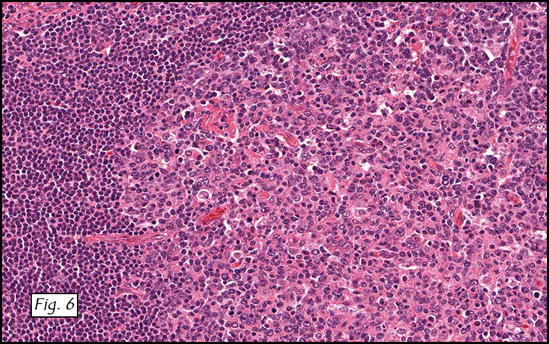
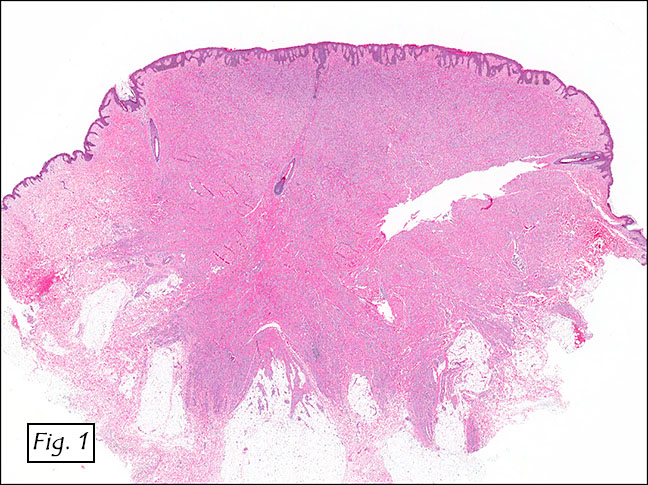
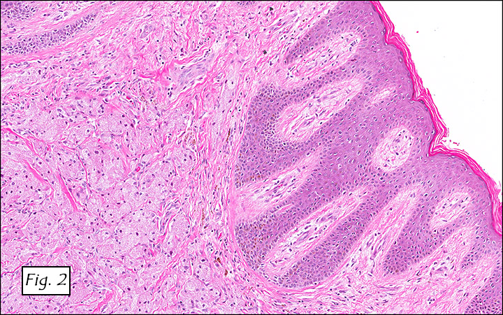
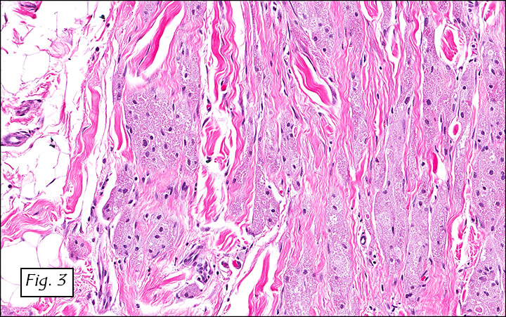
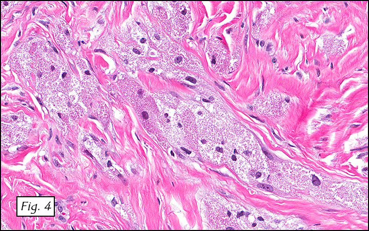
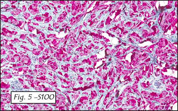
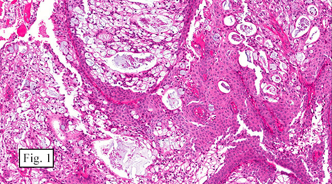
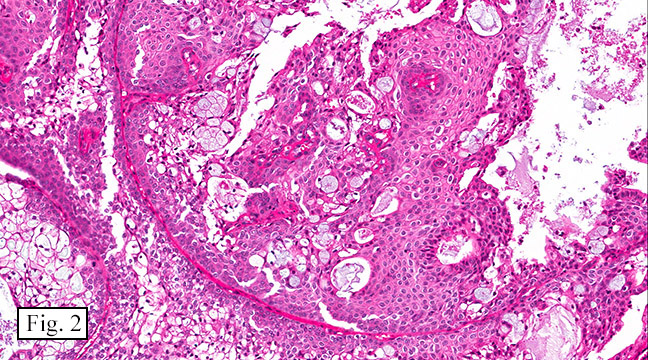
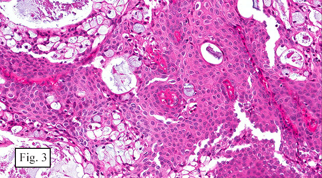
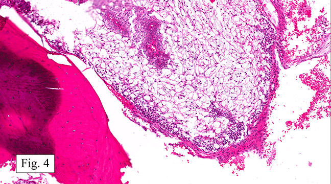
The excised specimen consisted of a yellow-tan, ill-defined, 2.5 x 1.4 x 0.1 cm fibrous subcutaneous nodule (Fig. 1). The overlying epidermis showed elongated hyperplastic rete ridges (Fig. 2). The tumor was composed of sheets of eosinophilic cells with infiltrative borders (Fig. 3). Higher power views demonstrated large polygonal cells with eosinophilic granular cytoplasm, indistinct cell borders, and centrally-situated, small, hyperchromatic nuclei (Fig. 4). No spindling, atypia, pleomorphism, mitoses, nor necrosis were identified. The tumor cells were positive for S-100 (Fig. 5).

Diagnosis: Granular cell tumor, vulva

Li Lei, PGY2 and Donald R. Chase, M.D.
Department of Pathology and Human Anatomy,
Loma Linda University Medical Center, Loma Linda, California
California Tumor Tissue Registry, Loma Linda, California

Discussion: Granular cell tumor (GCT) was first described by Virchow and Weber in 1854, and was later characterized as granular cell myoblastoma by Abrikossoff in 1926. Other names, such as Abrikossoff’s tumor, and granular cell schwannoma, have also been used. After decades of debate regarding the histogenesis, most believe that GCT is of neurogenic origin, most likely Schwann cells. Others advocate that GCT is a degenerative change resulting in a cytoplasmic accumulation of lysosomes that occur primarily in Schwann cells as well as in a variety of other cell types. Despite the unresolved controversy on the histogenesis, the simple term “GCT” is currently used by most pathologists.

GCT accounts for 0.5 – 1.3% of all soft tissue tumors. It occurs in patients of all age groups, with a peak incidence in the third to sixth decades. Women are affected twice as often as men. GCT can occur anywhere in the body, but most commonly involves the tongue. It typically presents as a solitary, usually asymptomatic nodule, though pruritus, pain, and ulceration have occasionally been reported. GCT involving critical anatomic sites such as trachea and neurohypophysis can cause serious complications. Between 3 to 20% of patients have multiple tumors that appear synchronously or metachronously, making it a challenge in some cases to determine if the tumor is primary or metastatic.. Multiple GCTs may be part of LEOPARD syndrome which is associated with PTPN11 mutation. Rarely, familial cases have been reported, suggesting that GCT may be inheritable.

Approximately 5 – 16% of GCTs occur in the vulvar region. For reasons unknown, it is more common in African-Americans than Caucasians. Vulvar GCT generally involves the labium majus, though clitoris, mons pubis, perineum and perianal region may also be affected. The male counterparts, GCTs of scrotum and/or penis, are extremely rare with only 22 cases reported in the literature. Vulvar GCT is usually slow-growing, but may enlarge rapidly during pregnancy. Although most vulvar mesenchymal tumors express estrogen receptors and/or progesterone receptors, it is still unclear if vulvar GCTs do.

An MRI may help to diagnose the tumor pre-operatively. GCT usually shows a peripheral hyperintense rim encircling a central hypointense core. These features are most appreciated on T1-weighted images, and may become more prominent on T2-weighted images. There is also variable peripheral enhancement in post-contrast images.

Grossly, GCT is a stellate-shaped or circumscribed, nonencapsulated, subcutaneous mass. The cut surface is tan to yellow, firm and homogeneous. It is usually less than 3 cm, but may grow up to 12 cm.

Microscopically, GCT displays either nodular or infiltrative growth pattern(s). It is composed of sheets, nests, or cords of large polygonal cells, which are characterized by indistinct cytoplasmic borders, abundant eosinophilic granular cytoplasm, and centrally-located small oval nuclei with hyperchromasia. Within the fine or coarsely granular cytoplasm, larger eosinophilic globules surrounded by clear halos are sometimes seen.

Benign GCTs may be slightly spindled, however prominent spindling is a clue of more aggressive behavior. Fibrosis or desmoplasia is variable. An important but nonspecific feature of these tumors is pseudoepitheliomatous hyperplasia of overlying squamous epithelium. This can be mistaken for well-differentiated squamous cell carcinoma if the tumor is only superficially sampled.

The cytoplasmic granules of tumor cells may contain large amounts of hydrolytic enzymes, which are positive for Luxol fast blue and Periodic Acid-Schiff (PAS) with diastase resistance. The cells show diffuse strong nuclear and cytoplasmic S-100 positivity, though some malignant GCTs may lose S-100 expression. Other ancillary positive markers include neuron specific enolase, myelin basic protein, laminin, calretinin, vimentin, osteopontin, inhibin-alpha, HLA-DR, protein gene product 9.5, CD68, carcinoembryonic antigen (CEA), and CD57 (Leu-7). The tumor cells are negative for cytokeratin, desmin, chromogranin, HMB-45, and glial fibrillary acidic protein (GFAP).

Ultrastructurally, the tumor cells are surrounded by replicated basal lamia suggestive of repeated cycles of cellular injury and repair. This is another feature supporting the degenerative theory of histiogenesis. The cytoplasmic granules are composed of membrane-bound, autophagic, vacuoles containing myelin figures and fragmented rough endoplasmic reticulum and mitochondria, consistent with phagolysosomes. There are also small interstitial cells with angulated bodies resulting in a Gaucher cell-like appearance.

Approximately 98% of the GCTs are benign. However, malignant transformation can develop as soon as 5 months after an initial benign diagnosis. Features suggestive of malignancy include large size (>4 cm), rapid growth, necrosis, spindling of tumor cells, vesicular nuclei with prominent nucleoli, high Ki67 labeling index (>10%), > 2 mitoses/10 high power fields at 200X magnification, high nuclear to cytoplasmic ratio, and pleomorphism. Note that mild to moderate nuclear atypia is not indicative of malignancy. Occasionally, granular cells may be found in in the wall or lumen of supporting blood vessels, but this phenomenon does not portend aggressive behavior. Malignant GCTs are usually very aggressive with local recurrence rates up to 70% and 3-year survival rates of less than 50%. They often metastasize to regional lymph nodes, lungs, liver, and bones. Since metastases may occur more than 10 years after treatment, long-term follow-up is necessary.

The differential diagnosis includes:

• Reactive histiocytic proliferation that occurs as nodular collections of histiocytes in response to a variety of stimuli such as trauma or foreign materials. The histiocytes typically have reniform vesicular nuclei, and are arranged to circumscribe the necrotic debris. A detailed clinical history also helps in making the correct diagnosis.

• Genital rhabdomyoma usually grows as a polypoid mass in younger patients. Tumor cells are characterized by strap-like or epithelioid morphology, cytoplasmic cross striations, large cigar-shaped nuclei, and immunoreactivity for muscle markers such as desmin.

• Genital leiomyoma usually shows myxoid change and an epithelioid phenotype. The tumor cells are positive for desmin and smooth muscle actin, and negative for S-100.

• Hibernoma also shows granular eosinophilic cells and strong S-100 positivity. The more unique features are multivacuolated cytoplasm with multiple small lipid droplets, which can be demonstrated by lipid stain such as Oil Red O. Moreover, vulvar hibernoma is exceedingly rare with only one case reported in the literature.

• Squamous cell carcinoma rarely has granular cells. In younger patients it is usually related to human papilloma virus (HPV) infection, and is positive for p16.

• Alveolar soft part sarcoma can be confused with malignant GCT on H&E. It is most commonly seen in patients 15 to 35 years of age but rarely involves the vulva. PAS staining with diastase reveals cytoplasmic glycogen and rhomboid or rod-shaped crystals. Most of cases show nuclear immunoreactivity for transcription factor E3 (TFE3).

• Melanoma may mimic malignant GCT if it is not pigmented. Both show strong nuclear and cytoplasmic S-100 staining. The diagnosis of melanoma can be confirmed by HMB-45 and Melan-A positivity as well as identification of melanosomes and premelanosomes on electron microscopy.

Treatment depends on the biologic behavior and growth pattern of the tumor. Benign GCTs with well-defined pushing borders are usually cured by simple local excision. Those with infiltrative borders need wide local excision to minimize the risk of recurrence. Some of them still recur even with negative surgical margins, and this may be the first sign of potential aggressive behavior. Malignant GCT warrants more aggressive intervention including radical local surgery with or without regional lymph node dissection. The role of radiation is controversial, however it seems that a dose of 60 Gy or higher may be necessary to be effective. The tumor does not respond to chemotherapy.

Suggested Reading:

Goldblum J, Folpe A, Weiss S. Enzinger & Weiss’ Soft Tissue Tumors, 6th ed: Philadelphia, Elsevier Inc, 2014; 43-44, 838-845.

Rosai J. Rosai and Ackerman’s Surgical Pathology, 10th ed: Philadelphia, Elsevier Inc, 2011; 2181-2182.

Abdullgaffar B, Keloth TR, Raman LG, Mahmood S, Almulla A, Almarzouqi M, Al-Hasani S. Unusual benign polypoid and papular neoplasms and tumor-like lesions of the vulva. Ann Diagn Pathol. 2014; 18(2):63-70.

Tawfiq N, Sabri S, Saiss K, Bouchbika Z, Benchekroun N, Jouhadi H, Sahraoui S, Benider A. Granular cell tumor: report of a complicated vulvar localization of pulmonary metastases. Cancer Radiother. 2013; 17(7):671-674.

Ramos PC1, Kapp DS, Longacre TA, Teng NN. Malignant granular cell tumor of the vulva in a 17-year-old: Case report and literature review. Int J Gynecol Cancer. 2000; 10(5):429-434.

Fanburg-Smith JC, Meis-Kindblom JM, Fante R, Kindblom LG. Malignant granular cell tumor of soft-tissue: diagnostic criteria and clinicopathologic correlation. Am J Surg Pathol 1998; 22:779-94.

January 2015: A 13 year old boy with a posterior maxillary sinus nodule

History: A 13 year-old boy presented with a left posterior maxillary nodule which had developed over a two year period.

The excised 2 x 2 x 1.5 cm oval, lobular nodule had a dark-red to pink-gray cut surface. Microscopically, it consisted of polygonal cells with eccentric nuclei and clear or mucin-containing cytoplasm that were admixed with nests of cells with eosinophilic cytoplasm, intercellular bridges and basal cells (Figs. 1 and 2). Pools of extracellular mucin were also seen. The cells failed to show mitotic figures and had only minimal nuclear atypia (Fig. 3). The nodule involved maxillary bone (Fig. 4).

Diagnosis: Central/intraosseous Mucoepidermoid Carcinoma of Maxilla

Tae Hun Kim, MSIV and Donald R. Chase, M.D.
Department of Pathology and Human Anatomy,
Loma Linda University Medical Center,
Loma Linda, California
California Tumor Tissue Registry, Loma Linda, California

Discussion: Mucoepidermoid carcinoma (MEC) is the most common malignant salivary gland tumor. It occurs in both minor and major salivary glands with a predominance in the latter, especially the parotid gland. MEC can rarely occur in sites that are normally devoid of salivary gland tissue, e.g. the mandible and maxilla. The term central or intraosseous MEC is utilized for these cases.

Central MEC is a rare tumor with an estimated incidence rate of 2-4% of all MEC. Like traditional MEC, central MEC affects females more than men. It is more common in the molar regions of the mandible than the maxilla. The clinical presentation varies from asymptomatic individuals with an incidental nodule on radiologic evaluation to symptomatic individuals with swelling, pain, facial asymmetry, or trismus. Radiology usually demonstrates a well-circumscribed unilocular/multilocular radiolucent nodule with evidence of bony destruction.

The exact origin of central MEC is unknown. Some authors proposed that it originates from ectopic salivary gland tissue, while others thought that it develops from existing odontogenic cysts. The latter theory is favored because metaplastic cells have been seen in odontogenic lesions. Half of central MEC are associated with cyst(s) or unerupted tooth, and other neoplasms have been shown the develop from odontogenic cysts.

Histologically, central MEC is similar to that of low-grade MEC. It consists of mucous, epidermoid, and basal cells invading in nests and solid sheets. Pleomorphism is absent or minimal.

Immunohistochemically, central MEC are positive for keratin (CK7 and CK14) and mucin markers (MUC1, MUC2, MUC4, MUC5AC, and MUC5B). Central MEC can also demonstrate the MAML2 gene rearrangement.

Accepted criteria for the diagnosis of central MEC includes:
• Presence of intact cortical plates
• Radiologic evidence of bony destruction
• Exclusion of another primary tumor
• Histopathological confirmation
• Detectable intracellular mucin

Brookstone and Huvos proposed a staging system for central MEC:
• Stage I – Intact cortical bone without clinical bony expansion
• Stage II – Intact cortical bone with clinical bony expansion
• Stage III – Cortical bone perforation, breakdown of overlying periosteum, or nodal spread

The differential diagnosis includes:
• Glandular odontogenic cysts – Locally aggressive developmental cyst that occurs mostly in the mandible with an anterior predilection; cyst lined by stratified squamous epithelium, cuboidal, or columnar ciliated cells. Small microcysts and clusters of mucous cells can also be present. Because of its histologic similarity with central MEC, invasive growth, negative immunohistochemical staining of Maspin (expressed in MEC), and genetic abnormality are helpful in distinguishing these entities.
• Keratocystic odontogenic tumor – Parakeratinized stratified squamous epithelium with uniform thickness. No mucous cells are present.
• Adenosquamous carcinoma – Central MEC does not have anaplastic nuclear features or squamous cell carcinoma in-situ.
• Metastatic lesions to mandible/maxilla – Presence of primary tumor elsewhere in body

Central MEC may be treated by either conservative or aggressive surgical resection. Recurrence rates are up to 40% and 13% for conservative and aggressive resections, respectively. Less than 10% of central MEC cases present with metastatic disease.

Suggested Reading:

Brookstone MS, Huvos AG. Central salivary gland tumors of the maxilla and mandible: a clinicopathologic study of 11 cases with an analysis of the literature. J Oral Maxillofac Surg. 1992; 50:229-36.

Moghadam SA, Moghadam FA. Intraosseous mucoepidermoid carcinoma: Report of Two Cases. J Dent. 2014; 15(2): 86-90.

Rosai J. Rosai and Ackerman’s Surgical Pathology, 10th ed. Edinburgh, Mosby, 2011; 831-832.

Spoorthi BR, Rao RS, Rajashekaraiah PB, Patil S, Venktesaiah SS, Purushothama P. Predominantly cystic central mucoepidermoid carcinoma developing from a previously diagnosed dentigerous cyst: case report and review of the literature. Clin Pract. 2013 Jun 20;3(2)e19.

Wenig B. Atlas of Head and Neck Pathology, 2nd ed: Philadelphia, Saunders/Elsevier, 2008; 615-622.

Scroll to top