 History: A 25 year old man with a 1 cm nodule in his arm had an excisional biopsy.
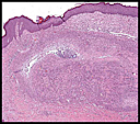
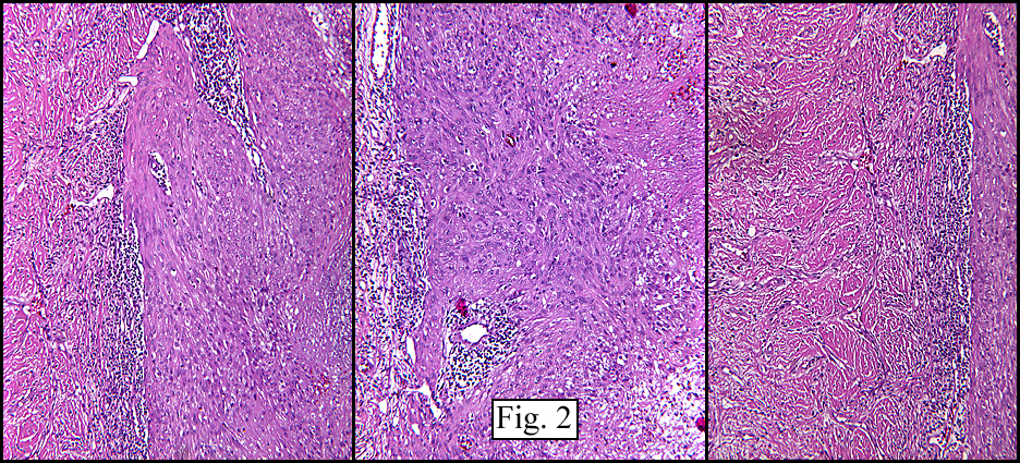
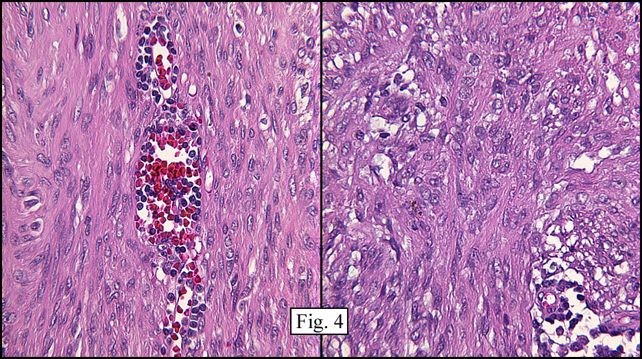
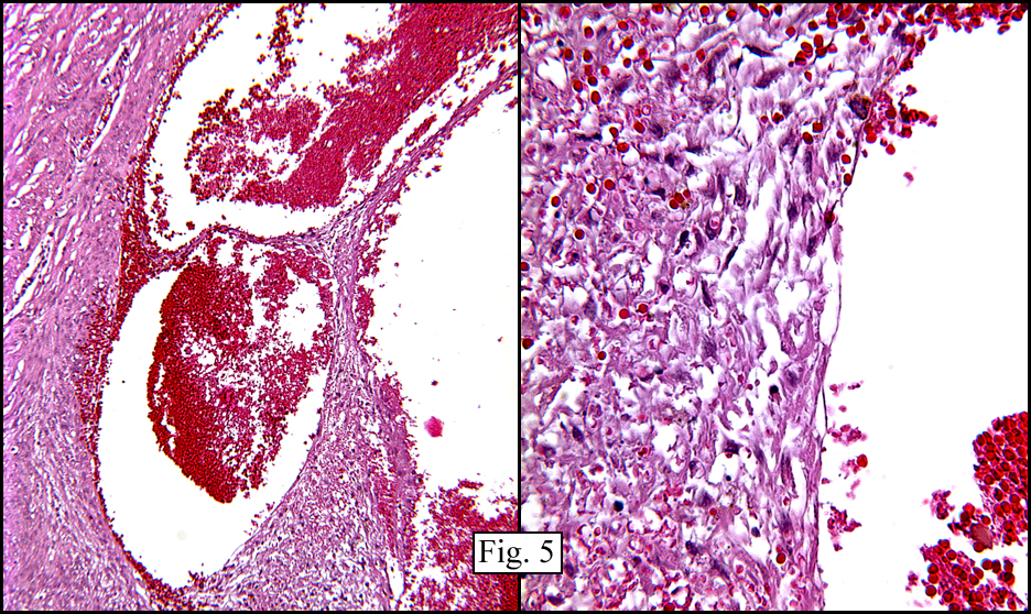
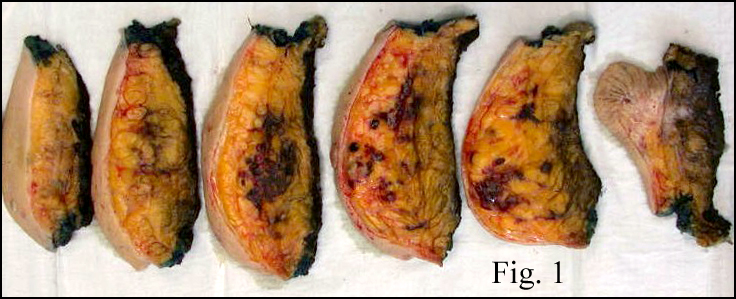
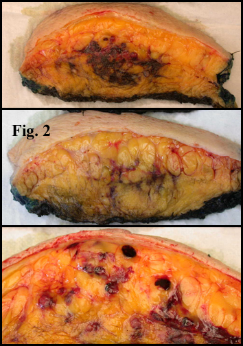
The superficial dermis resembled a simple acrochordon however the deeper dermis had sheets and nodules of spindled cells arranged in short fascicles (Figs. 1,2) These fusiform cells had ovoid nuclei and tapered eosinophilic cytoplasm (Figs. 3,4). At the periphery was a cuff of lymphocytes. There were also cystic spaces filled with erythrocytes (Fig. 5). Hemosiderin pigment was focally evident.“October 2007: A 25 year old man with a nodule in the arm”Continue reading
 History: A 41 year-old man presented in orthopedics clinic with a complaint of a slow-growing mass in his left calf. The patient was a long-haul trucker who stated that he first began to notice a gradual enlargement of his calf following minor trauma sustained while falling off his truck. He complained of a moderate amount of aching pain (rated 2-3/10 in intensity) without radiation. He indicated that the pain was not made worse with activity and was intensely felt with flexion of the knee.
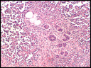 History: An 82 year old woman presented with multiple nodules in the left breast eight months following local resection of a cutaneous angiosarcoma in the same breast. A simple mastectomy was performed but she died the following day due to cardiac complications.
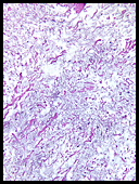 History: A 43-year-old male presented with a mass in his left mid-back that had been present for approximately two years. When excised, the mass was well-circumscribed and measured 5.5 x 4.0 x 2.5 cm. It was found to be superficial and did not involve bone or muscle.