April 2012: Hepatomegaly in a 14 month old girl

History: A 14 month old girl presented with hepatomegaly, with the palpable liver margin 5 cm below the inferior mid-clavicular line. Shortly after presenting, the patient died, and an autopsy described an enlarged liver weighing 1015 grams. Sectioning showed a yellow-green, 8 x 4 cm variegated mass with cystic, gelatinous, and focally calcified areas, located anteriorly between the left and right lobes.

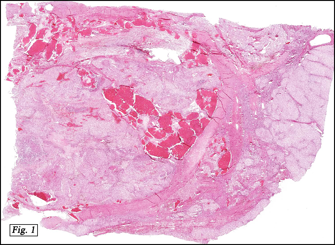
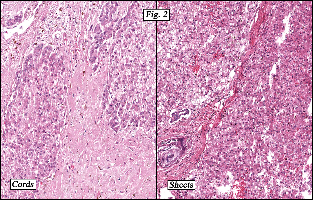
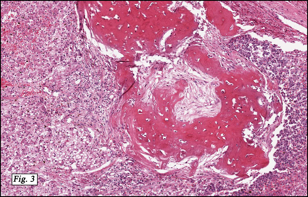
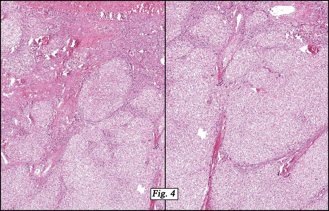
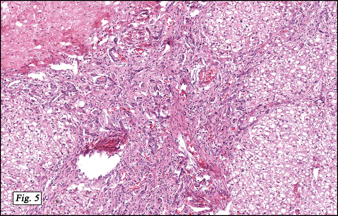
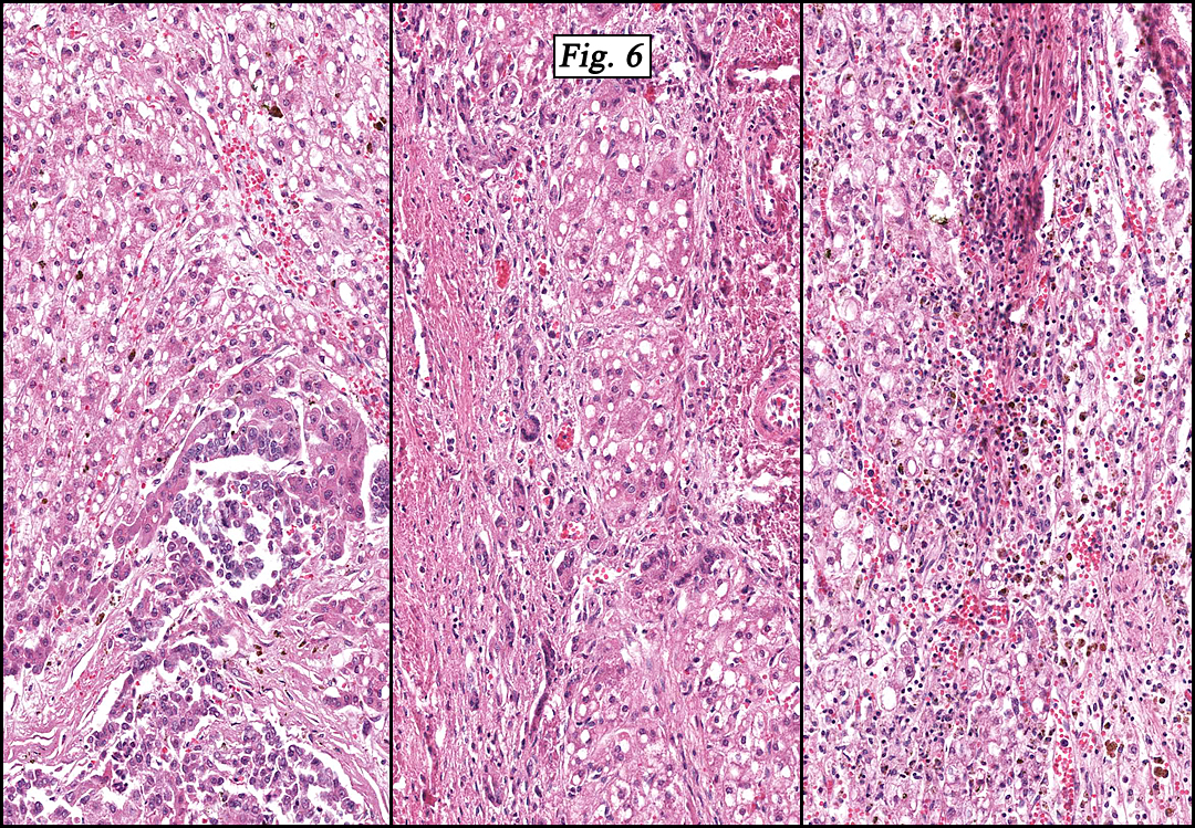
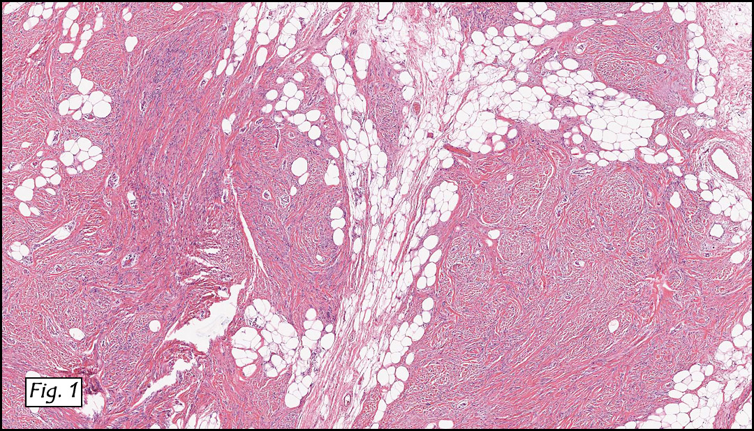
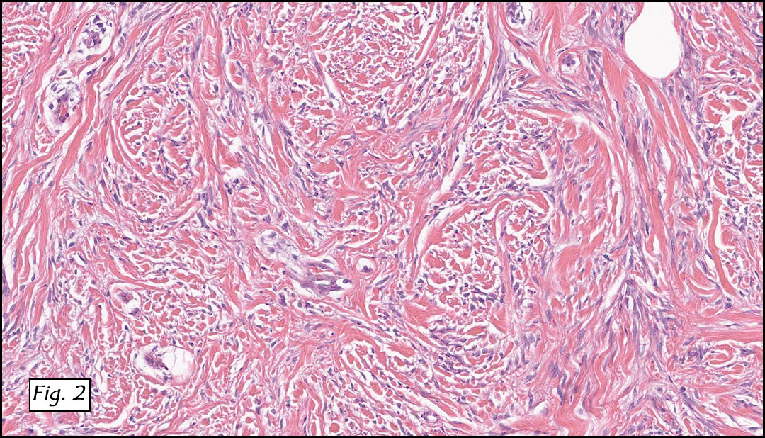
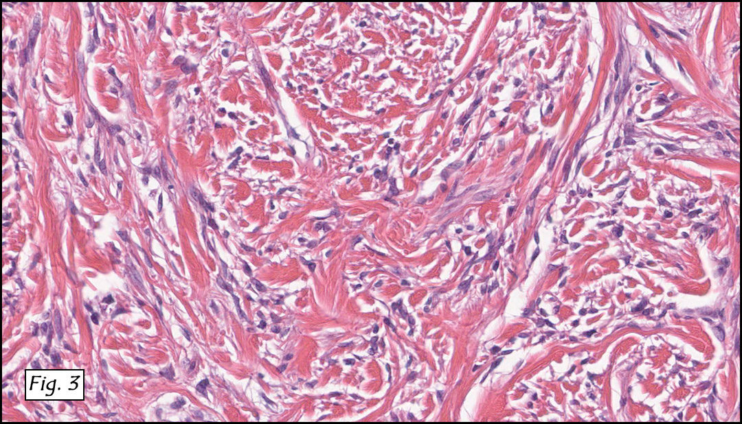
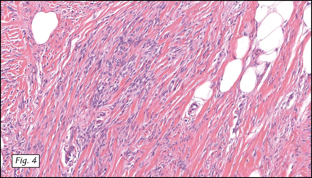
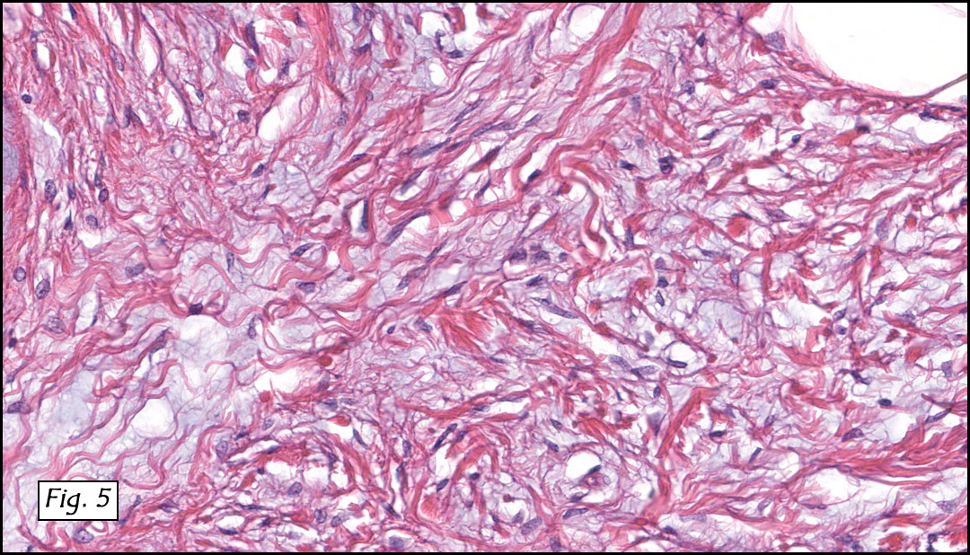
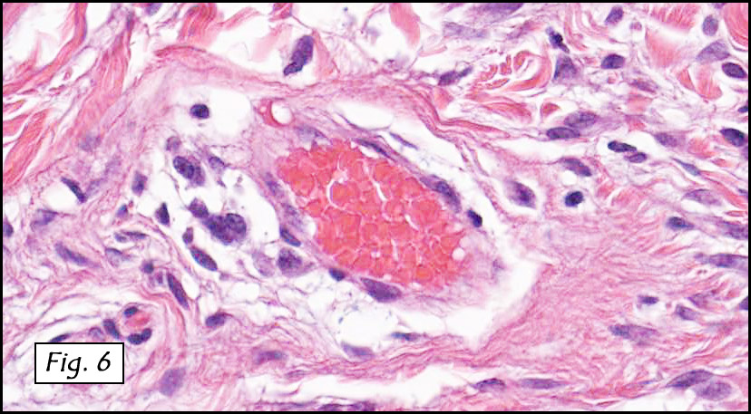
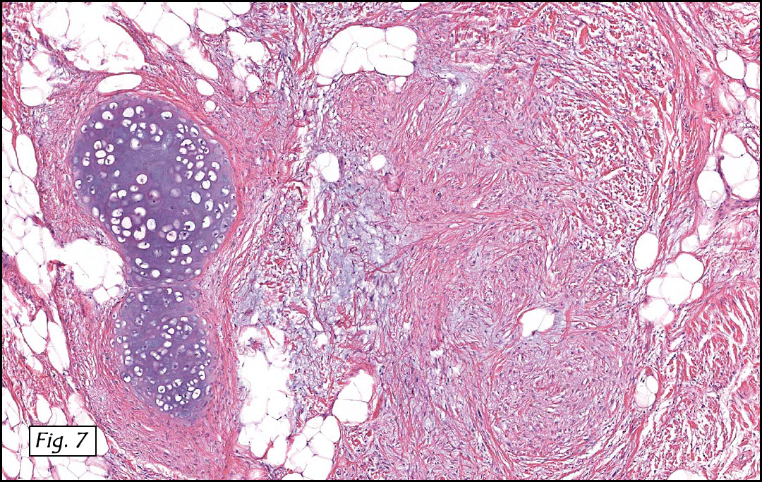
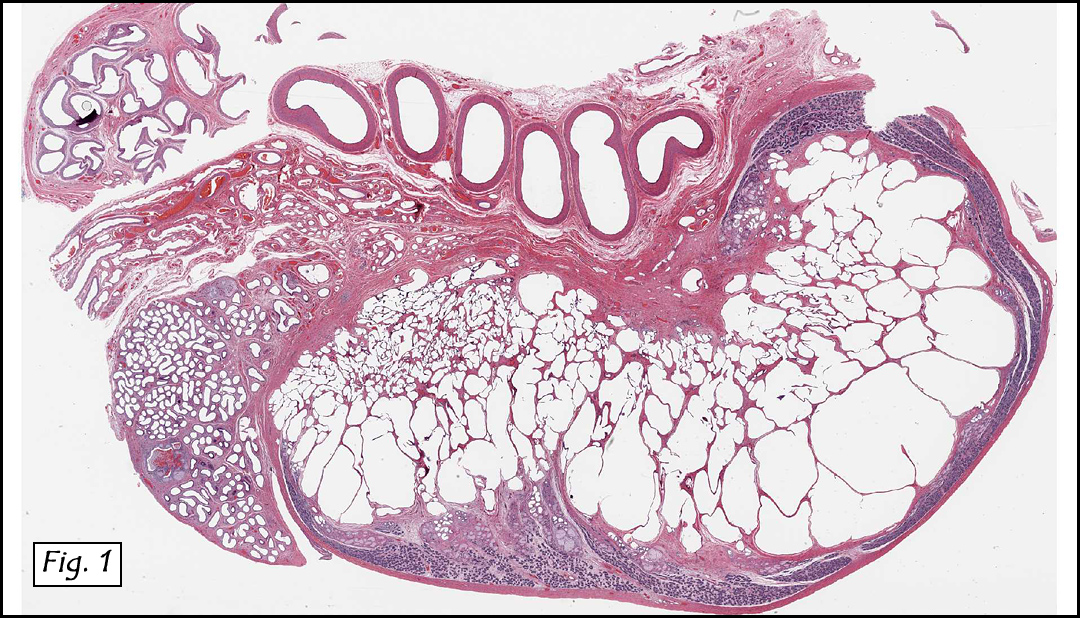
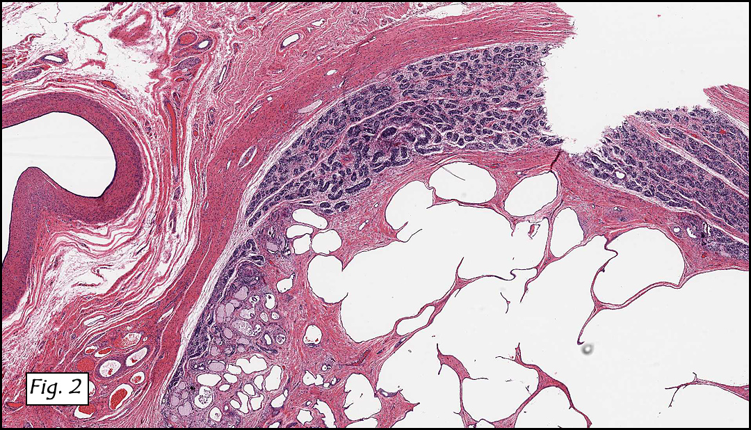
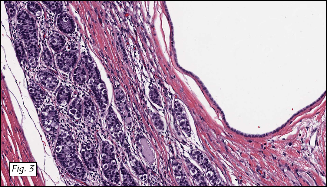
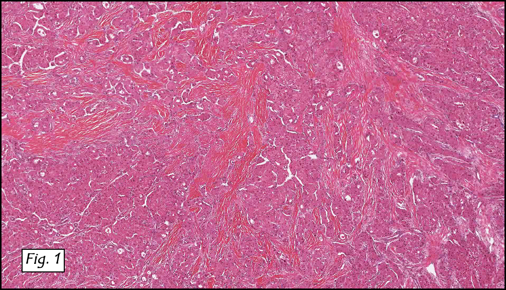
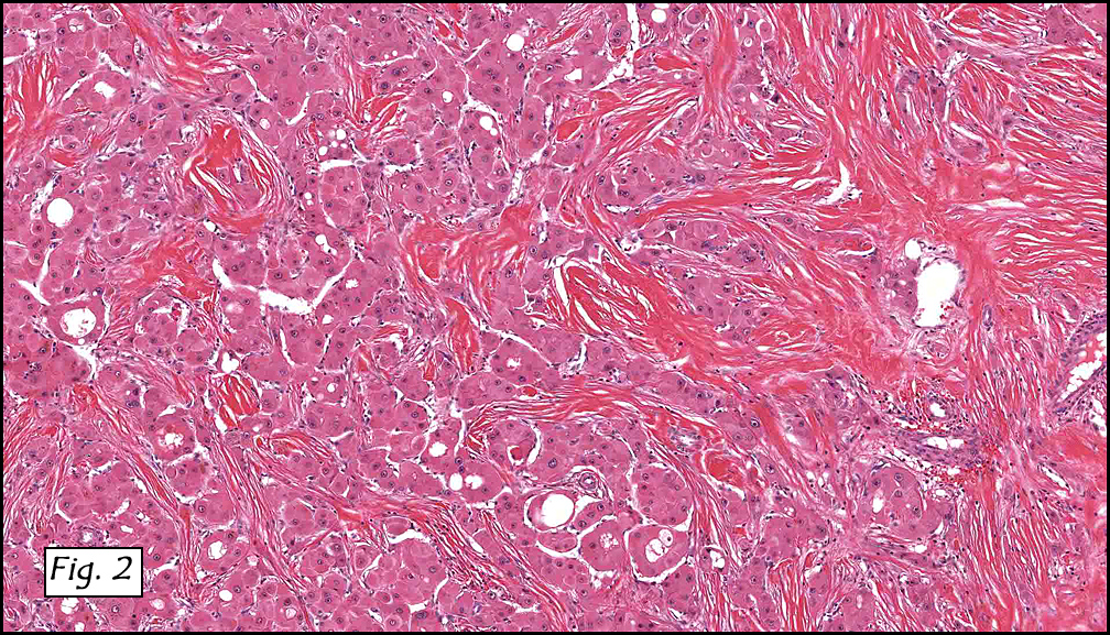
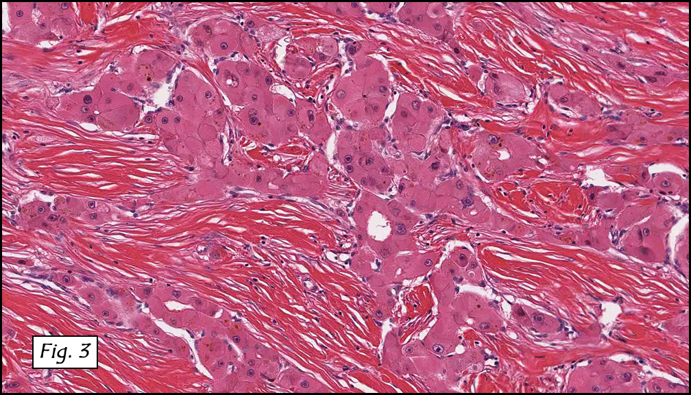
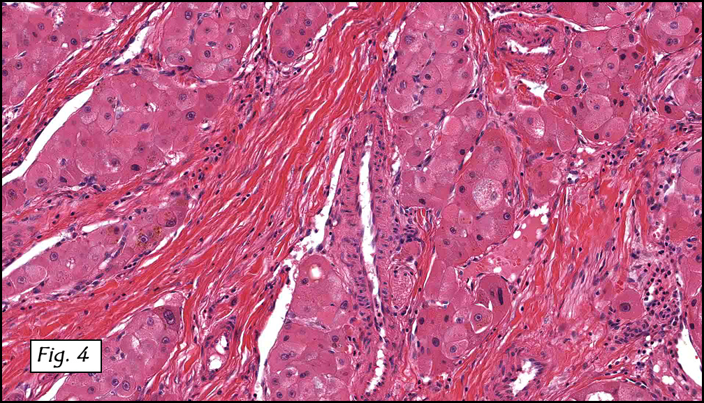
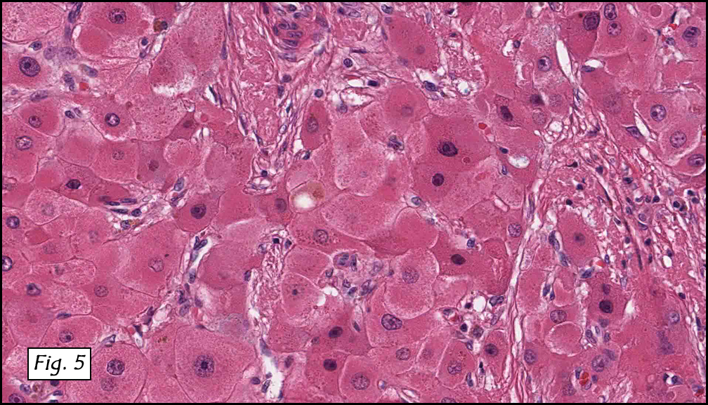
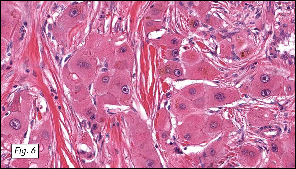
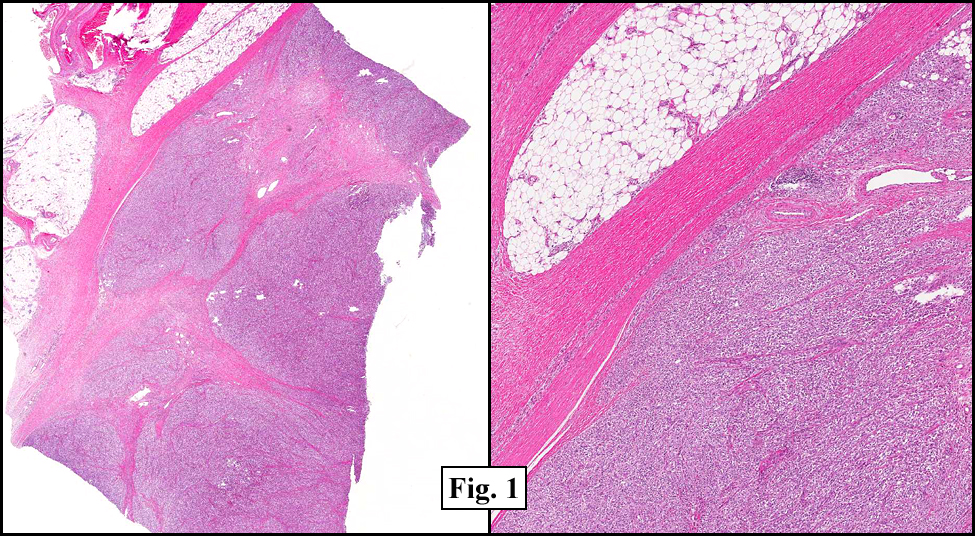
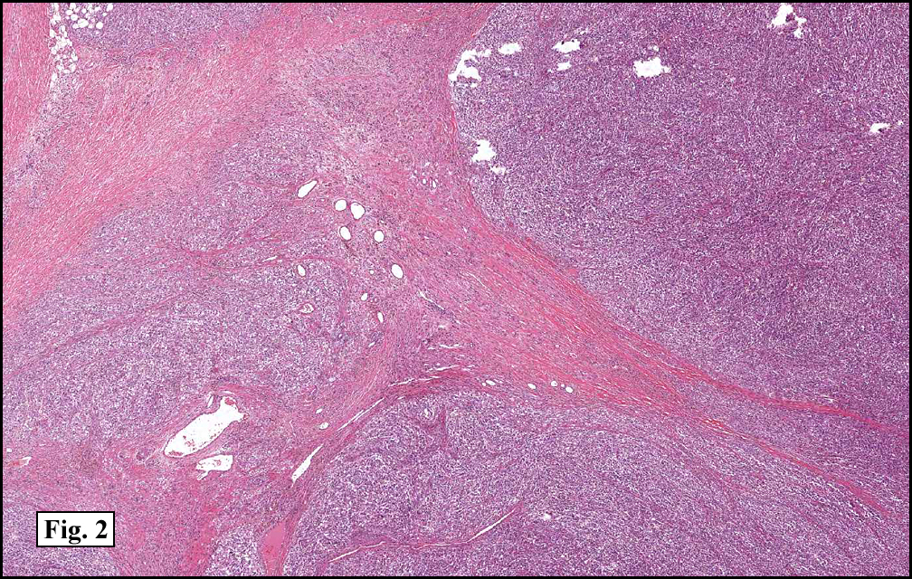
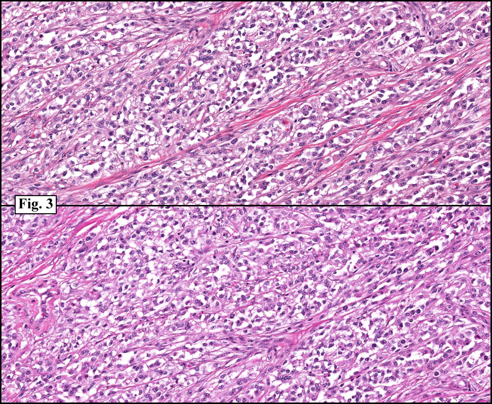
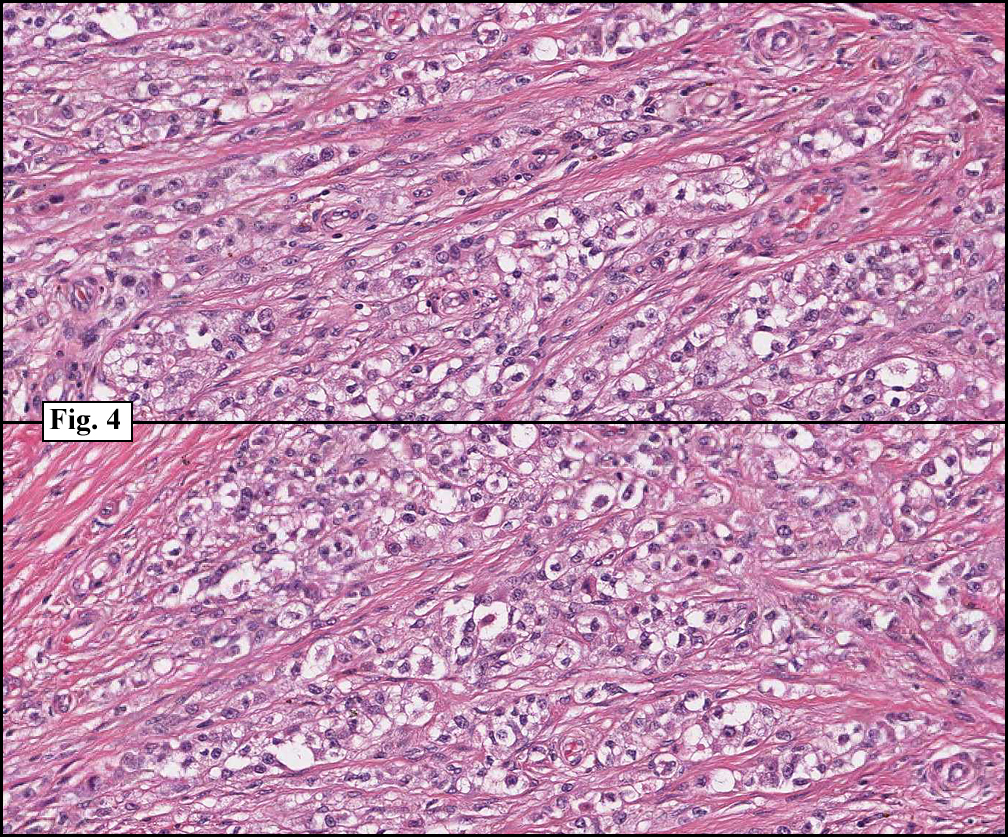
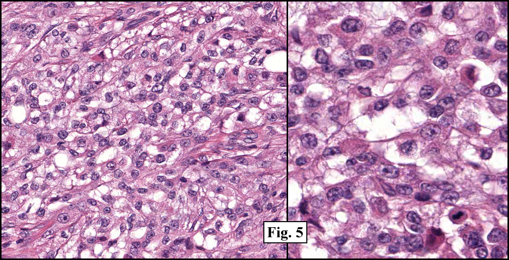
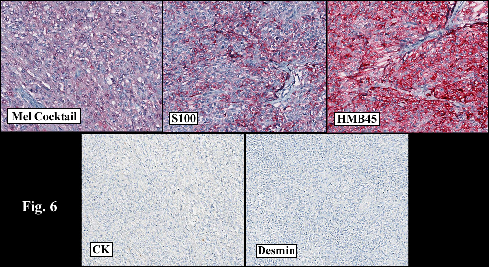
Microscopically, the tumor was partially encapsulated, lobulated, and exhibited patchy light and dark regions. Some areas were telangiectatic with numerous expanded blood vessels filled with erythrocytes (Fig. 1). Growth was usually as cords and sheets (Fig. 2). Abundant osteoid could be seen adjacent to fibrous septa and loose mesenchymal elements (Fig. 3). The lighter staining regions consisted of more mature hepatocytes growing in a lobular pattern separated by fibrous septa (Fig. 4). Proliferation of bile ducts could also be seen (Fig. 5). Also present were clusters of blastemal cells with an increased nuclear to cytoplasm ratio with greater nuclear pleomorphism and prominent nucleoli (Fig. 6, left). Rare multinucleated tumor giant cells were present (Fig. 6, center), and some regions were suggestive of extramedullary hematopoiesis (Fig. 6, right). There were significant areas of hemorrhage and hemosiderin deposition. Mitoses were rare.

Diagnosis: “Hepatoblastoma, Mixed Pattern”

Shino Magaki MSIV, Anwar Raza MBBS, and Donald R. Chase MD
Department of Pathology and Human Anatomy,
Loma Linda University and Medical Center
California Tumor Tissue Registry, Loma Linda, California

Discussion: Hepatoblastoma (HB) is the most common liver malignancy in children, accounting for approximately two-thirds of all primary pediatric hepatic malignancies and 1% of all pediatric malignancies. Two-thirds are diagnosed prior to the age of 2 years while 90% are diagnosed before the age of 4 years, with the mean age at diagnosis of 18 months. HB is rarely seen at birth or in young adults, and males are twice as likely as females to be affected. HB was probably first described by Misick in 1898. The etiology is unknown but 5% of cases are associated with a congenital abnormality such as Beckwith-Wiedemann syndrome or familial adenomatous polyposis. Common cytogenetic alterations include trisomy 2, 8 and 20. Activating mutations in the beta-catenin/Wnt pathway is seen in about half of the cases. HB has also been associated with low birth weight and has been increasing in incidence over the past few decades, possibly due to improved survival of low birth weight infants.

Clinically, patients often present with a rapidly enlarging abdominal mass, failure to thrive, and weight loss. Less common symptoms include vomiting, diarrhea, and jaundice. Virilization and precocious puberty from human chorionic gonadotropin secreted by the tumor have been reported. Serum alpha-fetoprotein (AFP) is elevated in 90% of cases and is a useful marker for tumor recurrence. Tumors that do not express AFP are thought to be more biologically aggressive, and serum levels less than 100 ng/mL at the time of diagnosis indicate a poorer prognosis. On ultrasound, HB usually appears as a well demarcated solitary mass although it can be multifocal. CT often shows contrast enhancement and calcification. On MRI the purely epithelial subtype appears homogenous and hypointense on T1 and hyperintense on T2 weighted images, whereas the mixed epithelial and mesenchymal subtype is more heterogeneous.

Grossly, HB typically occurs as a single mass on a background of non-cirrhotic liver and may contain areas of hemorrhage and necrosis. The tumor tends to be well circumscribed but unencapsulated or partially encapsulated and have a variegated appearance depending on the proportion of its components. In pure epithelial HB, the tumor is soft, fleshy and tan-white. Mixed tumors show a heterogeneous surface, tend to be firmer and often are calcified. Frequently large at detection, HB can measure up to 20-25 cm in diameter (average 10-12 cm), and usually weigh over 1 kg. They are most common in the right lobe.

Histologically, HBs are classified into pure epithelial or mixed epithelial-mesenchymal subtypes with six main histological patterns:

The fetal pattern (30%) demonstrates tumor cells which are uniform and similar in size to normal hepatocytes with a slightly increased nuclear:cytoplasm (N:C) ratio. Cells form trabeculae 2-3 cells thick that are separated by sinusoids. An alternating light and dark staining pattern due to lipid and glycogen, respectively, is often seen. Portal tracts and bile ducts and ductules are absent, and mitoses are rare (

In the embryonal pattern (20%), cells are less differentiated, have a higher N:C ratio with coarser chromatin, and may form sheets. More mitoses and necrosis are seen.

Cells are the least differentiated in the anaplastic small cell pattern (3%), forming sheets of small cells with scant cytoplasm and prominent nucleoli, resembling small blue cell tumors elsewhere. Tumor cells are positive for CK and negative for bile.

In the macrotrabecular pattern (3%), fetal or embryonal cells form trabeculae 10 or more cells thick throughout the tumor.

The mixed epithelial-mesenchymal pattern (44%) is characterized by the presence of both epithelial and mesenchymal cells. The mesenchymal component may show primitive stellate-shaped cells with little cytoplasm or demonstrate chondroid and rhabdomyoblastic differentiation. Osteoid is the most common heterologous element and is more frequent found after chemotherapy. The mixed pattern can be further subdivided into tumors with (10%) or without (34%) teratoid features such as skeletal muscle, keratinized squamous epithelium, and intestinal epithelium.

Differential Diagnoses:

Although Hepatocellular carcinoma (HCC) may be indistinguishable from a macrotrabecular pattern of HB, other features help differentiate them (Table 1).

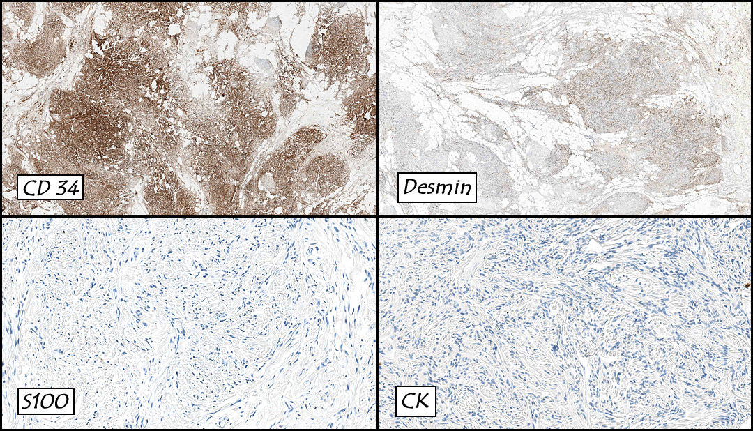
Since small cell tumors, including neuroblastoma, rhabdomyosarcoma, and lymphoma, resemble the anaplastic small cell pattern in HB, the presence of more typical, better differentiated foci should be looked for. Immunohistochemistry is useful, with HB showing CK positivity and neurofilament, desmin, leukocyte common antigen (CD45) negativity; neuroblastoma is positive for neurofilaments, rhabdomyosarcoma is positive for desmin, and lymphoma is CD45 positive.

Table 1. Hepatoblastoma and hepatocellular carcinoma in the pediatric age group (

Hepatoblastoma Hepatocellular carcinoma
Incidence Most common primary liver malignancy
in children (0.5-1.5 diagnoses per
million annually)
2nd most common primary liver
malignancy in children
Age 90%<5 yrs Most >10 years
M:F ratio 1.2-3.6:1 1.7:11:1
Association with genetic
syndromes
Familial adenomatous polyposis,
Beckwith-Wiedemann syndrome,
trisomy 18
Glycogen storage disease type
IA, hereditary tyrosinemia,
Fanconi syndrome
Other associations Parental exposure to environmental
toxins, low birth weight
Cirrhosis, viral hepatitis
Serum AFP elevations 90%, usually highly elevated 60-80%
Single mass 70-80% 15-40%
Trabeculae Usually 2-3 cells thick with occasional
macrotrabeculae
Usually>2-3 cells thick
Mitoses Rare Common
Tumor giant cells Rare Common
Cytoplasmic inclusions
(e.g. Mallory’s hyaline,
alpha-1-anti-trypsin)
Absent Common
Extramedullary
hematopoiesis
Common Absent


Treatment consists of complete surgical resection with or without neoadjuvant chemotherapy to decrease tumor size prior to surgery. Prognosis is dependent upon the extent of complete resection. At present, the 5-year survival rate is 75%. Liver transplantation is considered when the tumor is limited to the liver but unresectable. The pure fetal pattern is thought to predict a more favorable outcome while the pure anaplastic small cell pattern, increased mitoses, and vascular invasion suggest a poorer prognosis. Most frequent metastatic sites are the regional lymph nodes and lungs.

Suggested Reading:

Beach R, Betts P, Radford M, Millward-Sadler H. Production of human chorionic gonadotrophin by a hepatoblastoma resulting in precocious puberty. J Clin Pathol. 37:734-37, 1984.

Buccoliero AM, Castiglione F, Maio V, Moncini D, Sardi I, Taddei A, Martin A, Messineo A, Taddei GL. Teratoid hepatoblastoma. Fetal Pediatr Pathol. 27:274-81, 2008.

Das CJ, Dhingra S, Gupta AK, Iyer V, Agarwala S. Imaging of paediatric liver tumours with pathological correlation. Clin Radiol. 64:1015-25, 2009.

Litten JB, Tomlinson GE. Liver tumors in children. Oncologist. 13:812-20, 2008.

Maruyama K, Ikeda H, Koizumi T. Hepatoblastoma associated with trisomy 18 syndrome: a case report and a review of the literature. Pediatr Int. 43:302-5, 2001.

Mills SE, Carter D, Greenson JK, Oberman HA, Reuter V, Stoler MH. Sternberg’s Diagnostic Surgical Pathology (4th ed) Philadelphia: Lippincott. 1743-47, 2004.

Reynolds P, Urayama KY, Von Behren J, Feusner J. Birth characteristics and hepatoblastoma risk in young children. Cancer. 100:1070–76, 2004.

Sallam A, Paes B, Bourgeois J. Neonatal hepatoblastoma: two cases posing a diagnostic dilemma, with a review of the literature. Am J Perinatol. 22:413-19, 2005.

Schnater JM, Köhler SE, Lamers WH, von Schweinitz D, Aronson DC. Where do we stand with hepatoblastoma? A review. Cancer. 98:668-78, 2003.

March 2012: A breast mass in a 62 year old woman

History: A 62 year-old woman underwent surgical resection of a 55 gram, 6.2 x 5.4 x 4.2 cm encapsulated right breast mass. The cut surface was tan with variably-sized pink and tan soft tissue nodules ranging from 1.0 to 2.2 cm.

Microscopically the moderately cellular soft tissue tumor consisted of slender fibroblast-like spindle cells admixed with dense collagenous bands (Fig. 1). The cells were arranged in sheets and small fascicles which were intermixed with interweaving fibrous stroma and mature adipose tissue (Figs. 2, 3, 4). Mucoid changes were also seen (Fig. 5) as well as perivascular mast cells (Fig. 6). Chondroid metaplasia was focally present (Fig. 7). Mitoses were not increased in number and there was no evidence of significant atypia or necrosis. Immunostains showed decoration for CD31 (4+) and desmin (1+) and were negative for S100 and cytokeratin (Fig. 8).

Diagnosis: “Myofibroblastoma with focal chondroid metaplasia”

Kate Grogan, MD, and Donald R. Chase, MD
Department of Pathology & California Tumor Tissue Registry
Loma Linda University and Medical Center, Loma Linda, California

Discussion: First described by Wargotz et al. in 1987, myofibroblastomas (MFBs) are uncommon mesenchymal neoplasms which typically occur in variably aged patients (25-85 years) with an average of 63 years. Although originally reported as having a higher incidence in men, they are currently thought to occur in equal gender frequency. They traditionally present as a well-circumscribed, mobile unilateral mass of several months duration. A single case of synchronous bilaterality has been reported. Grossly, MFBs are well circumscribed and often have a delicate fibrous psuedocapsule. The cut surface is typically gray-pink and vaguely lobular.

Microscopically, MFBs are variably cellular and are comprised of myofibroblast-resembling spindle cells which have tapered nuclei with finely dispersed chromatin, small nucleoli and a moderate amount of eosinophilic to amphophilic cytoplasm. The spindle cells often have wavy contours and are arranged in short fascicles. The stroma contains coarse bands of hyalinized collagen and conspicuous mast cells, admixed with a variable amount of adipose tissue.

Mammographically, MFBs appear as lobulated, well-demarcated and homogenous densities. Microcalcifications are absent.

The majority of cases show staining for CD34, vimentin and ER/PR. Desmin, caldesmon and androgen receptors are variably positive, and S-100 and cytokeratin are typically negative.

Cytogenetic findings: Partial monosomy 13q has been detected in two cases, as well as partial monosomy 16q in one of these two cases. Curiously, these same rearrangements are characteristic of spindle cell lipomas.

Differential Diagnosis:

• Fibromatosis of the breast typically presents as an ill-defined mass which may be associated with skin dimpling, nipple retraction and a stellate mammographic appearance. These entities will also be CD34 negative, whereas the majority of myofibroblastomas demonstrate CD34 positivity.

• Nodular fasciitis, although CD34 positive, typically demonstrates a more infiltrative pattern and has stroma that is focally pushed apart by pools of mucinous material.

• Myoepitheliomas are rare spindled lesions, which usually showing mild to moderate nuclear pleomorphism. These lesions are traditionally keratin, vimentin and EMA positive, and CD34 and ER/PR negative.

• Well-differentiated metaplastic carcinoma of the breast and stromal sarcomas usually present as ill-defined infiltrating masses, whereas myofibroblastomas are generally well-circumscribed and rarely invade adjacent breast tissue.

• Myofibroblastomas are readily distinguished from fibroadenomas due to their lack of ductal structures.

Conservative, but complete surgical excision is the treatment of choice. MFBs rarely recur and do not have metastatic potential.

Suggested Reading:

Tavassoli F, Eusebi V. Tumors of the Mammary Gland (4th edition). Washington DC: American Registry of Pathology, 2009.

Weiss S, Goldblum J. Enzinger and Weiss’s Soft Tissue Tumors (5th edition). Philadelphia: Mosby/Elsevier Inc. 535-6, 2008.

Pauwels P, Sciot R, Croiset F, Rutten H, Van den Berghe H, Dal Cin P. Myofibroblastoma of the breast: genetic link with spindle cell lipoma. J Pathol 2000; 191 (3): 282-5.

Tavassoli F. Pathology of the Breast (2nd edition). Stamford, Connecticut: Appleton & Lange. 686-691, 1999.

Magro G. Mammary myofibroblastoma: a tumor with a wide morphologic spectrum. Arch Pathol Lab Med 2008; 132(11): 1813-20.

February 2012: A 3 year old boy with an enlarged testicle

History: A three-year-old boy underwent orchiectomy for a cystic scrotal mass. Microscopically, the rete testis was found to be markedly distorted by cystic dilatation which compressed the adjacent seminiferous tubules (Figs. 1, 2). The cystic rete lining was of low cuboidal epithelium (Fig. 3).

Diagnosis: Cystic dysplasia of the testis
Ellsworth BD, Zuppan C, Chase DR
Department of Pathology & California Tumor Tissue Registry
Loma Linda University Medical Center, Loma Linda, California

Discussion: Cystic dysplasia of the rete testis is a rare anomaly that usually presents in young boys as a unilateral painless scrotal mass. Approximately 50 cases have been documented. The cystic changes are well-appreciated by ultrasound and may be suspicious for malignancy. On ultrasound, variably-sized cysts are seen within the mediastinum testis with no changes in the testicular tissue and epididymis. Histologically, the rete testis is markedly dilated and cystic but lined by flat cuboidal epithelium. This process compresses and often obliterates the nearby testicular parenchyma which can render the testis infertile. Ipsilateral urinary tract anomalies are almost invariably present, most commonly renal agenesis or dysplastic kidney. For this reason, diagnosis of cystic dysplasia of the rete testis warrants further imaging of the urinary tract to look for additional anomalies.

The dysplastic changes are thought to occur as a result of improper development of the mesonephric duct. During normal embryogenesis, the mesonephric duct gives rise to the ureteral bud at its cephalic end which stimulates nephrogenesis of the metanephric blastema. From its caudal end, the extra testicular ducts (efferent ducts, epididymis, and vas deferens) derive. Defects in the mesonephric duct can result in anomalies in any of these structures. Normally, the rete testis and testicular cords (derived from the germinal epithelium) anastomose with the mesonephric-derived extra testicular ducts at the efferent ductules. Failure of this process causes anomalies and results in cystic distortion of the rete testis. With this relationship in mind, Nistal et al have theorized that inappropriate (pre-pubertal) secretory activity by the rete testis and fluid entrapment are responsible forecyst development, a process that occurs until puberty when the seminiferous tubules canalize and develop lumens allowing the fluid to relocate. This process seems to have occurred in two reported cases of resolved cystic dysplasia. In both cases, resolution was relatively sudden and occurred near the time of (reference – Thomas 2003).

Treatment commonly consists of unilateral orchiectomy. Testis-sparing enucleation is occasionally performed to spare hormonally active tissue, but yields a chance of recurrence. As no malignant behavior has been observed and two reported cases have regressed, conservative therapy or watchful waiting has been suggested.

Suggested Reading:

Keetch DW, McAlister WH, Manley CB, Dehner LP. Cystic dysplasia of the testis. Pediatr Radiol. 1991;21:501-503.

Nistal M, Regadera J, Paniagua R. Cystic dysplasia of the testis. Light and electron microscopic study of three cases. Arch Pathol Lab Med.1984;108:579-583.

Thomas AD, Wu H, Canning DA, et al. Spontaneous regression of cystic dysplasia of the testis. J Urol. 2003;169:645.

Wojcik LJ, Hansen K, Diamond DA, et al. Cystic dysplasia of the rete testis: a benign congenital lesion associated with ipsilateral urological anomalies. J Urol. 1997;158:600-604.

Zaragoza M, Buckler L, Parikh M. Cystic dysplasia of the testis: An unusual cause of a pediatric scrotal mass. Urology. 1996;47:244-247.

Robson W, Thomason M, Minette L. Cystic dysplasia of the testis associated with multicystic dysplasia of the kidney. Urology. 1998;51:477-479.

Camassei F, Francalanci P, Ferro F, et al. Cystic dysplasia of the rete testis: Report of two cases and review of the literature. Pediatr Dev Pathol. 2002;5:206-210.

Erberli D, Gretener H, Dommann-Scherrer C, et al. Cystic dysplasia of the testis: A very rare pediatric tumor of the testis. Urol Int.2002;69:1-6.

Fisher JE, Jewett Jr TC, Nelson SJ, et al. Ectasia of the rete testis with ipsilateral renal agenesis. J Urol 1982;128:1040-3.

January 2012: A 30 year old woman with a mass in a transplanted liver

History: A 30 year-old Caucasian woman, status-post hepatectomy for a liver malignancy, presented four years later with an enlarging 4.5 cm right-sided mass in her “new” transplanted liver. The tumor was 4.2 x 3.1 x 2.2 cm and was well-circumscribed, yellow-green, and had a central scar.

Microscopically, the tumor was solid, with prominent ramifying bands of fibrous tissue (Figs. 1, 2) which encased atypical hepatocytes arranged in medium to large-sized nests (Figs. 3, 4). The cells had large vesicular nuclei with prominent nucleoli and abundant granular eosinophilic cytoplasm (Figs. 5, 6). Bile pigment was focally present (Fig. 6). The tumor extended into perihepatic fat (Figs. 7). Neither hemorrhage nor necrosis were seen.

Diagnosis: Hepatocellular carcinoma, fibrolamellar variant

Kate Grogan, MD, and Donald R. Chase, MD
Department of Pathology & California Tumor Tissue Registry
Loma Linda University and Medical Center, Loma Linda, California

Discussion: First described by Edmondson in 1956, fibrolamellar hepatocellular carcinoma (also known as polygonal cell type HCC) is an uncommon variant of hepatocellular carcinoma which typically occurs in young patients mean age of 26 years) who usually lack the common risk factors associated with conventional HCC. Although it was previously thought that there was a slight female predominance, current studies suggest that it occurs in equal frequency among men and women. Almost half of liver cell carcinomas in the United States occur in patients under the age of 35, and most are of this type. It is associated with a better prognosis overall than patients with HCC and cirrhosis, and the prognosis appears to be similar to that in patients with typical HCC without cirrhosis. Since it was first described, more than 180 cases have been described in the literature. Grossly, the tumors are solitary, well-circumscribed, yellow-brown and vaguely nodular. Frequently, they may have a fibrous central scar (similar to the central scar seen in focal nodular hyperplasia). Serum AFP levels are typically within normal limits.

Microscopically, the tumor consists of well-differentiated polygonal cells with abundant granular eosinophilic cytoplasm, which are arranged in nests, sheets or cords. The cytoplasm often contains pale bodies or PAS positive hyaline globules. In the background are dense acellular collagen bundles. As in other forms of HCC, tumors may also have focal acinar structures, bile, multinucleated tumor cells, copper, or fat.

Fibrolamellar carcinomas typically express both hepatic (CK 8 and 18) and biliary (CK 7, 19) cytokeratins. Recent studies have shown that expression of Hep-Par-1 and CD99 may be supportive of the diagnosis. Neuroendocrine markers may be focally positive, but they are of uncertain significance.

Cytogenetic findings: These tumors are often diploid, and overall show fewer chromosomal abnormalities than do classic HCC. Tumors with no cytogenetic abnormalities appear to behave less aggressively.

Differential Diagnosis:

• Hepatocellular carcinoma, sclerosing variant may be histologically similar, however, the tumor cells are typically smaller and pseudoglandular formation is common. Also diffuse lamellar fibrosis combined with oncocytic cellular features is not typical of usual hepatocellular carcinoma
• Cholangiocarcinoma may show glandular formation, and are usually mucin positive.
• Metastatic tumors with extensive fibrosis may mimic the collagen seen in fibrolamellar carcinoma, but is usually arranged in a more haphazard manner and lacks the directional growth characteristic of fibrolamellar carcinoma.

Complete resection of the involved lobe is the therapy of choice. When tumor is deemed unresectable, liver transplantation is an option (as in this case). The tumor metastasizes primarily to regional lymph nodes, peritoneum and lung.

Suggested Reading:

Odze R, Goldblum J. Surgical Pathology of the GI Tract, Liver, Biliary Tract, and Pancreas (2nd edition). Philadelphia: Saunders/Elsevier Inc. 1306-7, 2009.

Liu s, Chan KW, Wang B, Qiao L. Fibrolamellar hepatocellular carcinoma. Am J Gastroenterol 2009; 104: 2617-24.

Kakar S, Burgart LJ, Batts KP et al. Clinicopathologic features and survival in fibrolamellar carcinoma: comparison with conventional hepatocellular carcinoma with and without cirrhosis. Mod Pathol 2005; 18: 1417-23.

MacSween R, Burt A, Portmann B, Ishak K, Scheuer P, Anthony P. Pathology of the Liver (4th edition). London: Churchill Livingstone/Harcourt Publishers Limited. 740-1, 2002.

Torbenson M. Review of the clinicopathologic features of fibrolamellar carcinoma. Adv Anat Pathol 2007; 14: 217-23.

Moreno-Luna LE, Arrieta O, Garcia-Leiva J et al. Clinical and pathologic factors associated with survival in young adult patients with fibrolamellar hepatocarcinoma. BMC Cancer 2005; 5: 142.

December 2011: A 36 year old man with an anterior leg mass

History: A 36 year-old man underwent surgical resection of a 10.0 x 5.5 x 4.0 cm ill-defined left anterior leg mass The excised specimen had a tan, vaguely lobulated cut surface with central hemorrhage It grossly appeared to abut the deep fascial resection margin.

Microscopically, the mass was well-circumbscribed (Fig. 1). It had lobules of nested spindled cells separated by fibrous septae (Fig. 2, 3). The cytoplasm was generally clear and the nuclei were vesicular, many times with prominent nucleoli (Fig. 4). Rare mitotic figures were seen (Fig. 5, right). Immunohistochemistry revealed that the tumor cells were positive for melanoma cocktail, S100, and HMB45, and were negative for cytokeratin and desmin (Fig. 6).

Diagnosis: Clear cell sarcoma

Kate Grogan, MD, and Donald R. Chase, MD
Department of Pathology & California Tumor Tissue Registry
Loma Linda University and Medical Center, Loma Linda, California

Discussion: Initially described by Franz Enzinger in 1965, clear cell sarcoma (CCS or “Malignant melanoma of soft parts”) is a rare aggressive neoplasm of adolescents and young adults (with a mean age of 31 years). It typically presents as a mass in the deep tissues of the lower extremities, usually adjacent to tendons, fascia or aponeuroses.

Histologically, the CCS consists of nests or fascicles of spindled cells with a vesicular chromatin pattern, prominent nucleoli and abundant clear to weakly eosinophilic cytoplasm. In general, CCS tends not to be highly pleomorphic. Mitotic figures are scarce, but multinucleated tumor giant cells are frequently identified. Melanin is present in approximately 50% of cases.

The great majority of cases show diffuse staining for S-100 and frequently also express HMB-45, MiTF and Melan-A.

Cytogenetic findings: One of the most defining diagnostic features of clear cell sarcoma is a reciprocal translocation of chromosomes 12 and 22, more specifically t(12;22)(q13;q12). This translocation results in the fusion of EWS with ATF-1, with the resultant protein mimicking the action of melanocyte stimulating hormone (MSH).

Differential Diagnosis:

  • Malignant melanomas typically involve the dermis, whereas clear cell sarcomas traditionally originate in deep structures, with rare dermal involvement. In ambiguous cases, FISH analysis and RT-PCR can be performed to evaluate for the hallmark EWS-ATF1 gene fusion, which is not seen in malignant melanoma.
  • Cellular Blue Nevi often occur in similar age groups and locations. The blue nevi cells tend to be smaller and nuclei have a less vesicular pattern. Often recurrent cases have pronounced cytologic atypia, and molecular genetic analysis may be indicated to differentiate atypical blue nevus from clear cell sarcoma.
  • Fibrosarcoma, synovial sarcoma and peripheral nerve sheath tumors also have a fascicular growth pattern, however are usually easily distinguished from CCS with immunohistochemistry.
  • Poorly preserved or degenerating specimens may be confused with a round cell sarcoma, particularly alveolar rhabdomyosarcoma, but immunohistochemistry should help delineate them.

In general, complete surgical excision with generous tumor-free margins is the treatment of choice. Chemotherapy is typically ineffective in these patients. Recurrences, which reflect the adequacy of initial excision, range from 14% to 39%. Up to one half of these patients will develop lung or lymph node metastases within 2-8 years. Late metastases (up to 20 years post surgical intervention) have been reported. Prognostic factors include size, necrosis, and nodal metastases.

Suggested Reading:

Chase DR, Rosai J, Argani, P. Clear Cell Sarcoma of Tendon Sheath. CTTR 129th Semi-Annual Slide Seminar. 37-9, 2010.

Antonescu CR, Dal Cin P, Nafa K, Teot LA, Surti U. Fletcher CD, Ladanyi M. EWSR1-CREB1 Is the Predominant Gene Fusion in Angiomatoid Fibrous Histiocytoma. Genes, Chromosomes & Cancer 2001; 46: 1051-1060.

Antonescu CR, Tschernyavsky SJ, Woodruff JM, Jungbluth AA, Brennan MF, Ladanyi M. Molecular Diagnosis of Clear Cell Sarcoma. Detection of EWS-ATF1 and MITF-M Transcripts and Histopathological and Ultrastructural Analysis of 12 cases. Journal of Molecular Diagnostics 2002 4: 44-52.

Enzinger FM, Clear Cell Sarcoma of Tendons and Aponeuroses. An Analysis of 21 cases. Cancer 1965; 18: 1163-74.

Chung EB, Enzinger FM. Malignant melanoma of soft parts. A reassessment of clear cell sarcoma. Am J Surg Pathol 1983; 7: 405-13.

Meis-Kindblom JM. Clear Cell Sarcoma of Tendons and Aponeuroses: A historical Perspective and Tribute to the Man Behind the Entity. Adv Anat Pathol 2006; 13: 286-292.

Weiss S, Goldblum J. Enzinger and Weiss’s Soft Tissue Tumors (5th edition). Philadelphia: Mosby/Elsevier Inc. 926-34, 2008.

Zucman J, Delattre O, Desmaze C, et al. EWS and ATF-1 gene fusion induced by t(12:22) translocation in malignant melanoma of soft parts. Nature Genetics 1993; 4: 341-345.

Scroll to top