November 2012: A five month old girl with a right cheek mass

History: A healthy Caucasian girl was delivered vaginally at full term, with no complications. At two weeks, a soft, non-discrete, non-tender right cheek mass was noted. The mass was non-pulsatile, did not bleed or interfere with feeding or respiration, nor did the size of the mass fluctuate with crying or movement. There were no abnormalities of the overlying skin or mucosa. The mass was closely observed for the next few months. Ultrasound demonstrated a 4 x 3 x 1.4 cm solid whorled mass anterior to the parotid; MRI revealed a well-circumscribed mass involving the right masseter muscle, adjacent to the mandible with increased T2-signal within the bone marrow of the mandible. At five months of age, she underwent intraoral excision of the mass under anesthesia. The patient, now 16 months old, recovered from surgery without complications. Post-operative MRI demonstrated a residual and/or recurrent 3 cm right masseter mass with no other nodules or masses have been observed. No family history of similar lesions or syndromic conditions was elicited.

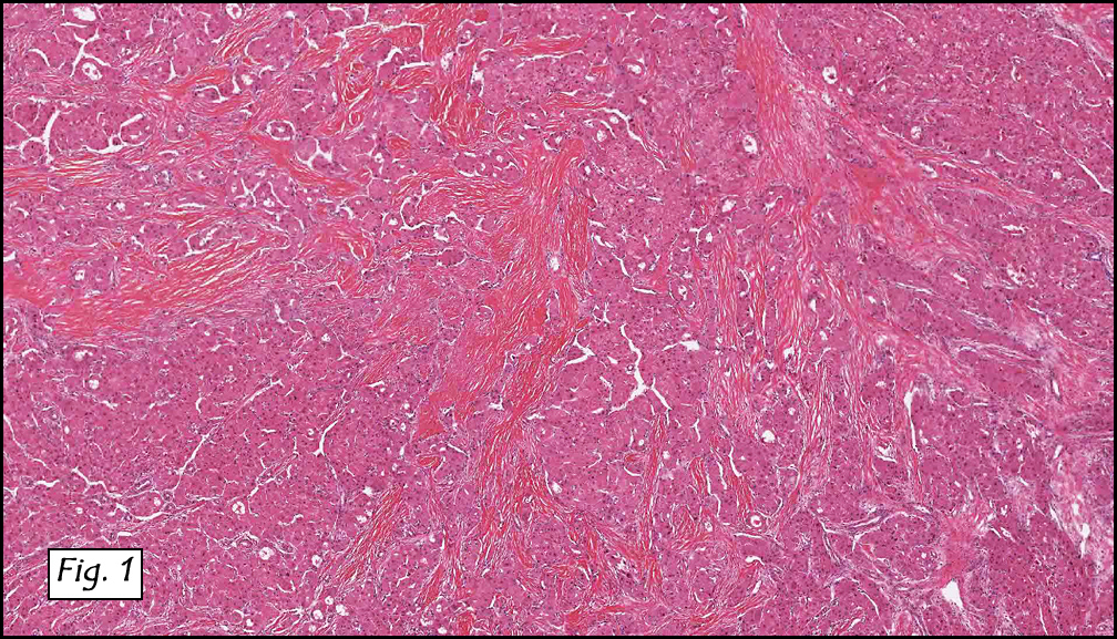
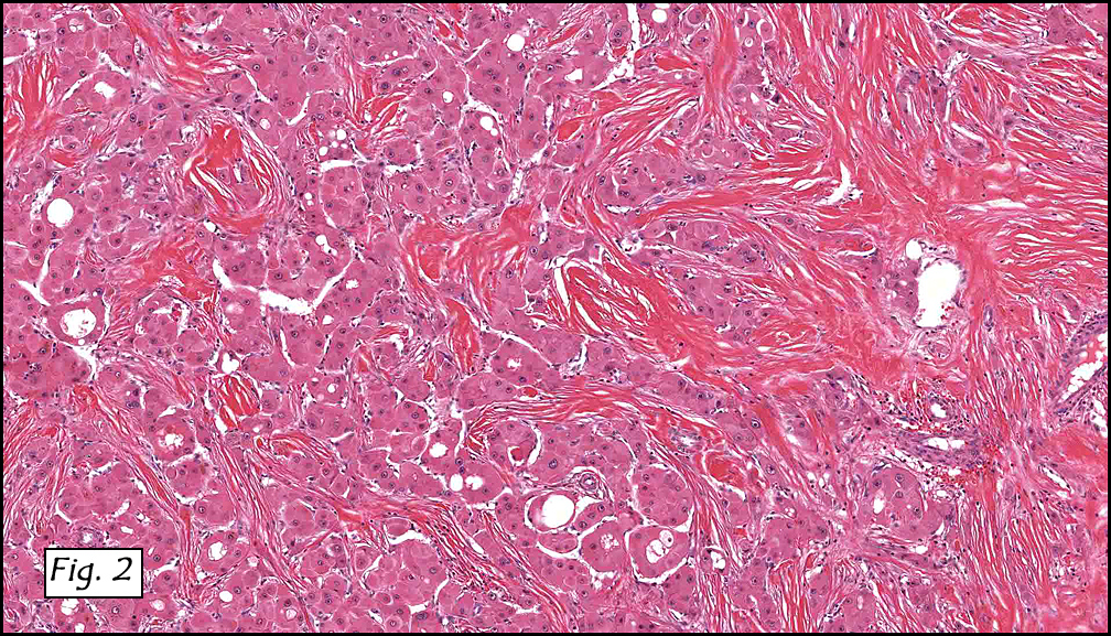
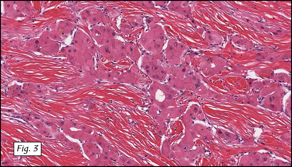
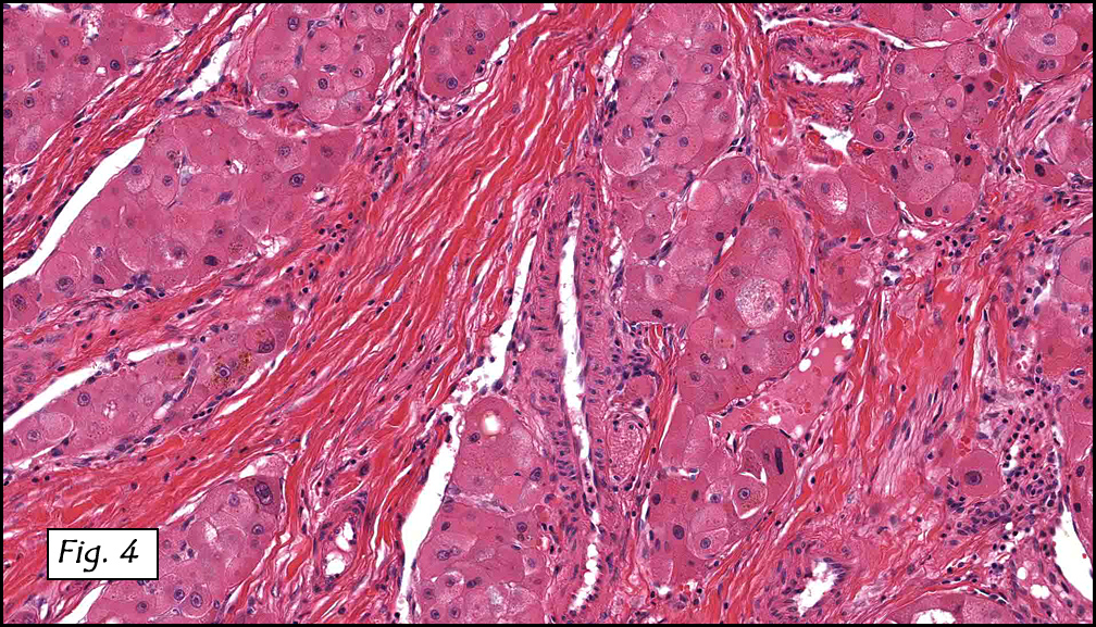
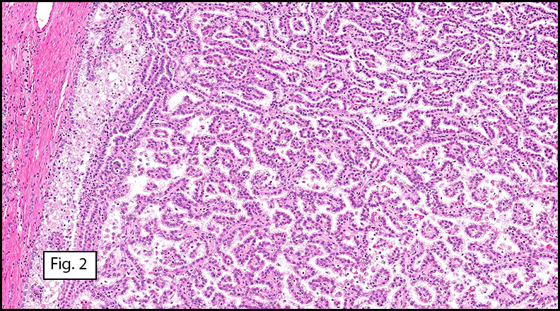
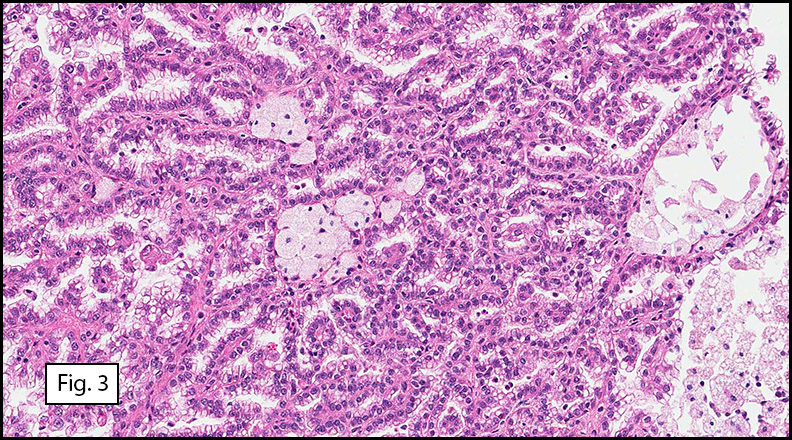
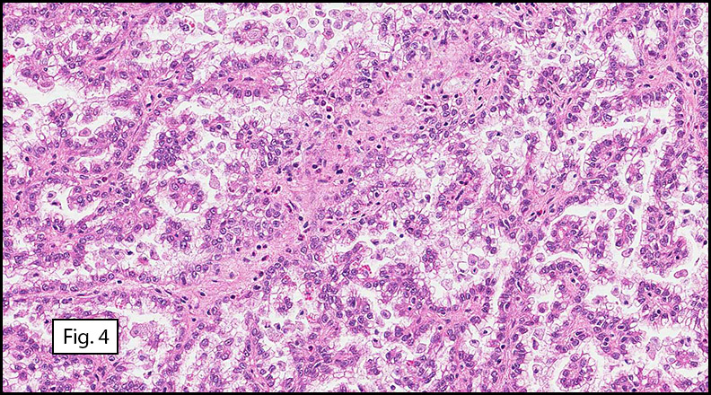
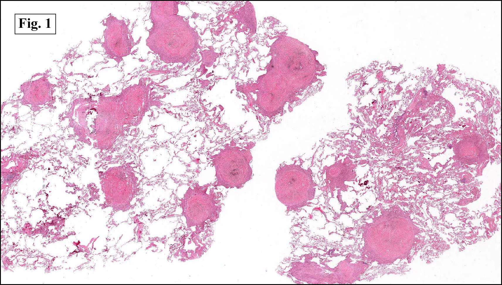
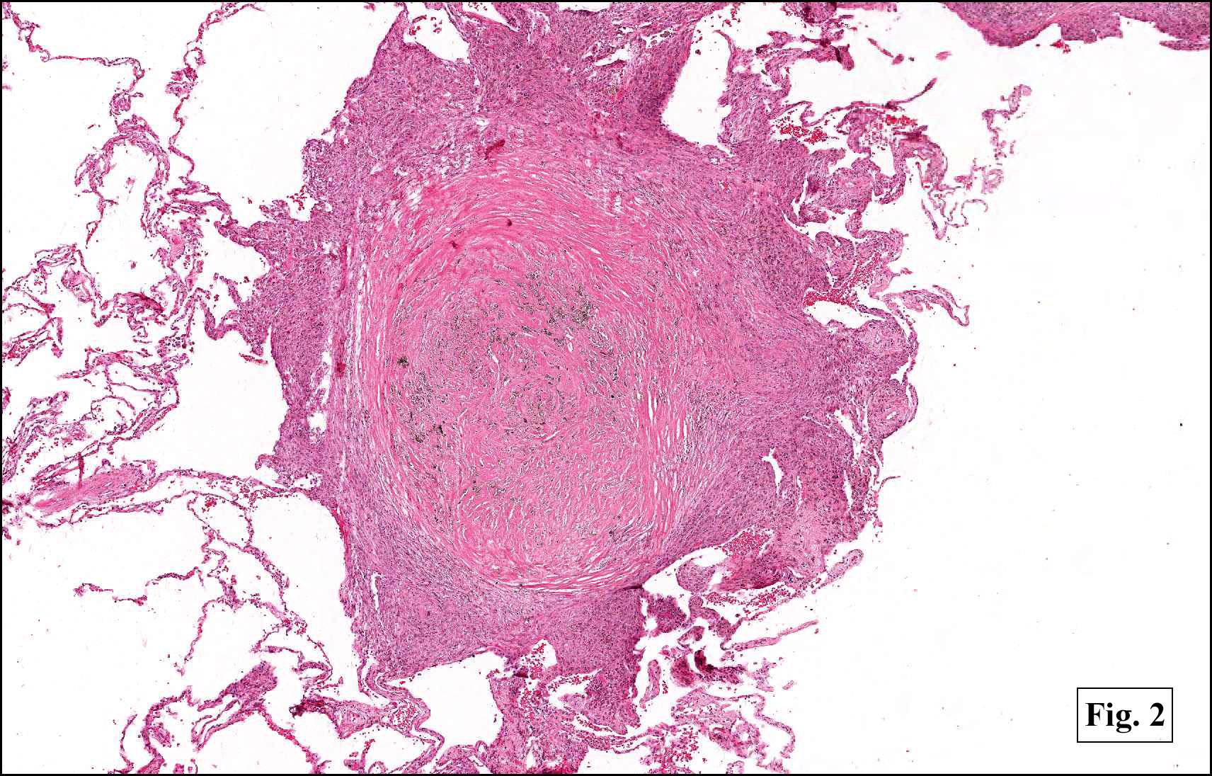
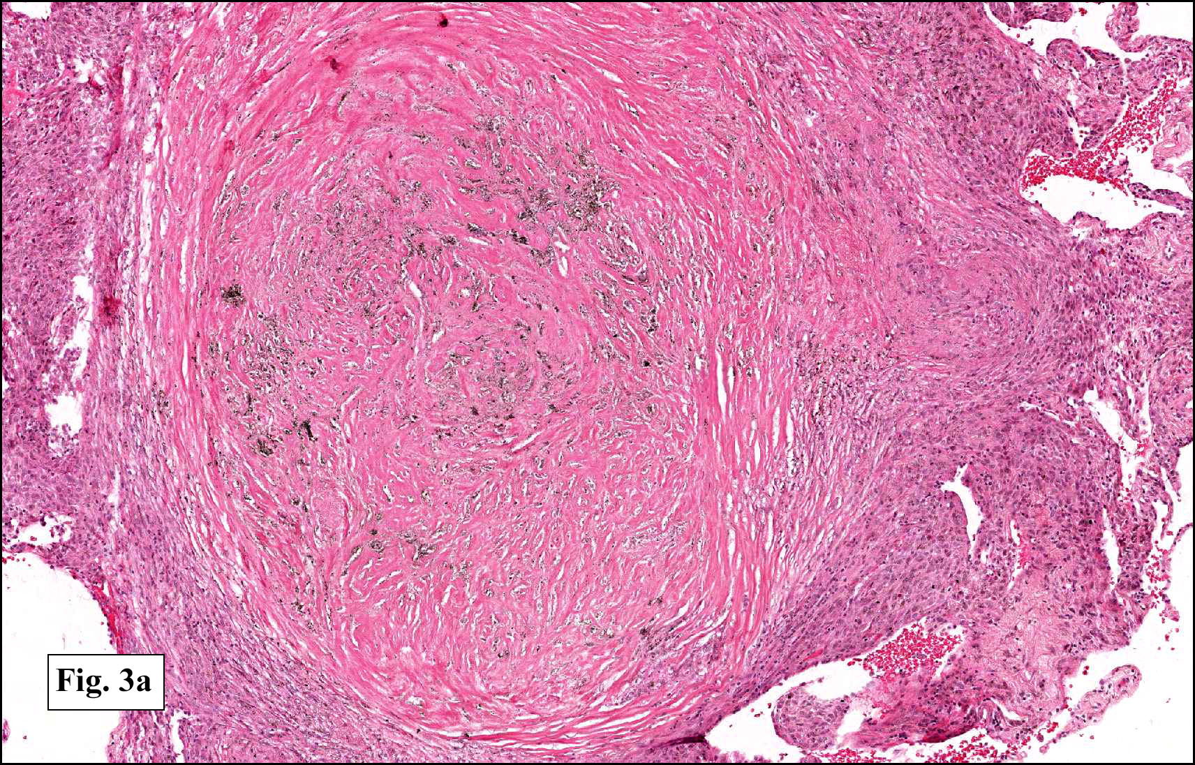
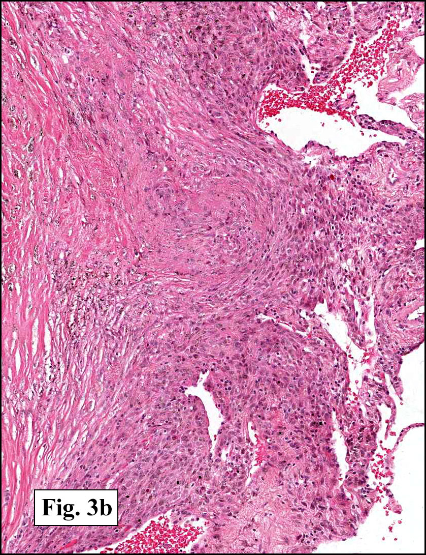
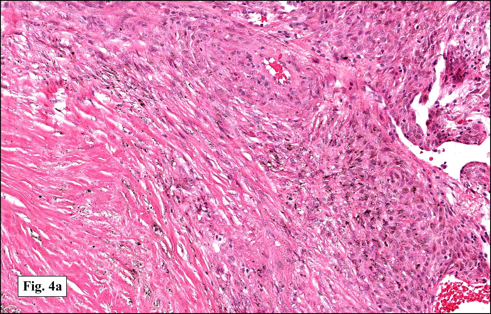
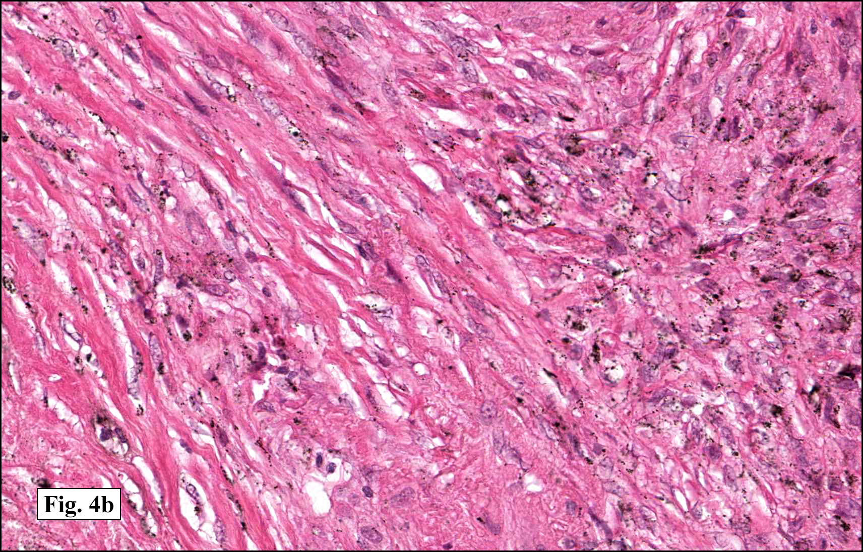
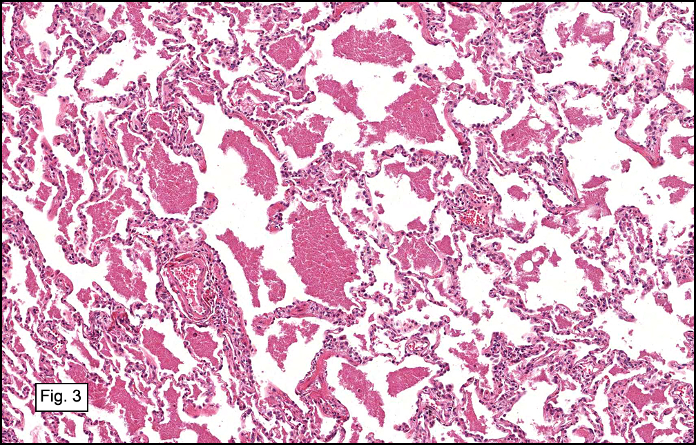
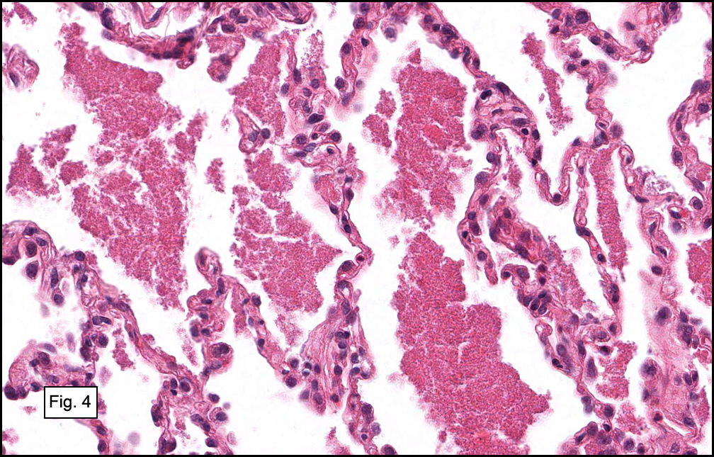
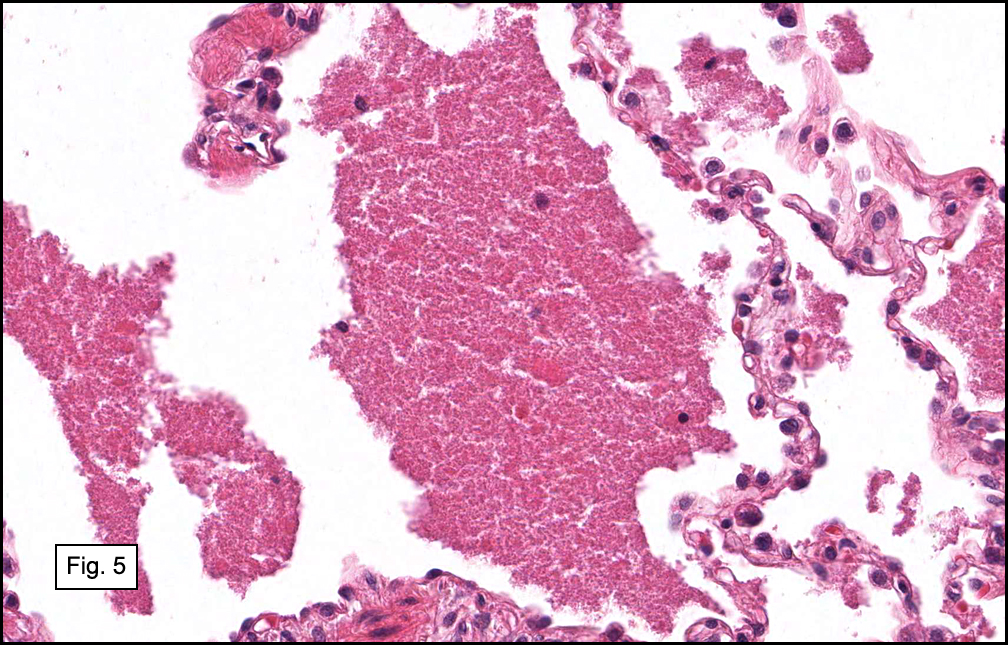
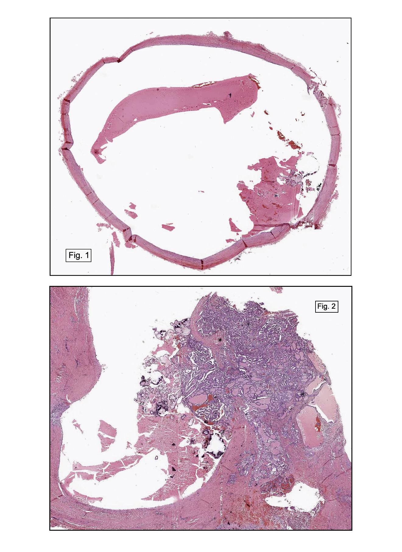
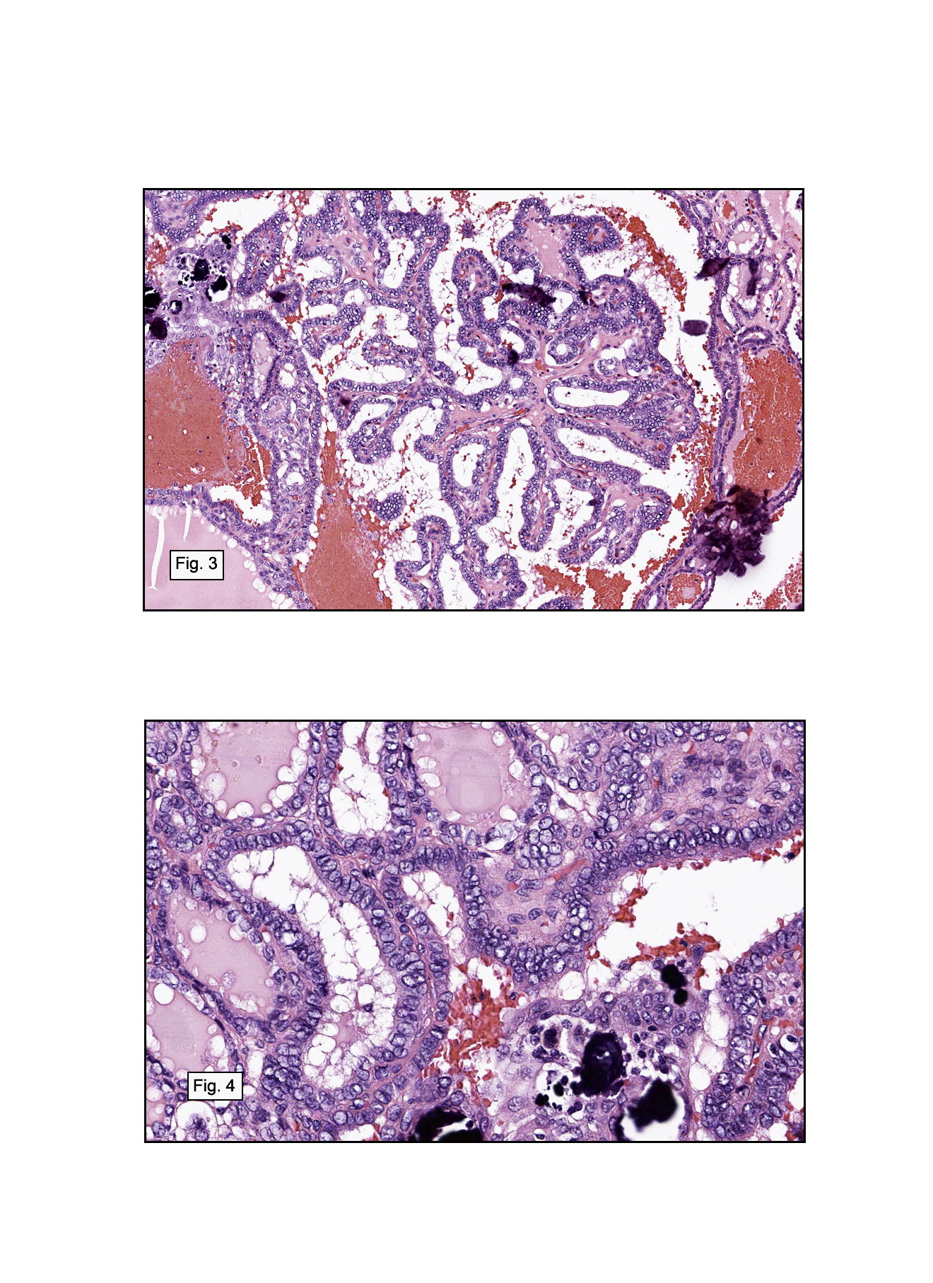
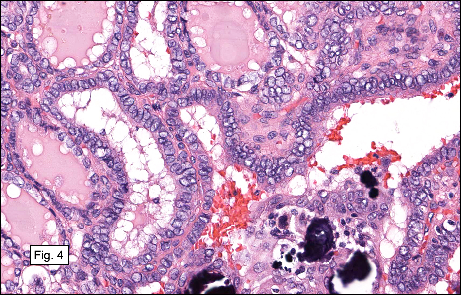
Resection of the lesion yielded an aggregate of approximately 2.0 x 1.5 x 0.4 cm homogenous brown soft tissue. Microscopically, the lesion appeared to be well-circumscribed but extended to the inked margins (Fig. 1). Skeletal muscle differentiation and maturation were observed with cross striations and haphazard arrangement of irregular fascicular bundles of muscle cells with scant to moderate eosinophilic cytoplasm (2,3,4). No definite immature cells were identified, and no myxoid stroma was noted. No areas of hypercellularity, increased mitotic activity, pleomorphism or necrosis were observed.

Diagnosis: “Fetal rhabdomyoma, intermediate (cellular) type, cheek”

Evelyn Choo MD1, Rachel Conrad MD1, Anwar Raza, MD1, Donald Chase MD1,2

1. Department of Pathology and Human Anatomy, Loma Linda University and
Loma Linda Medical Center, Loma Linda, California
2. California Tumor Tissue Registry, Loma Linda, California

Discussion: Fetal rhabdomyoma is a rare but benign skeletal muscle neoplasm, first described by Dehner in 1972. It typically occurs in the head and neck region of young children and demonstrates varying degrees of skeletal muscle differentiation.

The term rhabdomyoma encompasses a complex classification system of several distinct benign skeletal muscle neoplasms. It is organized by location (cardiac versus extracardiac), and the extracardiac type is divided into adult, genital and fetal categories based on tissue differentiation and clinical presentation. The adult form tends to occur in head and neck region of 40-60 year old males and displays prominent eosinophilic polygonal cells with vacuolated cytoplasm. The genital form typically presents as a polypoid lesion in vulva and vagina of middle-aged women; microscopically, long strap-like muscle fibers with prominent cross-striations are seen in a collagenous and myxoid matrix. The fetal form shows immature skeletal muscle differentiation and is more common in the head and neck region of children below age four.

The fetal form is further subdivided into a myxoid type and an intermediate type. The myxoid subtype (also known as “classic”) is comprised almost entirely of immature primitive spindle cells in a myxoid stroma and tends to appear in postauricular soft tissue. In contrast, a wider spectrum of myocyte maturation is seen in the intermediate subtype (also known as “cellular” or “juvenile”), initially described by Di Sant’Agnese and Knowles in 1980. This subtype of rhabdomyoma appears more often in soft tissue of face or mucosal tissue and represents a more differentiated lesion than classic fetal subtype.

Clinically, fetal rhabdomyoma presents in children less than four years old, may often be congenital and is more common in males than females (2.4:1). The lesion consists of a solitary, well-defined, non-tender mass involving soft tissue or mucosa. It grows very slowly and seldom ulcerates the overlying skin or mucosa. Multiple extracardiac rhabdomyomata have been associated with nevoid basal cell carcinoma syndrome (also known as Gorlin-Goltz syndrome) and may show a mutation in PTCH on chromosome 9q22. However, no correlation is seen with tuberous sclerosis as in cardiac rhabdomyoma.

The gross appearance of fetal rhabdomyoma consists of a well-circumscribed, smooth, polypoid mucosal lesion, typically two to six centimeters in diameter. Lesions are typically solitary. Cross-sectioning reveals a grey to pink surface, often with a myxoid appearance.

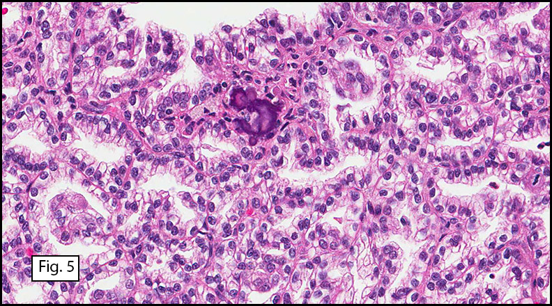
Microscopically, these lesions display either a myxoid or an intermediate pattern. The myxoid version is comprised of a myxoid matrix surrounding scattered or short bundles of immature spindle-shaped or oval cells with immature skeletal muscle fibers. The small uniform nuclei contain delicate chromatin and are enveloped by tapered eosinophilic cytoplasmic processes. Rare cross-striations are present. The intermediate type closely resembles classic adult head and neck rhabdomyoma, and contains a wider spectrum of myocyte differentiation with few immature spindle-shaped muscle precursors and numerous prominent strap-shaped muscle cells. The myocytes have central vesicular nuclei, scant to moderate eosinophilic cytoplasm, and frequent cross-striations with occasional vacuolation and glycogen. They are haphazardly arranged in irregular fascicular bundles. Ganglion-like rhabdomyoblasts with prominent nucleoli may be present. In both myxoid and intermediate types, the absence of significant atypia, necrosis, or mitotic activity is noted. Invasion and destruction of surrounding tissues should not be seen.

Immunohistochemical staining shows positivity for muscle-specific actin (MSA), myoglobin, and desmin; focal positivity may be seen for smooth muscle actin (SMA), S100, glial fibrillary acidic protein (GFAP), and vimentin.

Electron microscopy will show thick and thin myofilaments in the more mature myocytes; z-bands and glycogen may also be demonstrated in the cytoplasm. The immature myocytes may lack specific ultrastructural features of myocyte differentiation.

The recommended therapy for fetal rhabdomyoma is complete excision. Prognosis is excellent. Local recurrence is rare and is typically associated with incomplete resection. Metastases have never been described in the literature. Only two controversial cases of possible malignant transformation have been reported but may have been misdiagnosed as benign initially, and recurrent cases should be carefully evaluated for a misdiagnosis of rhabdomyosarcoma.

The differential diagnosis of fetal rhabdomyoma includes rhabdomyosarcoma (both embryonal and spindle cell types), adult rhabdomyoma, genital rhabdomyoma, Triton tumor (neuromuscular hamartoma), rhabdomyomatous mesenchymal hamartoma of the skin, and infantile fibromatosis.

1. Rhabdomyosarcoma consists of pronounced diffuse cellular immaturity, necrosis, mitotic figures, and infiltration of adjacent tissues with frank destruction. The presence of nuclear atypia in rhabdomyosarcoma is the most important criteria to distinguish it from fetal rhabdomyoma.
2. Adult rhabdomyoma has distinctive polygonal “spider” cells with vesicular nuclei, central nucleoli, and eosinophilic cytoplasm. It tends to occur in the head and neck region of adult males.
3. Genital rhabdomyoma demonstrates predominately mature long, strap-like striated muscle cells and occasional immature myocytes in a background of myxoid material and collagen. It typically presents as an asymptomatic, slow-growing, small polypoid lesion in the vulvo-vaginal region of middle-aged women. Rare occurrences in the male urogenital tract have also been described.
4. Triton tumor is composed of nerve fascicles mixed with mature striated skeletal muscle. It is associated with neurofibromatosis in younger patients and typically affects the axial skeleton.
5. Rhabdomyomatous mesenchymal hamartoma consists of normal dermal elements with mature striated skeletal muscle.
6. Infantile fibromatosis is not as well-circumscribed as fetal rhabdomyoma. It tends to involve regions deeper than the subcutis, has a fasciculated spindle cell pattern, lacks cytoplasmic striations, and contains interspersed fat cells.

Fetal rhabdomyoma is a rare neoplasm of immature skeletal muscle. Distinguishing it from the more common, more aggressive rhabdomyosarcoma is important. Clinical characteristics (location, behavior) and microscopic traits (mitoses, necrosis, and especially nuclear atypia) may be helpful. Prognosis is excellent after surgical excision. Recognition of this benign neoplasm is important in communicating accurate prognostic information and in preventing over-aggressive treatment.

Suggested Reading:

1. Dehner LP, Enzinger FM, Font RL. Fetal rhabdomyoma: an analysis of nine cases. Cancer. 1972;30:160-166.
2. Di Sant’Agnese PA, Knowles DN. Extracardiac rhabdomyoma: a clinicopathologic study and review of the literature. Cancer. 1980;56:780-789.
3. Kapadia SB, Barr FG. Rhabdomyoma. In: Fletcher CDM, Unni K, Mertens F, eds. World Health Organization Classification of Tumours. Pathology and Genetics of Tumors of Soft Tissue and Bone. Lyon, France: IARC Press, 2002:142-145.
4. Premalata CS, Kumar RV, Saleem KM, Fathima LJ, Das K. Fetal rhabdomyoma of the lower extremity. Pediatr Blood Cancer. 2009;52(7):881-883.
5. Walsh SN, Hurt MA. Cutaneous fetal rhabdomyoma: a case report and historical review of the literature. Am J Surg Pathol. 2008;32(3):485-491.
6. Weiss SW, Goldblum JR. Enzinger & Weiss’s Soft Tissue Tumors. 5th ed. Philadelphia: Mosby-Elsevier, 2008:583-592.
7. Yang S, Zhao C, Zhang Y, Liao S. Mediastinal fetal rhabdomyoma in nevoid basal cell carcinoma syndrome: a case report and review of the literature. Virchows Arch. 2011; 459:235-238.

October 2012: A 61 year old woman with a left kidney mass

History: A sixty-one year old woman underwent a hand-assisted laparoscopic nephrectomy. The 256.0 gram, 12.0 x 6.5 x 2.5 cm left kidney contained a single 4.3 x 4.0 x 3.2 cm homogeneous tan-white, pseudo-encapsulated mass in the mid ante-hilar pole. The mass was adjacent to but did not penetrate the renal capsule. No tumor involvement of the perirenal adipose, hilar vessels, renal pelvis or ureter was noted. The residual renal parenchyma had a normal red-brown appearance and showed a distinct cortico-medullary junction. No other lesions were identified.

Microscopically, the well-circumscribed tumor approached but did not extend through the renal capsule (Fig. 1). A distinctive tubulo-papillary architecture was observed (Fig. 2), with fibrovascular cores lined by a single layer of tumor cells. The tumor cells had moderate to focally abundant basophilic cytoplasm. The low-grade nuclei displayed open chromatin and rare nucleoli, consistent with Fuhrman Nuclear Grade 2. Occasional foamy histiocytes (Fig. 3), clear cells (Fig. 4), and psammoma bodies were seen (Fig. 5). Sarcomatoid features, tumor necrosis, and lymphovascular invasion were absent. The adjacent non-neoplastic kidney demonstrated chronic inflammation and hyalinized glomeruli, consistent with mild benign nephrosclerosis.

Diagnosis: “Papillary Renal Cell Carcinoma, Type 1 (Basophilic)”

Rachel Conrad, MD1; Pamela Boswell, DO2; Manju Aron, MD3; Mahul Amin, MD3; Donald R. Chase, MD1,4.

1. Department of Pathology and Human Anatomy, Loma Linda University and
Loma Linda Medical Center, Loma Linda, California
2. Scripps Clinic Medical Laboratories, La Jolla, California
3. Cedars Sinai Medical Center, Los Angeles, California
4. California Tumor Tissue Registry, Loma Linda, California

Discussion: Papillary renal cell carcinoma is a relatively rare type of renal cell carcinoma with unique morphologic, immunohistochemical, and genetic features. It tends to occur in the sixth to seventh decades with a male predominance (3:1).

Papillary renal cell carcinoma has been associated with several different clinical syndromes. Hereditary/familial papillary renal cell carcinoma (7q31 c-MET mutation) results in multiple bilateral type 1 papillary renal tumors. Hereditary leiomyomatosis and renal cell cancer (1q42-43 mutation) leads to a solitary unilateral type 2 papillary renal cell carcinoma, usually around age thirty, along with cutaneous and uterine leiomyomas and leiomyosarcomas. Birt-Hogg-Dube syndrome (17p11.2 mutation) has multiple chromophobe and oncocytoma renal cell tumors with rare clear cell or papillary renal cell carcinomas. Fibrofolliculomas, lung cysts, and a history of spontaneous pneumothorax are frequently noted. Familial paraganglioma syndrome displays multiple renal tumors with benign and malignant paragangliomas and papillary thyroid carcinoma. PTEN hamartoma tumor syndrome results in unifocal papillary RCC and chromophobe RCC with gastrointestinal polyposis, tumors of the breast, thyroid and uterus, and frequently, lipomas and trichilemmomas.

The gross appearance of papillary renal cell carcinoma consists of a well-circumscribed nodule with a fibrous pseudocapsule. Lesions are often bilateral and multifocal, and hypovascularity may be seen on radiographic studies. Cross-sectioning reveals a yellow-brown surface with regions of cystic degeneration, hemorrhage, and necrosis.

Microscopically, the tumors contain fibrovascular cores lined by neoplastic cells containing foamy histiocytes and occasional neutrophils. Solid-papillary and tubulopapillary growth patterns may also be observed. Sarcomatoid dedifferentiation is seen in five percent of papillary renal cell carcinomas and confers a poor prognosis. Additional findings may include stromal and intracellular hemosiderin deposition, occasional psammoma bodies, necrosis, and clear cell change in areas of hemorrhagic degeneration.

Papillary renal cell carcinoma has been classified by Delahunt and Eble (1997) into two subtypes. Type 1 (also known as “basophilic”) shows a single layer of cells with scant basophilic-to-amphophilic finely granular cytoplasm and low-grade nuclei arranged along the papillary basement membrane. Type 2 shows pseudostratified cells with abundant eosinophilic granular cytoplasm and large nuclei displaying a higher Fuhrman grade. As expected, the higher grade tumors have a worse prognosis.

Immunohistochemical staining shows membranous positivity for CK7, with stronger expression in Type 1 papillary renal cell carcinoma. A granular cytoplasmic pattern is seen for alpha methylacyl CoA-racemase (AMACR). Other positive stains include AE1/AE3, CAM5.2, EMA, CD10, PAX2, PAX8, RCC, and PN15/gp200. Negative staining is seen for CAIX, Cathepsin-K, TFE3, and 34-beta-E12.

Cytogenetic studies often show trisomy 7, trisomy 17, and loss of Y without abnormalities of 3p. Additional genetic anomalies may be observed.

The recommended therapy for papillary renal cell carcinoma is complete extirpation. The overall prognosis is better than for conventional renal cell carcinoma, with a 5 year survival rate of 45-70%. Survival drops to 15-20% if extension to the renal vein or perinephric fat is observed. Metastases are frequently seen in tumors larger than 3 cm and tend to involve the lungs, lymph nodes, bones, and liver.

The differential diagnosis of papillary renal cell carcinoma includes metanephric adenoma, conventional clear cell renal cell carcinoma with papillary foci, clear cell papillary renal cell carcinoma, oncocytic papillary renal cell carcinoma, Xp11 translocation renal cell carcinoma, collecting duct carcinoma, and mucinous tubular and spindle cell carcinoma of the kidney.

1. Metanephric adenoma tends to occur in young to middle aged females. The microscopic appearance is consistent with developing metanephric tubular epithelium and displays tiny tubules and papillae with bland nuclei and scant stroma. This lesion is usually small in size and benign in nature. Immunostaining shows rare focal CK 7 positivity and negativity for EMA and AE1/AE3.
2. Conventional clear cell renal cell carcinoma with papillary foci is fairly common. It may be distinguished from papillary renal cell carcinoma by its hypervascularity on radiographic imaging, golden yellow color on sectioning, and solid or acinar architecture with clear-to-eosinophilic cells separated by thin vascular septae. True papillae are rarely seen; instead, small papillations protrude into necrotic or cystic spaces. Chromosome studies frequently show deletion of the von Hippel-Lindau gene (3p), and inactivation of this gene can be detected by diffuse strong membranous staining for CAIX. Negative immunohistochemical staining is observed with CK7 and AMACR.
3. Clear cell papillary renal cell carcinoma is a small indolent tumor associated with end-stage renal disease. Microscopically, the papillary fronds are lined by clear cells with low Fuhrman nuclear grade and characteristic subnuclear vacuoles. Smooth muscle metaplasia and cystic changes are common. No hemosiderin deposition, foamy histiocytes, or psammoma bodies are seen. Immunohistochemistry shows a cup-like pattern for CAIX and CK7, with patchy positivity for 34-beta-E12; CD10 and AMACR are negative.
4. Oncocytic renal cell carcinoma with papillary features has a distinctive mahogany-brown color with a central scar on sectioning. Microscopic features include abundant granular eosinophilic cytoplasm and a linear arrangement of low-grade apical nuclei. Pseudostratification is absent, and papillary architecture is focal, not diffuse. IHC staining is positive for parvalbumin and negative for RCC and CK7.
5. Xp11 translocation renal cell carcinoma is the most common pediatric renal cell carcinoma subtype and may be associated with previous chemotherapy. Extensive psammomatous calcification may be seen radiographically and microscopically. Papillary architecture with clear-to-eosinophilic cells and vesicular high-grade nuclei is common, but cystic, solid, or nested architecture can also be seen. Immunohistochemical staining is positive for Cathepsin-K, TFE3, and PAX8, with focal staining for melanocytic markers. Weak expression of cytokeratins and epithelial markers may be seen. Cytogenetic studies often show translocation of ASPL and TFE3, t(X;17)(p11.2q25).
6. Collecting duct carcinoma tends to display prominent desmoplasia, frequent invasion, and high grade nuclear features. IHC stains are negative for CD10, AMACR, and PN15/gp200.
7. Mucinous tubular and spindle cell carcinoma of the kidney can be distinguished due to its mucinous stroma and overall architectural pattern. It may share focal papillary growth, mucin production, foam cells, and AMACR positivity with papillary renal cell carcinoma, but it will lack the classic diffuse papillary pattern.

Due to its better prognosis, associated familial syndromes, and tendency toward multiple bilateral hypovascular tumors, papillary renal cell carcinoma is important to separate from other lesions with papillary architecture. Characteristic microscopic traits include true fibrovascular cores containing foamy histiocytes and hemosiderin. Immunohistochemical stains (CK7, AMACR) and cytogenetic studies (trisomy 7/17, loss of Y) can be useful in challenging cases. Treatment consists of surgical excision and careful assessment for metastases. The astute observer will be alert to the unique aspects of this rare tumor.

Suggested Reading:

1. Delahunt B, Eble JN. Papillary renal cell carcinoma. In: Eble JN, Sauter G, Epstein JI, Sesterhenn IA, eds. World Health Organization Classification of Tumours. Pathology and Genetics of Tumors of the Urinary System and Male Genital Organs. Lyon, France: IARC Press, 2004:15-22.
2. Merino MJ, Eccles DM, Linehan WM, et al. Familial renal cell carcinoma. In: Eble JN, Sauter G, Epstein JI, Sesterhenn IA, eds. World Health Organization Classification of Tumours. Pathology and Genetics of Tumors of the Urinary System and Male Genital Organs. Lyon, France: IARC Press, 2004:15-22.
3. Mester JL, Zhou M, Prescott N, Eng C. Papillary renal cell carcinoma is associated with PTEN hamartoma tumor syndrome. J Urol. 2012; 79(5):1187.e1-7
4. Sukov WR, Lohse CM, Leibovich BC, Thompson RH, Cheville JC. Clinical and pathological features associated with prognosis in patients with papillary renal cell carcinoma. J Urol, 2012; 187(1):54-59.
5. Ross H, Martignoni G, Argani P. Renal cell carcinoma with clear cell and papillary features. Arch Pathol Lab Med. 2012;136(4):391-9.
6. Rosai J. Rosai and Ackerman’s Surgical Pathology. China: Mosby, 2004:1251-1264.
7. Delahunt B, Eble JN. Papillary renal cell carcinoma: a clinicopathologic and immunohistochemical study of 105 tumors. Mod Pathol. 1997;10:537.
8. Murphy WM, Grignon DJ, Perlman EJ. AFIP Atlas of Tumor Pathology Series 4: Tumors of the Kidney, Bladder, and Related Urinary Structures. Washington, D.C.: American Registry of Pathology, 2004.

September 2012: A 70 year old sandblaster with pulmonary nodules

History: A 70-year-old retired sandblaster underwent a left lower lobectomy for lung cancer. He had stopped smoking one year earlier, after smoking 1.5 packs per day for approximately 50 years. Four years later, two new nodules were discovered, resulting in a right lower lobectomy.

Gross examination revealed a dominant, ill-defined, marbled, gray-and-white tan 6 cm tumor which involved the pleural surface and showed central cystic degeneration, as well as numerous smaller satellite lesions of similar appearance. Three hilar lymph nodes were also identified.

In some sections (not illustrated), squamous cell carcinoma was identified. The point of interest for us, however, was the background lung. Scattered throughout the parenchyma were numerous dense, homogenous nodules composed of brightly-eosinophilic, concentrically-layered material (Fig. 1, 2, 3a, 3b). Dust-filled macrophages were observed, as was emphysematous change (Fig. 4a, 4b). The eosinophilic nodules also involved the accompanying lymph nodes.

Diagnosis: “Silicosis Co-Existent With Pulmonary Adenocarcinoma”

Christina M. Birsan M.D., and Donald R. Chase, M.D.
Department of Pathology and Human Anatomy, Loma Linda University and
Medical Center, Loma Linda, California
California Tumor Tissue Registry, Loma Linda, California

Discussion: Silicosis, a chronic lung disorder, is characterized by progressive development of parenchymal nodules and pulmonary fibrosis as a result of inhalation of crystalline silica. Most cases are related to exposure to quartz, the most abundant mineral in the earth’s crust and also the most abundant form of crystalline silica. Identified in Egyptian mummies, silicosis is the oldest recognized occupational lung disease, with exposure occurring in various rock-cutting, sand-blasting, and ceramics industries.

Clinically, silica-induced lung disease may be acute, accelerated, or chronic. Acute silicoproteinosis is similar to pulmonary alveolar proteinosis, and occurs in cases of heavy exposure. Patients are generally symptomatic within 3 years of exposure, and most cases are fatal. When the disease manifests between 3 to 10 years after exposure, accelerated silicosis is the suggested term. Chronic (classic or nodular) silicosis is the most common form, usually occurring decades after exposure to relatively low levels of silica. Classically, it requires at least 20 years after onset of exposure. It may manifest as simple silicosis (nodules 10 mm or less) or complicated silicosis (nodules greater than 1 cm). Simple silicosis is generally asymptomatic until advanced stages when dyspnea develops. Complicated silicosis results from coalescing smaller nodules over time (also called conglomerate silicosis). Cough and sputum production may occur, but are often the result of chronic bronchitis or concurrent infections. Crackles and digital clubbing are rare in silicosis, and suggest alternate diagnoses. Infections may complicate the process (mycobacterial infections in particular) and can cause fever and weight loss. Severe, longstanding silicosis and fibrosis may cause signs of cor pulmonale. Rheumatoid factor, antinuclear antibodies, serum immune complexes, and polyclonal increase in immunoglobulins may be detected in some cases. In fact, connective tissue disorders (most commonly scleroderma, rheumatoid arthritis, and systemic lupus erythematosus) have been associated with silica exposure. Signs and symptoms in those cases are more specific to the individual entities, and pulmonary silicosis is not always radiologically detectable. Significant associations between silicosis and lung cancer have been reported as well.

Radiologically, classic silicosis manifests as multiple nodules which mostly involve upper lobes. Best seen on CT scans, these nodules are well-circumscribed, uniform in size, and usually less than 5 mm in diameter. They may progress to form conglomerate masses, resulting in larger size. Complicated silicosis is usually bilateral and causes marked loss of lung volumes in the upper lobes, expansion and hyperinflation of the lower lobes, and superior retraction of the hila. Hilar lymph nodes are usually involved and may show a peripheral “eggshell” type calcification. Nodules have also been described in liver, spleen, bone marrow, and abdominal lymph nodes.

Grossly, chronic silicosis demonstrates firm, rounded, well-demarcated nodules, usually about 3 to 6 mm and slate gray to black. A cuff of pigmentation may be seen surrounding nodules on the pleural surface.

Microscopically, the same discrete nodules are apparent. Their formation begins with the accumulation of dust-filled macrophages along lymphatic routes, bronchovascular bundles, and in the pleura and septa. Early stages of disease may demonstrate only these accumulated macrophages. Increased reticulin and fibrosis in the sheets of macrophages represent developing nodules, which then enlarge and become discrete and round, replaced by dense lamellar collagen. They may become calcified, hyalinized, or may undergo central degeneration including cavitation and necrosis, in which case infection (especially tuberculosis) should be carefully ruled out. Polarized light reveals weakly birefringent silica and strongly birefringent round or oval silicate particles within nodules and in surrounding macrophages. If exposure stops, nodules may become fibrous with a few surrounding histiocytes. Emphysematous change is common in surrounding lung. Acute silicosis (silicoproteinosis) resembles pulmonary alveolar proteinosis (PAP), with an eosinophilic, granular, PAS-positive material filling the alveolar spaces. Unlike PAP, however, interstitial inflammation and fibrosis and irregular hyaline scars may be present. Silicotic nodules are usually poorly-formed or absent in such cases.

The pathogenesis of silicosis involves the inhalation of particles less than 10 micrometers in diameter, with the most pathogenic particles being approximately 1 micrometer. The intensity and duration of exposure are the most important factors, as the damage occurs once the silica particles are deposited in the lung. Direct cytotoxicity or the production of oxidants and other mediators may be involved, however the exact sequence of events is unknown. It appears that alveolar macrophages ingest the silica particles and die, perhaps liberating fibroblast-stimulating factors which promote fibrosis. Evidence also suggests that silica interferes with the ability of macrophages to inhibit the growth of mycobacteria, hence the association with tuberculosis. Toxic effects of silica on alveolar type 2 cells has been implicated in silicoproteinosis (acute silicosis).

The diagnosis of silicosis rests upon an appropriate exposure history, consistent radiologic findings, and the absence of other diseases which could explain the radiologic evidence. Observing silica, silicates, silicotic nodules, or increased dust in lymph nodes alone is not sufficient for the diagnosis, as virtually all adults have a small amount of silica in their lungs.

Unfortunately, no effective therapy exists once the inhalation of silica dust has occurred. Most affected patients are asymptomatic, however, and have a normal lifespan, with only a small percentage experiencing progressive respiratory disability.

Suggested Reading:

Travis W, Colby T, Koss M, et al. Non-neoplastic disorders of the lower respiratory tract. In King D, ed. Atlas of Nontumor Pathology. Washington, DC: American Registry of Pathology, Armed Forces Institute of Pathology; 2002.

Katzenstein AL III. Katzenstien’s and Askin’s Surgical Pathology of Non-neoplastic Lung Disease. 3rd edition. Philadelphia, PA: WB Saunders Company; 1997.

Hammar S. Pleural diseases. In: Dail D, Hammar S, eds. Pulmonary Pathology. 2nd ed. New York, NY: Springer-Verlag; 1994.

Erren TC, Morfeld P, Glende CB, et al. Meta-analyses of published epidemiological studies, 1979-2006, point to open causal questions in silica-silicosis-lung cancer research”. Med Lav 2011 Jul-Aug; 102(4):321-35.

August 2012: A 34 year old man with fever and a lung infiltration.

History: A 34-year-old Caucasian male accountant for a powder factory and brass foundry experienced low-grade fever, debility (fatigue?), and weakness. He was found to have a slight infiltration in the right lower and mid lung fields. Cultures were negative, however he continued to have progressive dyspnea for the next two years. During that time he also developed a chronic cough productive of a copious amount of white sputum which was occasionally greenish or streaked with blood. He was admitted with severe dyspnea on exertion.

A physical examination revealed no significant findings, however the chest x-ray showed diffuse bilateral mottling in all lung areas, mostly in the lower lobes. Hemoglobin levels were slightly high at 18.2, and sputum cultures grew alpha streptococcus and staphylococcus epidermis. A left thoracotomy was performed. No pleural adhesions were noted, however the surgeons observed that the lung did not expand normally. Biopsies were taken from the left lingular segment and the left upper lobe.

Two tissue samples were examined. One was soft, fluffy, red-pink, and well aerated. The other contained firm, gray nodules up to 0.8 cm in greatest diameter.

Histologic sections showed focal, nodular areas (Fig. 1) comprised of alveoli which were filled with an intensely eosinophilic material made up of tiny granules and amorphous debris (Figs. 2,3, 4, 5). These areas were immediately adjacent to essentially normal lung tissue, without a surrounding inflammatory response. No other significant abnormalities were identified.

Diagnosis: “Pulmonary Alveolar Proteinosis”

Christine M. Birsan M.D., and Donald R. Chase, M.D.
Department of Pathology and Human Anatomy, Loma Linda University and Medical Center, Loma Linda, California
California Tumor Tissue Registry, Loma Linda, California

Discussion: Pulmonary alveolar proteinosis (PAP) is a rare condition in which a lipid-rich, granular proteinaceous eosinophilic material fills the alveoli. “Primary” cases are found in isolation, while “secondary” PAP may occur in the setting of infection (tuberculosis, Pneumocystis jirovecii), malignancy (especially leukemia and lymphoma), immune deficiency (chemotherapy, congenital alymphoplasia, hypogammaglobulinemia, juvenile dermatomyositis), environmental dust exposure (wood, aluminum, silica, kaolin), and lysinuric protein intolerance. A history of smoking is present in many, but not all cases. Most patients present between the ages of 20-50, however 18% of cases occur in infants and children. A congenital form is described by Leslie and Wick as a lethal disease caused by defects in surfactant production and metabolism, some of which are in association with mutations of the surfactant protein B gene and genes related to secretion of surfactant proteins. “Secondary” PAP has also been observed in infants, most commonly seen with viral infections (respiratory syncytial virus, cytomegalovirus, and parainfluenza virus). “Primary” PAP is unusual in infants and children, but may be seen in adolescents. Males are affected more often than females at a ratio of approximately 2:1-4:1, and the disease may be more common in Caucasians.

The pathogenesis of PAP is unknown. Studies have suggested that macrophage dysfunction may play a role, in which there is a reduced ability to process surfactant (a significant component of the accumulated intra-alveolar material). Mutant mice lacking the gene for granulocyte-macrophage-colony-stimulating factor (GM-CSF) develop a similar disease process which is reversed when GM-CSF is replaced. In addition, antibodies against GM-CSF have been discovered in human cases of PAP (especially “primary” PAP), further supporting this hypothesis. Alternatively, defective production of surfactant may be a consideration, as the pathologically accumulated surfactant lacks its usual surface-active properties, while surfactant extracted from uninvolved areas of the patient’s lung shows normal activity. It has been shown experimentally that the material from PAP produces macrophage dysfunction in normal human blood monocytes and reduces activity of lymphocytes, raising the possibility that the macrophage dysfunction may be a secondary result rather than a primary cause. The wide variety of clinical situations in which this process is observed suggests that PAP represents a common tissue reaction to a broad range of insults.

Clinically, the onset of PAP is often insidious. About one-third of patients are asymptomatic at presentation despite having extensive radiologic abnormalities. Patients who are symptomatic may have a nonproductive cough, a cough productive of chunky, gelatinous material, and/or streaky hemoptysis. Dyspnea on exertion, fatigue, weight loss, chest pain, and low-grade fever may also be present. Occasionally clubbing and cyanosis are observed. Crackles are sometimes heard on auscultation however they are often absent.

Radiographic findings usually include bilateral and symmetric areas of vaguely nodular airspace consolidation or hazy ground-glass opacity. Peri-hilar regions and lower lobes are most severely affected. Interlobular septal thickening and ground-glass opacities seen on CT scans produce a characteristic “crazy paving” appearance. This pattern, although suggestive of the diagnosis, is also seen in other conditions.

Pulmonary function testing most commonly demonstrates a restrictive process, and a decrease in the diffusion capacity for carbon monoxide (DLCO) out of proportion to the reduced lung volume is sometimes seen. Hypoxemia and compensated respiratory alkalosis are frequent and are made worse by exercise. An elevated shunt fraction is usually present.

Lab values may reveal polycythemia, hypergammaglobulinemia, and increased lactate dehydrogenase. Serum levels of lung surfactant proteins A and D (SP-A, SP-D) have been found to be markedly high which can help narrow the diagnosis, however, other lung processes can have similar findings. Elevated levels of several tumor markers have been identified in the BAL fluid from some patients, including carcinoembryonic antigen (CEA), carbohydrate antigens sialyl Lewis (CA19-9), and sialyl SSEA-1 (SLX). KL-6, a mucin-like glycoprotein may also be useful for diagnosis when found in serum or BAL specimens. Cultures may reveal Nocardia or other opportunistic mycobacterial, fungal, and viral agents thought to represent secondary infection.

Cytologic preparations may suggest the diagnosis of PAP. In BAL specimens, abundant lipoproteinaceous material causes an opaque appearance, and large, acellular eosinophilic bodies may be seen in a background of eosinophilic granules. PAS-positive proteinaceous material as well as macrophages engorged with PAS-positive material point to the diagnosis.

Tissue examination is frequently necessary for definitive diagnosis. Grossly, lung tissue is heavy and viscid, containing yellow fluid which leaks from cut surfaces. Firm, yellowish-white nodules scattered throughout the parenchyma range from a few millimeters to 2 cm. Microscopically, eosinophilic proteinaceous granular material fills the alveoli and occasionally involves bronchioles and alveolar ducts, leaving the interstitial architecture of the lung generally intact. A diffuse pattern is usually seen, however focal or patchy involvement also exist, as seen in this case. Hyperplastic, cuboidal, type 2 pneumocytes are often line the alveolar septae. Sharply demarcated round, empty spaces, cholesterol clefts, and small, dense, globular eosinophilic clumps are distinctive findings within the accumulated material which may also contain cellular debris, foamy macrophages, ghosts of degenerated cells, and detached type 2 pneumocytes. It is usually PAS-positive and diastase-resistant, while staining for antibody to surfactant apoprotein may be positive especially in “primary” disease. Alcian and mucicarmine stains are negative. Frozen sections shows abundant lipid, highlighted with Oil Red O staining. Examination by electron microscopy demonstrates concentrically laminated myelin figures and lamellar bodies within the proteinaceous material which look identical to the cytoplasmic inclusions of type 2 pneumocytes, suggestive of surfactant. Polarization microscopy may reveal birefringent needle-like particles if there has been an exposure to dust. Interstitial fibrosis or inflammation are typically not prominent and if present may indicate an associated infection, but may also indicate long-standing or recurrent PAP.

The differential diagnosis for PAP includes pulmonary edema, alveolar mucinosis, and Pneumocystis jirovecii pneumonia. Pulmonary edema and alveolar mucinosis lack the granularity of PAP, as well as the intense eosinophilic staining, cholesterol clefts, and engorged macrophages containing PAS-positive debris. The intra-alveolar exudate seen in Pneumocystis jirovecii may be distinguished from PAP by the cysts or organisms which appear as “bubbles” within the eosinophilic material which stain positive for Gomori methenamine silver (GMS).

Treatment for PAP should be initiated when the patient becomes sufficiently symptomatic, as many patients have little or no impairment and spontaneous remission may occur. Severe dyspnea and hypoxemia at rest or with exercise warrant therapeutic whole lung lavage via a double-lumen endotracheal tube, the most widely accepted and effective treatment. Patients often feel dramatically better after this treatment, although potential complications include malpositioning of the endotracheal tube, saline spillover into unlavaged and ventilated lung, and hydropneumothorax. Thirty to forty percent of patients require lavage only once, while some patients require repeat lavages at intervals of 6 to 12 months. Corticosteroids or other immunosuppressives should not be used, as there is concern that they may increase mortality by aggravating or inducing secondary opportunistic infections. Further study is necessary to determine the potential use of GM-CSF as a therapeutic option.

Suggested Reading:

Travis W, Colby T, Koss M, et al. Non-neoplastic disorders of the lower respiratory tract. In King D, ed. Atlas of Nontumor Pathology. Washington, DC: American Registry of Pathology, Armed Forces Institute of Pathology; 2002.

Katzenstein AL III. Katzenstien’s and Askin’s Surgical pathology of non-neoplastic Lung disease. 3rd edition. Philadelphia, PA: WB Saunders Company; 1997.

Hammar S. Pleural diseases. In: Dail D, Hammar S, eds. Pulmonary Pathology. 2nd ed. New York, NY: Springer-Verlag; 1994.

Leslie K, Wick M. Practical pulmonary pathology. A diagnostic approach. 1st edn. Philadelphia: Churchill-Livingstone; 2005.

July 2012: A 50 year old man with an anterior neck mass

History: A 50 year old man presented with a 2 cm fluctuant midline anterior neck mass. The removed specimen was a unilocular cyst which exuded serous fluid. A papillary nodule was noted on its luminal surface.

Microscopically, the cyst had a denuded epithelial lining supported by fibrous tissue with varying numbers of chronic inflammatory cells (Fig. 1). It also showed a papillary excrescence protruding into the cyst wall (Fig. 2). Tumor cells were crowded and arranged in papillary fronds and follicular patterns (Fig. 3). They had enlarged nuclei and occasional calcifications. Some showed longitudinal clefts. Nucleoli were inconspicuous and peripherally located. Many of the cells showed central cytoplasmic clearing giving the cells an “Orphan Annie” appearance (Fig. 4).

Diagnosis: “Classic Papillary Thyroid Carcinoma” arising in thyroglossal duct cyst”

David Panther, MSIV, and Donald R. Chase, M.D.
Department of Pathology and Human Anatomy, Loma Linda University and Medical
Center, Loma Linda, California
California Tumor Tissue Registry, Loma Linda, California

Discussion: Thyroid tissue has its embryologic origin at the foramen cecum at the base of the tongue from which it migrates into the lower anterior neck. The tract then degenerates. However, incomplete regression of this tract, can accumulate fluid and form a cyst (thyroglossal duct cyst [TDC]) usually lined with columnar epithelium. Occasionally the epithelium can undergo squamous metaplasia, or become denuded and/or inflamed. During its descent, the thyroid may leave ectopic tissue anywhere along the path and up to 60% of TDCs are associated with residual thyroid tissue. These elements are physiologically similar to those in the lower neck, however they lack parafollicular C cells. The remnant tissue can be responsible for any traditional thyroid disease, except medullary carcinoma, a tumor of C cell origin.

Rarely (<1%) residual thyroid tissue give rise to a primary malignancy, mostly papillary thyroid carcinoma (80%), (PTC). Other malignancies include follicular carcinoma and anaplastic carcinoma

Questions have been raised over the origin of PTC in a TDC, namely whether this entity represents a TDC-related primary versus a thyroid primary with local metastasis to a TDC. However, a substantial number of cases have been reported where the thyroid was removed and carefully examined. Roughly 20-50% of the lower neck thyroids have had concurrent tumor, implying that at least half of the upper neck tumors are most likely to be primary TDC tumors. In addition, occult thyroid malignancies are often found at autopsy in asymptomatic individuals, indicating that a significant portion of concurrent TDC and thyroid gland tumors are coincidental. It is generally accepted that the thyroid should not be automatically excised unless clinical evidence of a mass or abnormal scintiscan is/are present in the lower neck thyroid gland.

The differential diagnosis includes other thyroid epithelial neoplasms, as well as squamous cell carcinoma. Although metastatic disease has not yet been reported in TDCs, possible look-alikes could include metastases from clear-cell variants of lung and renal carcinoma, melanoma, and mucinous gynecologic malignancies.

– Clear Cell (“Sugar”) Tumor of the Lung: cells have a large cytoplasmic glycogen droplet, displacing the nucleus peripherally. In addition, it stains positive for HMB-45.
– Clear Cell Renal Cell Carcinoma: central nuclei are surrounded by clear cytoplasm, with cells often arranged in nests surrounded by branching fibrovascular tissue.
– Melanoma: features include haphazard arrangement of cells, nuclei that are pleomorphic and readily seen nucleoli and mitoses. Key immunohistochemical markers include HMB-45, MART-1, Melan A, and S-100.
– Mucinous Cystadenocarcinoma: the cyst may be multiloculated. The epithelial component may be several cells thick, with atypical nuclei containing prominent nucleoli, pale cytoplasm, and extracellular mucin. CEA is the most important immunohistochemical marker.

Suggested Reading:

DeLellis RA, Nikiforov YE. Thyroid and Parathyroid. In: Gnepp, DR. Diagnostic Surgical Pathology of the Head and Neck. Saunders Elsevier, Philadelphia, PA. 2009.

Luna MA, Pfaltz M. Cysts of the Neck, Unknown Primary Tumor, and Neck Dissection. In: Gnepp, DR. Diagnostic Surgical Pathology of the Head and Neck. Saunders Elsevier, Philadelphia, PA. 2009.

LiVolsi, VA. Surgical Pathology of the Thyroid. Volume 22 in the series: Bennington, JL, ed. Major Problems in Pathology. WB Saunders Co., Philadelphia, PA. 1990.

Hartl DM, Al Ghuzlan A, Chami L, et al. High rate of multifocality and occult lymph node metastases in papillary thyroid carcinoma arising in thyroglossal duct cysts. Ann Surg Oncol. 2009;16:2595-601.

Peretz A, Leiberman E, Kapelushnik J, Hershkovitz E. Thyroglossal duct carcinoma in children: case presentation and review of the literature. Thyroid. 2004;14:777-85.

Motamed M, McGlashan JA. Thyroglossal duct carcinoma. Curr Opin Otolaryngol Head Neck Surg. 2004;12:106-9.

Scroll to top