July 2013: A 37 year old man with splenomegaly

History: A 37-year-old healthy man presented with fever and mild abdominal discomfort and was subsequently found to have an enlarged spleen. Upon removal, the spleen was noted to have a spongy cut surface with multiple, variably sized hemorrhagic nodules.

Microscopically, the nodules were comprised of anastomotic narrow and dilated vascular channels (Figs. 1a, 1b) with some areas forming pseudopapillary fronds (Figs. 2, 3). The vascular channels and pseudopapillary formations were lined with cytologically bland, tall endothelial cells, some of which had exfoliated into the vascular lumina (Fig. 3). The cells usually had vesicular to reniform nuclei, with occasional cells demonstrating hemophagocytosis and/or cytoplasmic eosinophilic globules (Fig. 4). Neither significant nuclear atypia nor significant mitotic activity was seen. Necrosis was not encountered. The tumor was positive for vascular marker CD31 and histiocytic marker CD68.

Diagnosis: Littoral Cell Angioma, Spleen

Elif L. Akin, M.D. and Donald R. Chase, M.D.
Department of Pathology & Human Anatomy
Loma Linda University Medical Center, Loma Linda, California

Discussion: Originally described in 1991 by Falk et al, littoral cell angioma (LCA) is a benign vascular neoplasm that occurs exclusively in the spleen. It is thought to arise from the spleen’s littoral cells which line the sinuses of the red pulp. LCA has no penchant for either gender or age and has been described in infants as well as in adults. Although LCA is occasionally diagnosed incidentally, patients typically present with fevers, anemia, and/or thrombocytopenia along with splenomegaly. Symptoms, when present, typically resolve following splenectomy.

Grossly, LCA usually forms well-circumscribed spongy, hemorrhagic nodules in the splenic red pulp. These nodules may be solitary but are most often multiple and range in size from 0.1 to 9.0 cm. The nodules of LCA may compress and even replace adjacent normal splenic parenchyma.

Microscopically, LCA is characterized by variably sized anastomosing vascular spaces with occasional cystic spaces and pseudopapillary fronds projecting into lumina. The cells lining the vascular spaces are mostly tall with vesicular and sometimes reniform nuclei. Nucleoli, when seen, are inconspicuous. The tumor cells contain ample cytoplasm, some with eosinophilic globules, and may project and exfoliate into the vascular lumina. Hemophagocytosis is often present. No significant nuclear pleomorphism, mitotic activity, or necrosis is present.

LCA has a distinct immunophenotype, demonstrating dual reactivity for endothelial and histiocytic markers. Generally, it expresses Factor VIII, CD31, CD68, CD163, and CD21. Of note, LCA does not express CD34 or CD8. That LCA lacks expression of CD8 is rather curious, as this marker has been shown to be positive in normal littoral cells. Because of this, some authors have suggested that LCAs actually do not arise from littoral cells, however, no convincing alternative has been presented.

Interestingly, in spite of the belief that LCA is a benign neoplasm, some authors have noted a possible association with malignancy in up to a third of patients, suggesting that LCA may be a part of a clinical syndrome. At this time, further studies are required.

The differential diagnosis includes a variety of benign and malignant vascular neoplasms:

• Angiosarcoma is rare in the spleen and is characterized by poorly demarcated and infiltrative nodules. It shows a more haphazard growth pattern than LCA and has significant cytologic atypia, increased mitotic activity and necrosis.
• Hemangioendothelioma has features somewhere in-between hemangioma and angiosarcoma. Although this intermediate grade malignancy has an immunophenotype virtually identical to that of LCA, it may be distinguished by epithelioid cells which are not as vesicular as LCA. It may also show intracytoplasmic vacuoles containing red blood cells.
• Hemangioma is a benign tumor characterized by vessels lined by flat endothelial cells with inconspicuous nuclei.
• Although both lymphangioma and LCA may have vascular spaces that contain proteinaceous material, lymphangioma has flatter endothelium and occasionally a tufted growth pattern.
• Vascular hamartoma is comprised of cytologically bland cells surrounded by red pulp-like stroma with lymphocytic aggregates associated with vessels. Unlike LCA, splenic vascular hamartomas express CD8.

In summary, littoral cell angioma is a benign neoplasm that occurs exclusively in the spleen. Patients usually present with multinodular splenomegaly which may be associated with hypersplenism and is cured by splenectomy. This neoplasm is thought to arise from the sinus lining cells of the splenic red pulp and it is characterized by anastomosing vascular channels with tall endothelial cells, pseudopapillary projections, hemophagocytosis, and cells sloughed into the vascular lumina. The neoplastic cells express dual reactivity for endothelial and histiocytic markers (CD31, CD68) but do not express CD34 or CD8. LCA has been associated with visceral malignancies; therefore, close clinical follow-up is advised for patients with this diagnosis.

Suggested Reading:

Arber DA, Strickler JG, Chen Y-Y, Weiss LM. Splenic vascular tumors: a histologic, immunophenotypic, and virologic study. Am J Surg Pathol 1997; 21:827-835.

Bisceglia M, Sickel JZ, Giangaspero F, Gomes V, Amini M, Michal M. Littoral cell angioma of the spleen: an additional report of four cases with emphasis on the association with visceral organ cancers. Tumori 1998; 84: 595-9.

Dascalescu CM, Wendum D, Gorin NC. Littoral-Cell Angioma as a Cause of Splenomegaly. N Engl J Med 2001; 345(10): 772.

Falk S, Stutte HJ, Frizzera G. Littoral cell angioma: a novel splenic vascular lesion demonstrating histiocytic differentiation. Am J Surg Pathol 1991; 15(11): 1023-1033.

Fernandez S, Cook GW, Arber DA. Metastasizing splenic littoral cell hemangioendothelioma. Am J Surg Pathol 2006; 30(8): 1036-1040.

Fletcher CD. Diagnostic Histopathology of Tumors (3rd ed). Churchill Livingstone Elsevier. 2007: 1305-1307.

Veillon DM, Williams RB, Sardenga LJ, Harrison GK, Cotelingam JD. ‘Little’ Littoral cell angioma of the spleen. Am J Surg Pathol 2000; 24(2): 306-307.

June 2013: An elderly woman with pelvic discomfort

History: An 85-year-old woman presented with pelvic discomfort of unknown duration. She had undergone a hysterectomy in the remote past. Physical examination revealed a pelvic hernia, with loops of bowel protruding through the introitus.

The subsequent surgery found an unexpected 245 gram, 9.0 x 3.0 x 0.4 cm mass in the right ischiorectal fossa. It was covered with gray-white mucosa that was glistening, gelatinous, and gray-white to gray-yellow.

Microscopically, the tumor was hypocellular, comprised of uniform spindle cells evenly distributed in an abundant myxoid stroma (Fig. 1). Within the myxoid stroma were numerous medium to large, thick-walled, occasionally hyalinized vessels (Fig. 2, 3a, 3b, 3c). Adjacent to some of the vessels were clusters of loose fibrillary collagen and mucin (Fig. 4). The tumor showed no significant pleomorphism, mitotic activity, nor necrosis. Immunohistochemical stains were positive for vimentin, and negative for desmin (except normal positivity in vessel walls).

Diagnosis: Aggressive Angiomyxoma, Pelvis

Elif L. Akin, M.D. and Donald R. Chase, M.D.
Department of Pathology & Human Anatomy
Loma Linda University Medical Center, Loma Linda, California

Discussion: Aggressive angiomyxoma (AAM) is a rare mesenchymal neoplasm originally described in 1983 by Steeper and Rosai. Their study of nine female patients described the tumor as a benign-appearing myxoid gynecologic neoplasm with prominent vascular features. In sharp contrast to conventional myxomas, which have a similar myxoid stroma, this new entity was distinctive with its increased vascularity and propensity for local invasion and frequent recurrence.

While AAM has now been described in patients ranging from 6 to 77 years of age, most reported cases tend to occur in reproductive-age women during their third and fourth decades. With a female to male ratio of about 6:1, this tumor generally arises in the vulva, vagina, pelvis, and perineum. In males, AAM occurs in analogous anatomic sites, including inguinal-scrotal and perineal regions.

Patients usually present with an asymptomatic perineal or vulvar mass that is discovered on routine physical exam or radiographic imaging. Symptoms, when present, are typically related to the tumor’s mass effect, which may result in the displacement of other viscera (e.g., bowel, bladder, etc.). Clinically, these tumors have been mistaken for labial cysts including Gartner and Bartholin cysts.

Grossly, aggressive angiomyxomas are usually large, often greater than 10 cm. Of note, Steeper and Rosai described one tumor measuring 60 cm in greatest dimension and mentioned another that reportedly weighed over five kilograms! AAMs generally have a smooth, unencapsulated outer surface and a soft, lobulated, gelatinous blue-gray cut surface with focal areas of hemorrhage.

Microscopically, AAM is characterized by a bland, hypocellular myxoid stroma with uniformly distributed fibroblasts and myofibroblasts. Vessels are prominent, variably sized, and usually show thick muscular walls with occasional hyalinization. Extravasated red blood cells are often seen. While the tumor may entrap soft tissue elements, significant nuclear pleomorphism, mitotic activity, and necrosis are not typically present. Immunohistochemical stains reveal diffuse positivity for vimentin and desmin. AAM may also show reactivity for estrogen receptors, progesterone receptors, smooth muscle actin and muscle-specific actin. Staining for S100 is typically negative but may highlight entrapped neural elements.

At this time, wide local excision is the mainstay therapy for AAM. Despite a bland microscopic appearance, AAM has an infiltrative growth pattern and lacks a well-defined capsule. Complete surgical excision is therefore difficult and many of the tumors are incompletely excised. Local recurrence rates approximate 30%. Although this neoplasm has historically been regarded as benign, there have been two documented cases of metastatic aggressive angiomyxoma (to the lung and mediastinum). Recent studies indicate that there may be a role for gonadotropin-releasing hormone agonists as neoadjuvant therapy. Indeed, these tumors are usually positive for estrogen and progesterone receptors, suggesting that there may be a hormonal influence on tumorigenesis. While surgery remains the main therapy for aggressive angiomyxoma, further exploration of hormonal therapy shows promise.

The differential diagnosis for aggressive angiomyxoma includes a broad spectrum of mesenchymal entities ranging from benign to aggressive neoplasms.

• Angiomyofibroblastoma: Although this benign genital stromal tumor occurs in a similar location and shares some histologic features with aggressive angiomyxoma, most angiomyofibroblastomas are small (usually < 3 cm), well-circumscribed neoplasms that generally do not have myxoid features. In contrast to the muscular, thick-walled vessels of aggressive angiomyxoma, the vasculature in angiomyofibroblastoma is often thin-walled, hyalinized, and surrounded by epithelioid cells, imparting an appearance of perivascular hypercellularity. Additionally, multinucleated giant cells are frequently identified in angiomyofibroblastomas. Distinguishing these tumors is important because, unlike aggressive angiomyxoma, angiomyofibroblastoma rarely recurs after excision. • Cutaneous myxoma (superficial angiomyxoma): This neoplasm, like aggressive angiomyxoma, is characterized by a hypocellular, myxoid stroma with evenly-spaced stellate or spindled cells. Although cutaneous myxoma may also demonstrate prominent vasculature, in contradistinction to AAM, cutaneous myxoma has thin-walled, arborizing vessels, a predilection for more superficial locations (usually dermal or subcutaneous), and characteristic stromal inflammation with increased numbers of neutrophils. • Myxoid leiomyoma: While this tumor can also be large and occur in the pelvic region, it lacks the vascularity of an aggressive angiomyxoma. Furthermore, the smooth muscle cells in a myxoid leiomyoma tend to be arranged in a loosely fascicular pattern, are larger, and have more eosinophilic cytoplasm than the cells of an aggressive angiomyxoma. • Myxoid neurofibroma: This lesion may be distinguished from aggressive angiomyxoma by its wavy or “buckled” nuclei, lack of prominent vasculature, and diffuse S100 positivity. • Pelvic fibromatosis: Though this infiltrative condition also occurs in reproductive-age women and may demonstrate prominent areas of myxoid change, pelvic fibromatosis does not have large, thick-walled vessels and is generally characterized by a spindle cells and collagen bundles in a fascicular arrangement. In summary, aggressive angiomyxoma is a large, infiltrative tumor that typically occurs in the pelvic and vaginal areas of reproductive-age females. It is comprised of spindled and stellate cells that are uniformly distributed in a myxoid stroma, which demonstrates thick-walled and hyalinized vessels. While this tumor appears to be cytologically bland, appropriate identification is important to warrant aggressive surgical intervention to prevent local recurrence. Suggested reading:

Kurman RJ. Blaustein’s Pathology of the Female Genital Tract: 4th ed. Springer-Verlag: New York. 1994: 114-115.

Blandamura S, Cruz J, Faure Vergara L, et al. Aggressive angiomyxoma: a second case of metastasis with patient’s death. Hum Pathol. 2003; 34: 1072-1074.

Fletcher CD. Diagnostic Histopathology of Tumors 3rd ed. Churchill Livingstone Elsevier. 2007: 742-743.

Robboy SJ, Mutter GL, Prat J, et al. Robboy’s Pathology of the Female Genital Tract: 2nd ed. Churchill Livingstone Elsevier. 2008: 103-105.

Sereda D, Sauthier P, Hadjeres R, Funaro D. Aggressive Angiomyxoma of the Vulva: A Case Report and Review of the Literature. J Lower Gen Tract Disease 2009: 46-50.

Siassi RM, Papadopoulos T, Matzel KE. Metastasizing Aggressive Angiomyxoma. N Engl J Med. 1999; 341 (23): 1772.

Skalova A, Michal M, Husek K, et al. Aggressive Angiomyxoma of the Pelvioperineal Region: Immunohistological and Ultrastructural Study of Seven Cases. Am J Dermatopathol. 1993; 15: 446-451.

Steeper TA, Rosai J. Aggressive Angiomyxoma of the Female Pelvis and Perineum: Report of Nine Cases of a Distinctive Type of Gynecologic Soft-Tissue Neoplasm. Am J Surg Pathol. 1983; 7: 463-475.
Sutton BJ, Laudadio J. Aggressive Angiomyxoma. Arch Pathol Lab Med. 2012; 136 (2): 217-221.
Weiss S, Goldblum J. Enzinger & Weiss’s Soft Tissue Tumors: 5th ed. Mosby/Elsevier Inc: Philadelphia. 2008: 1081-1088.

May 2013: A 33 year old man with an inguinal mass – Epithelioid sarcoma: current views

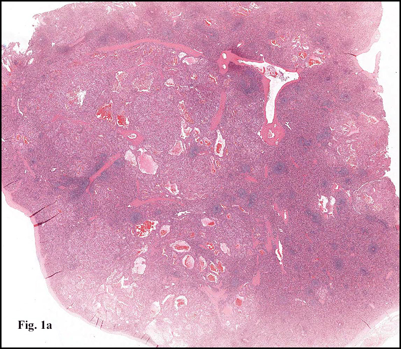
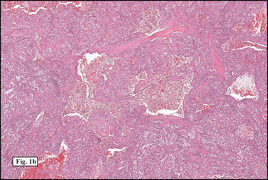
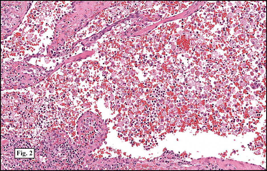
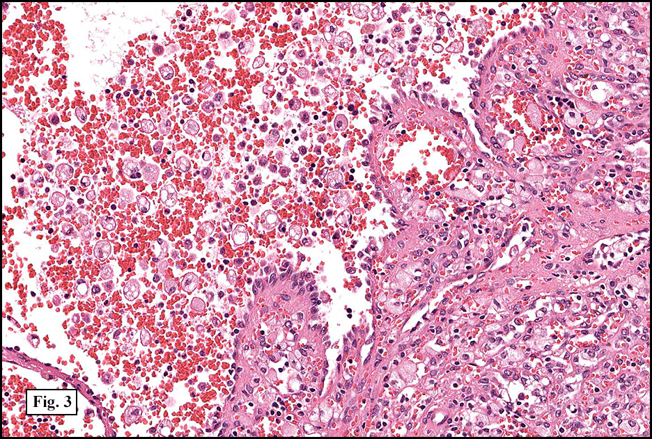
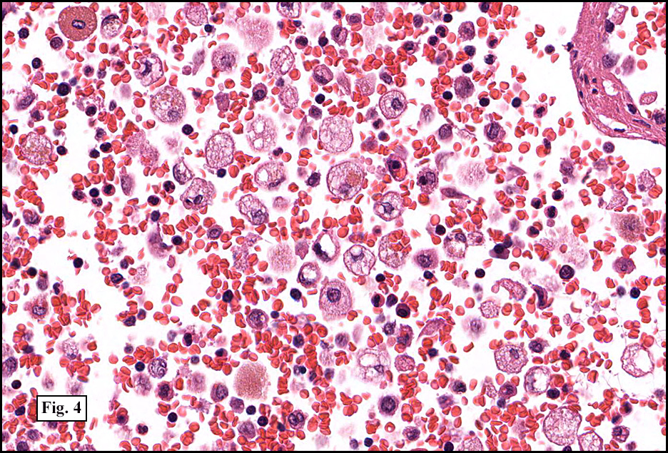
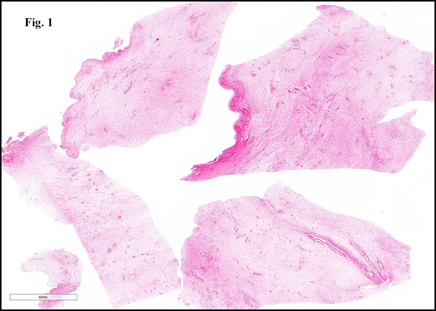
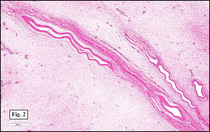
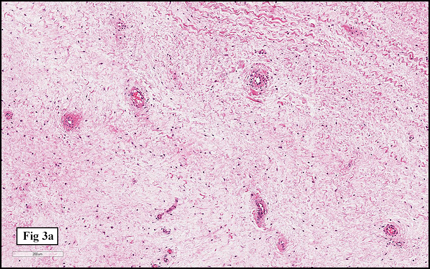
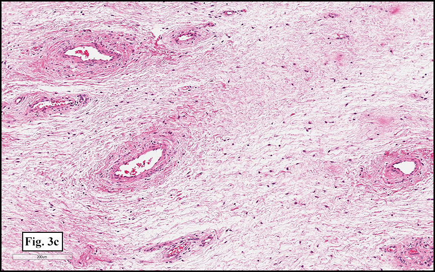
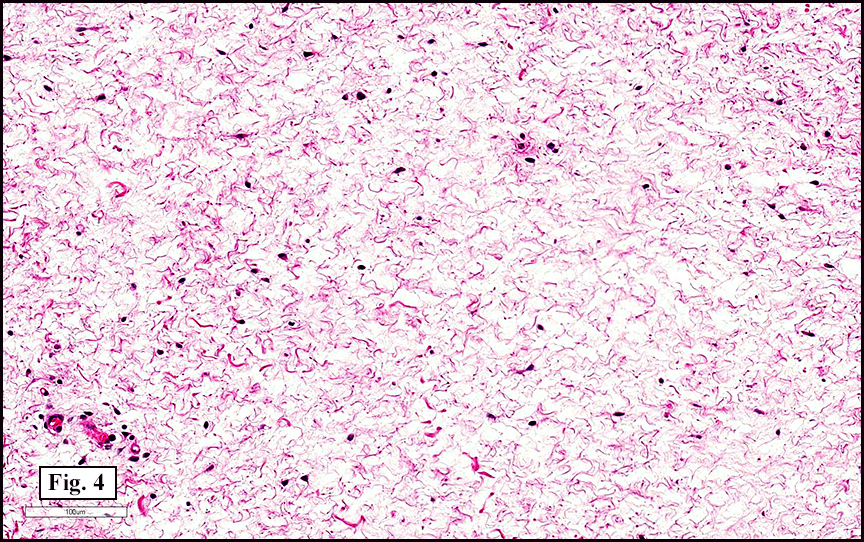
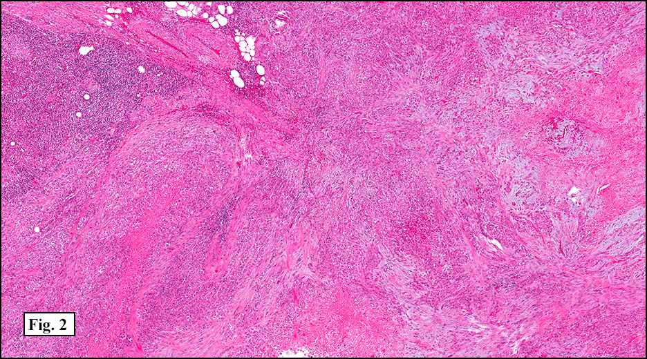
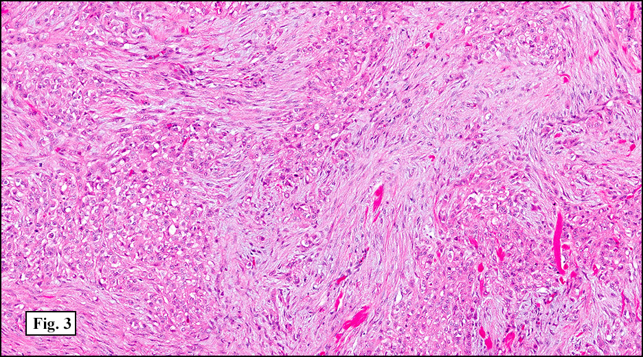
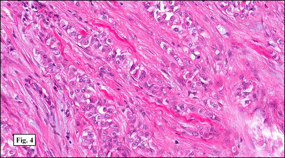
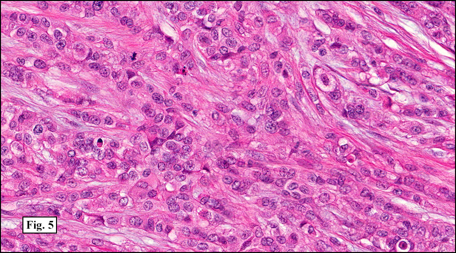
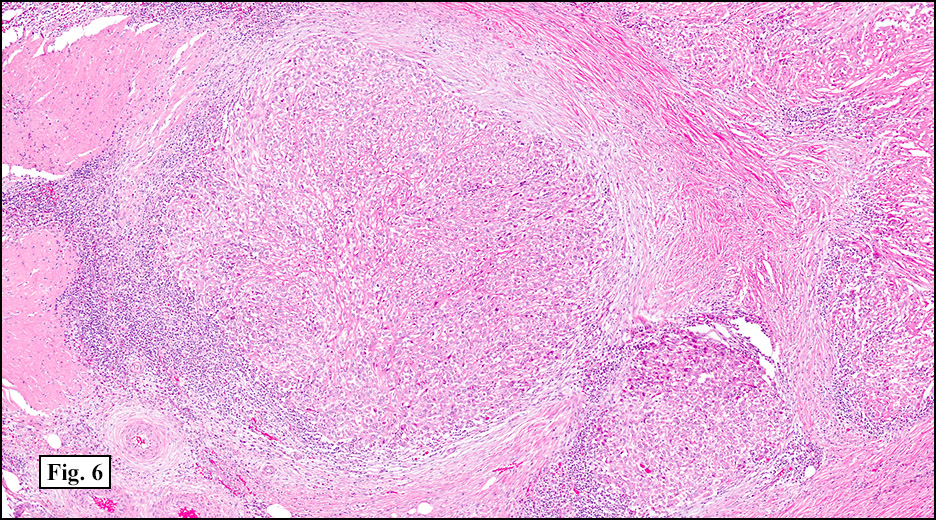
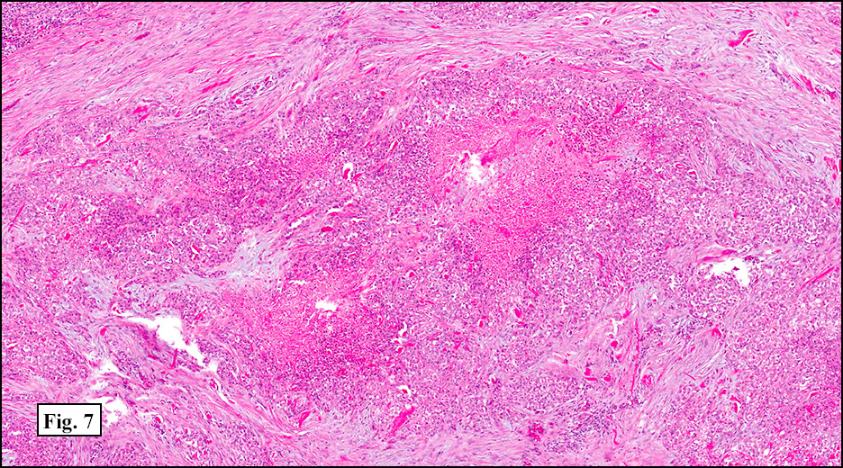
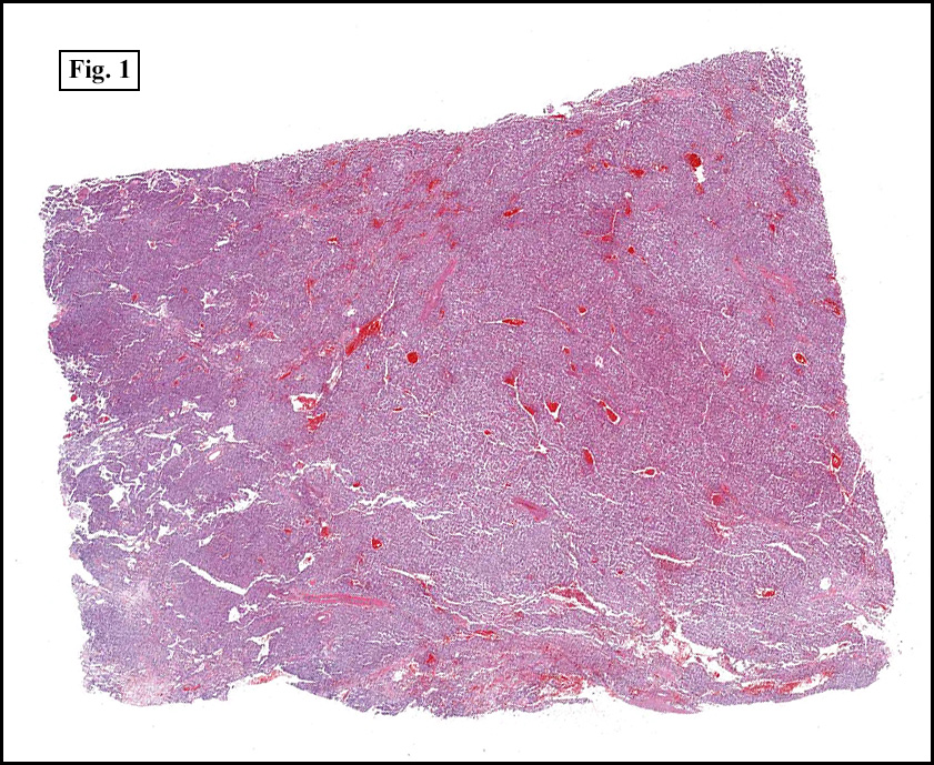
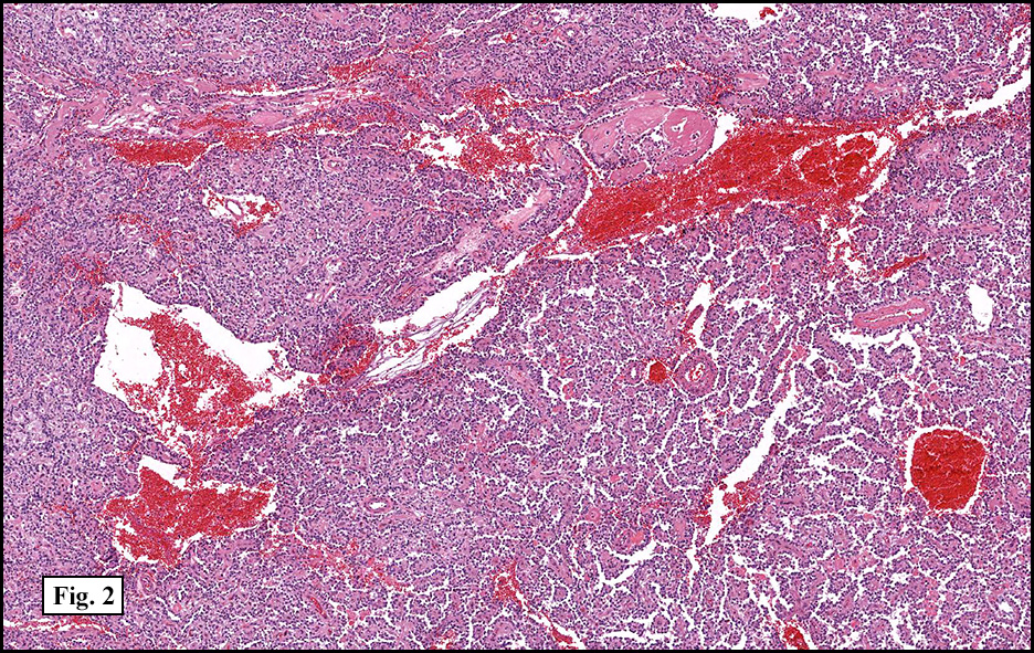
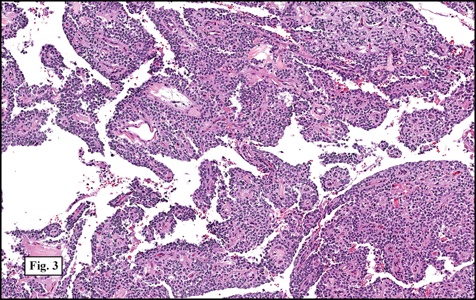
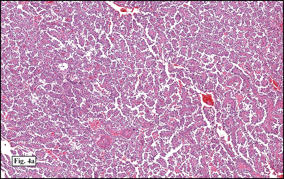
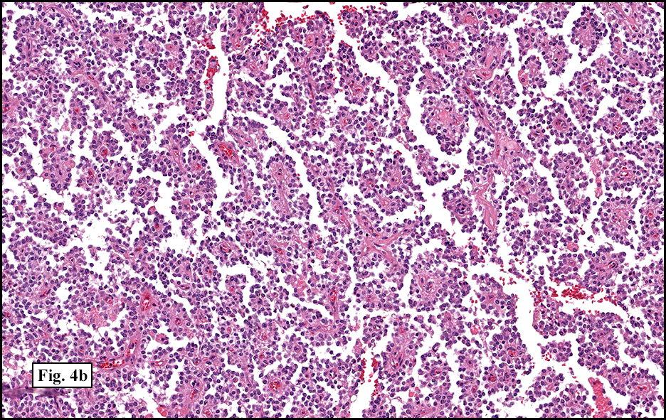
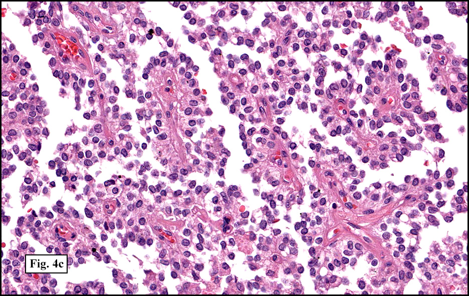
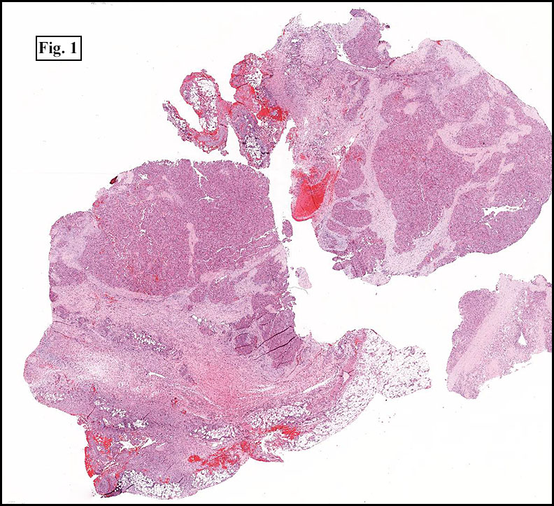
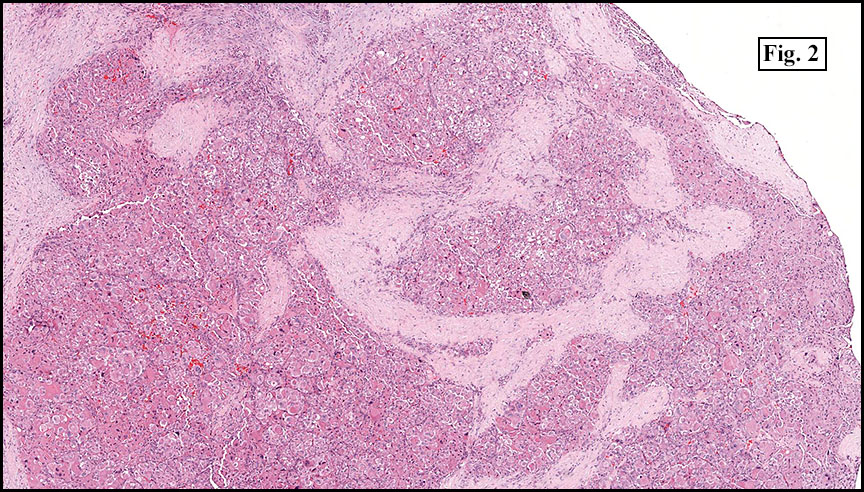
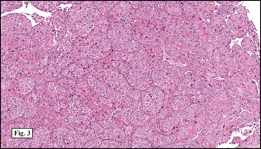
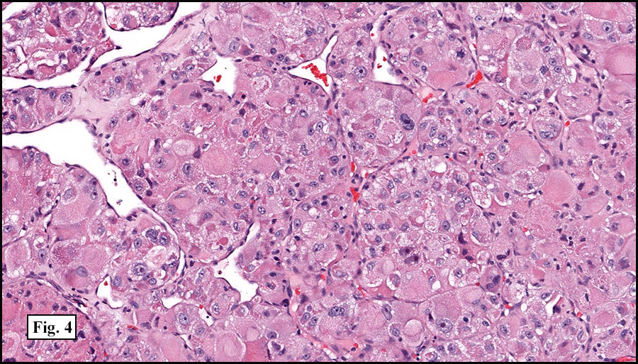
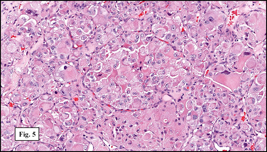
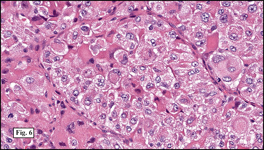
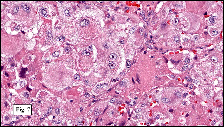
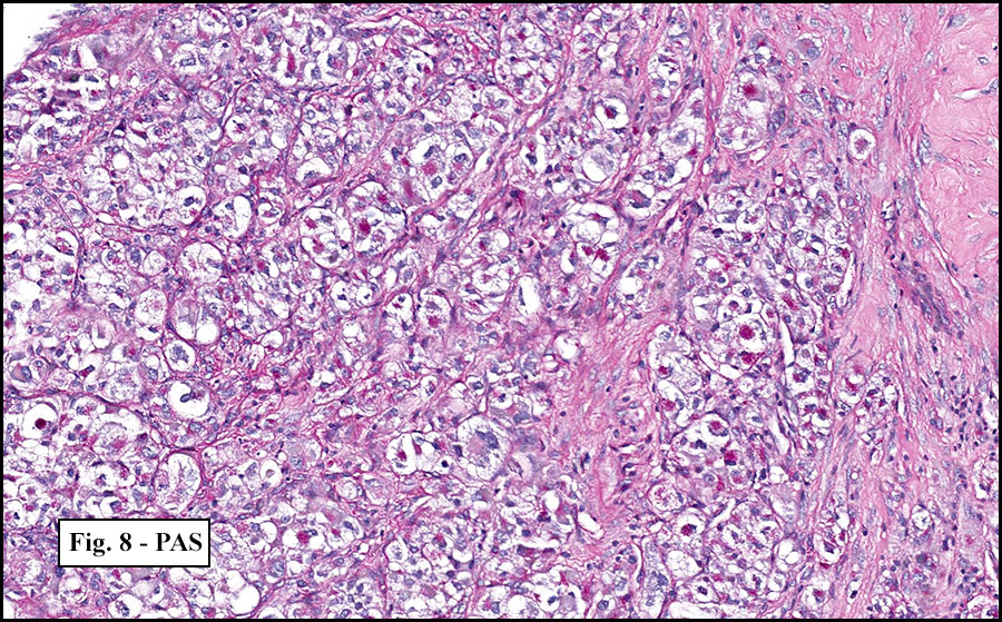
History: A 33-year-old man underwent resection of a 3.5 x 2.0 cm, inguinal, gray-white tumor with regional necrosis (Fig. 1). The neoplastic cells were eosinophilic and mostly epithelioid or spindloid (Figs. 2, 3). Their nuclei were vesicular and generally had one to two small nucleoli and showed occasional mitotic figures (Figs. 4, 5). They focally formed granuloma-like patterns with merging central cores of necrosis mimicking “caseous necrosis” seen in various infectious processes (Figs. 6, 7). Minimal pleomorphism was present. Neither glandular structures nor squamous differentiation was encountered. Immunostains were interpreted as follows:

Diagnosis: Epithelioid Sarcoma, Large Cell (“Proximal”, “Rhabdoid”) Variant

Rachel Conrad, MD1; Scott G. Silveira, MD2; Donald R. Chase, MD1,3

1. Department of Pathology and Human Anatomy, Loma Linda University and Loma
Linda Medical Center, Loma Linda, California
2. Department of Pathology, Oakwood Hospital and Medical Center, Dearborn,
Michigan
3. California Tumor Tissue Registry, Loma Linda, California

Discussion: First described by Franz Enzinger in 1970, epithelioid sarcoma (ES) has been accepted as a unique soft tissue sarcoma which typically pursues an indolent but relentless course with numerous recurrences. It generally occurs in young adults (10-35 years of age), but may occur in the very young as well as in the elderly. Males are affected twice as often as females.

The distal upper extremities are the most common sites, followed by the distal lower extremities, the upper proximal extremities, the trunk, and the head and neck. Case reports have also described rare lesions in the spine, gingiva, orbit, tongue, esophagus, jejunum, iliac bone, and intra-articular joints. Unlike most sarcomas, ES has a tendency to initially metastasize to regional lymph nodes. Lung metastases are common, and the tumor has a predilection for scalp and brain metastases.

Most ES’s present as painless, slow-growing, palpable firm masses in the soft tissue or dermis. Although the average size is 3-6 cm, deeper lesions are likely to be larger, multinodular, and attached to tendons or nerves which may result in localized neural symptoms including carpal tunnel syndrome or median nerve palsy. Antecedent trauma has been reported in up to 20% of cases. Rare reports also describe ES occurring in conjunction with squamous cell carcinoma, lung adenocarcinoma, neurofibromatosis type 2, acute lymphoblastic leukemia, and exposure to hydrazine fuel or Agent Orange. Elevated serum CA-125 has been reported in a few cases and may prove useful in tracking disease progression.

Radiographic findings often reveal a nonspecific solid or cystic multilobulated lesion with small speckled calcifications. Adjacent bone erosion and thinning may be seen, but invasive osseous destruction is rare.

The usual gross appearance of ES is of firm, non-encapsulated tan-white nodules with indistinct, infiltrating margins. Superficial tumors may have skin ulceration or sinus tract formation. Hemorrhage and/or necrosis is usually present.

As originally described, the tumor is nodular with central degenerative necrosis. The tumor is comprised of eosinophilic epithelioid cells admixed with polygonal to plump spindloid cells and dense intercellular collagen. Minimal pleomorphism is seen. Mitotic figures range from 1-14/10 HPF. Vascular invasion is much more common than neural invasion. Some tumors are lymphotrophic, showing a chronic inflammatory cell infiltrate around their peripheries. Calcification and osseous metaplasia may be present, but cartilaginous change is rare.

The classically defined subtypes, “necrobiotic” and “solid,” were extensively studied in the 1985 paper by Chase and Enzinger. Additional variants such as “fibrotic”, “angiomatoid” and “large cell” were also noted at that time, and have received substantial recent attention

1. The necrobiotic (“granulomatous”) growth pattern consists of coalescent cystic nodules lined by undulating ribbons of polygonal and spindled cells. The spaces are filled with necrotic debris or hyalinized collagen. The epithelial-appearing cells are mostly centrally located and the spindled elements are toward the periphery. No abrupt transition is seen. Nuclei are rounded and usually show small nucleoli. Minimal pleomorphism is seen.

2. The solid (“nodular”, “lobular”) growth pattern has a multinodular lobular growth pattern with polygonal and spindled cells similar to those seen in the necrobiotic form; however, no significant necrosis is seen. Nuclei typically are eccentrically located, pushed aside by the intermediate filaments vimentin and cytokeratin. As with the necrobiotic variant, the cells have distinct cytoplasmic borders.

3. Unlike the “classic” necrobiotic and solid growth patterns, the fibrotic (“fibroma-like”) variant of ES consists mostly of spindled cells, with a lesser epithelioid cell component. Cells reside in a fibrous stroma and show a vague storiform growth pattern resembling a cellular fibrous histiocytoma.

4. The large cell (“proximal,” “rhabdoid”) variant usually occurs in deep tissues of limb girdles or buttocks, vulva and perineum, and consists of polygonal cells which are larger and more pleomorphic than the other variants of ES. The cells have increased numbers of mitoses. Some cells may resemble rhabdomyoblasts. This variant may express smooth muscle actin; however, desmin positivity is lacking. The rhabdoid phenotype is more commonly found in tumors that are “proximal” or “truncal” but is not unique to the proximal form of epithelioid sarcoma. Since similar cytomorphology may be seen in other tumors (synovial sarcoma, extraskeletal myxoid chondrosarcoma, malignant mesothelioma, bladder carcinomas), it may be viewed simply as a pattern, similar to the patterns of pleomorphic “MFH” and HPC. In view of the increased risk of recurrence and metastasis that are associated with “rhabdoid cells” in general, and with an ES in a proximal location, this variant may have a faster progression.

5. The angiomatoid type is the rarest of the ES variants. Histologically the tumors show cystic, blood-filled spaces bordered by epithelioid and spindled cells. An endothelial lining to the spaces is not seen; neither is vasoformative activity observed. CD34 may be positive in up to 50% of the cases. Occasional CD31 positivity may occur, however, ES has never been shown to stain for HFVIII.

Cytology specimens usually are hypercellular, and show discohesive or loosely clustered medium-to-large epithelioid or spindled cells. The cytoplasm is well-defined and abundant, with occasional vacuoles and dense globoid paranuclear condensations. The eccentric nucleus shows occasional multinucleation, atypia and nucleoli. Closely associated with the cells is a characteristic hyaline fibrillar eosinophilic material that resembles basement membrane and stains magenta pink with May-Grunwald-Giemsa stain. Background collagen fragments, necrosis, or inflammation may also be observed.

Electron microscopy typically demonstrates striking paranuclear whorls or masses of intermediate filaments, along with prominent Golgi and free ribosomes.

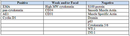
Staining patterns are similar for all variants of epithelioid sarcoma. The dense collagenous extracellular matrix highlights with Alcian blue and hyaluronidase. Trichrome stain yields a strong red hue to the cytoplasm. ES strongly stains with low and high-molecular weight cytokeratins, EMA, and vimentin. They are often positive for CD34 (~50%), nuclear cyclin D1, CD7, and CA-125. They may also be positive for muscle-specific actin and GLUT-1. Unlike other soft tissue sarcomas, ES often demonstrates intratumoral lymphatics by Lyve-1/podoplanin staining. Staining for p63 and cytokeratin 5/6 tends to be negative, as are S-100, neurofilaments, carcinoembryonic antigen, von Willebrand factor, factor VIII, and CD31. Frequently, loss of normal INI1 (SMARCB1) expression is observed.

A specific cell of origin has not yet been identified, although mesenchymal, endothelial, synovial, and fibrohistiocytic origins have been proposed. Genetic mutations in 11q, 18q11, and 22q11 have been occasionally observed; however, recent studies have identified inactivating mutations of the tumor suppressor gene INI1 (SMARCB1) at 22q11 in >90% of epithelioid sarcomas. This mutation can also be seen in some epithelioid malignant peripheral nerve sheath tumors, myoepithelial carcinomas, atypical teratoid/rhabdoid tumors of the central nervous system, and 80% of renal and malignant extrarenal rhabdoid tumors (MERT). The original definition of MERT was not always clearly defined in earlier studies and reports; thus, some adult cases of MERT may overlap with the large cell (‘rhabdoid’, ‘proximal-type’) variant of epithelioid sarcoma. Further studies are needed to establish more precise definitions based on the recent genetic findings.

Despite its usual appearance as a low to medium grade tumor, ES prognosis is very poor. It is exceptionally bad when occurring in axial (“proximal”) or deep locations. Increased aggressiveness is also seen in males, a tumor size (>5 cm), increased mitotic rates, and abnormal expression of dysadherin. The presence of hemorrhage, necrosis, vascular invasion, or an inadequate initial excision also portend poor prognosis. There is a high risk of tumor recurrence, often within the first year of diagnosis; however, recurrence may be seen as many as twenty years later. Metastases are frequent and often involve regional lymph nodes or lung. The overall 5 year survival is 50-85%, and the 10 year survival is 40-55%.

Because of ES known proclivity for relentless proximal progression, frequent recurrences, and metastases, the recommended therapy is early, complete excision by amputation or radical local excision. A few studies have reported equivocal results in limb-sparing versus amputation of localized lesions, but these study sizes were small and follow-up was limited. Regional lymph node dissection or at least sentinel lymph node biopsy should be performed, especially if radiographic findings are suspicious. Radiation and chemotherapy may provide some benefit, but cannot replace surgery. Recent chemotherapy have utilized include sunitinib malate, navelbine, anthracycline with ifosfamide, trabectedin, paclitaxel with 5-FU, and erlotinib (EGFR) with rapamycin. Long term follow-up is essential.

Chase and Enzinger’s 1985 paper highlighted a better prognosis in females and recommended investigation into hormone effects on tumor therapy. Recent case reports have described a connection to hormones in certain cases, including human chorionic gonadotropin expression with placenta metastases and the presentation and diagnosis of vulvar epithelioid sarcomas during pregnancy. Further research into hormone therapy may prove useful.

The differential diagnosis of ES includes benign inflammatory processes, epithelioid malignant peripheral nerve sheath tumor (MPNST), melanoma, epithelioid angiosarcoma, epithelioid sarcoma-like hemangioendothelioma, synovial sarcoma, ulcerating squamous cell carcinoma, and malignant extrarenal rhabdoid tumor (MERT).

• Numerous benign inflammatory processes may be considered in the differential for ES, including necrotizing infectious granuloma, necrobiosis lipoidica, granuloma annulare, rheumatoid nodule, nodular fasciitis, fibrous histiocytoma, and fibromatosis. Compared to these processes, the individual cells in epithelioid sarcoma are more sharply defined, larger, and more eosinophilic with prominent epithelioid features and architectural nodularity. The nuclei are more vesicular than are seen in palisading histiocytes. When occurring in the skin, the necrobiotic variant of epithelioid sarcoma mimics granuloma annulare and rheumatoid nodule, but may easily be distinguished by the expression of cytokeratin and by CD34 positivity (~50%). Nodular fasciitis tends to be GLUT-1 negative, while ES may show positivity (~50%).

• ES may grow along nerve bundles, mimicking epithelioid malignant peripheral nerve sheath tumor (MPNST). However, MPNSTs tend to be positive for S100 (approximately 50%) and are uniformly negative for cytokeratins. EMA, however, is only rarely expressed in MPNST.

• Melanoma may also be confused with ES, especially the large cell (“rhabdoid”, “proximal”) variant. Staining for S100 and other melanocyte markers (HMB-45, Melan A) will likely be positive in melanoma but are negative in ES.

• Epithelioid angiosarcoma may share ES’s loss of cellular cohesion, secondary hemorrhage, and large epithelioid cells with cytoplasmic vacuolation. Although epithelioid angiosarcoma may express cytokeratins and CD34; it strongly stains for endothelial markers such as von Willebrand factor and CD31.

• Epithelioid sarcoma-like hemangioendothelioma (EH) occurs in the extremities of young to middle aged adults and microscopically has vascular lumen formations, which are mimicked by ES’s occasional intracellular lipid droplets. Although EH may also express cytokeratin and vimentin, it marks for vascular markers CD31, HFVIII, and FLI-1, and unlike ES, CD-34 is uniformly negative.

• Biphasic synovial sarcoma shows an abrupt transition between epithelial and spindle patterns, unlike ES’s smoother transition. Synovial sarcoma tends to affect the proximal portion of extremities and is less likely to involve superficial structures and skin. It may share positive immunohistochemical staining for cytokeratin and EMA with ES, but stains negatively for CD34 and shows a characteristic (X;18) translocation.

• Ulcerating squamous cell carcinoma may share a similar clinical presentation as ES; however, it may demonstrate keratin pearls and dyskeratosis in adjacent epithelium, along with strong positivity for CD5/6 and p63. It lacks cyclinD1 (nuclear) and CD34 expression.

• Malignant extrarenal rhabdoid tumor (MERT) in soft tissue should be reserved for tumors with prominent rhabdoid features but lacking a clear line of differentiation. This aggressive tumor typically presents in children as a fleshy, necrotic lesion. It may overlap with the large cell (‘proximal’, ‘rhabdoid’) variant of ES, sharing similar rhabdoid cells, PAS-positive hyaline inclusions of intermediate filament whorls, staining patterns, and INI1 gene mutations (22q11).

Suggested Reading:

1. Weiss SW, Goldblum JR. Enzinger & Weiss’s Soft Tissue Tumors. 5th ed. Philadelphia: Mosby-Elsevier, 2008:1191-1203, 1208-1213.
2. Chase DR. Do “rhabdoid features” impart a poorer prognosis to proximal-type epithelioid sarcomas? Adv Anat Pathol. 1997;4(5):293-299. Regarding: Guillou L, Wadden C, Coindre JM, Krauz T, Fletcher CDM. ‘Proximal-type’ epithelioid sarcoma, a distinctive aggressive neoplasm showing rhabdoid features: clinicopathologic, immunohistochemical, and ultrastructural study of a series. Am J Surg Pathol. 197;21(2):130-146.
3. Chase DR, Enzinger FM. Epithelioid sarcoma: diagnosis, prognostic indicators, and treatment. Am J Surg Pathol. 1985;9(4):241-263.
4. Enzinger FM. Epithelioid sarcoma: a sarcoma simulating a granuloma or a carcinoma. Cancer. 1970;26:1029-1041.
5. Chase DR. Letters to the editor: rhabdoid versus epithelioid sarcoma. Am J Surg Pathol. 1990;14(8):792-793. Regarding: Perrone T, Swanson PE, Twiggs L, et al. Malignant rhabdoid tumor of the vulva: is distinction from epithelioid sarcoma possible? Am J Surg Pathol. 1989;13:848-858.
6. Chase DR. Epithelioid sarcoma. California Tumor Tissue Registry. Berlin, Germany. September 12, 2001. Seminar Presentation of Epithelioid Neoplasms.
7. Sur M, Nayler SJ. Proximal epithelioid sarcoma – a misnomer. Histopathology. 2001;39(6):641-643.
8. Rakheja D, Wilson KS, Meehan J, Schultz RA, Gomez AM. “Proximal-type” and classic epithelioid sarcomas represent a clinicopathologic continuum: case report. Pediatr Dev Pathol. 2005;8(1):105-114.
9. Boutilier R, Walsh N. Pathologic quiz case: cutaneous nodule of 3-year duration: Epithelioid sarcoma. Arch Pathol Lab Med. 2002;126(5):625-626.
10. Gupta H, Davidoff AM, Rao BN, Jenkins JJ, Spunt SL. Neonatal epithelioid sarcoma: a distinct clinical entity? J Pediatr Surg. 2006;41(7):e9-e11.
11. Keelawat S, Shuangshoti S, Kittikowit W, Lerdlum S, Thorner PS. Epithelioid sarcoma of the parotid gland of a child. Pediatr Dev Pathol. 2009;12(4):301-306.
12. Bento A, Baptista H, Pinheiro C, Martinho F. Jejuno-jejunal invagination caused by epithelioid sarcoma: a case report. J Med Case Rep. 2009;3:89.
13. Maggiani F, Debiec-Rychter M, Ectors N, Lerut A, Sciot R. Primary epithelioid sarcoma of the oesophagus. Virchows Arch. 2007;451(4):835-838.
14. Raoux D, Peoc’h M, Pedeutour F, Vaunois B, Decouvelaere AV, Folpe AL. Primary epithelioid sarcoma of bone: report of a unique case, with immunohistochemical and fluorescent in situ hybridization confirmation of INI1 deletion. Am J Surg Pathol. 2009;33(6):954-958.
15. Kosemehmetoglu K, Kaygusuz G, Bahrami A, Raimondi SC, Kilicarslan K, Yildiz Y, Folpe AL. Intra-articular epithelioid sarcoma showing mixed classic and proximal-type features: report of 2 cases with immunohistochemical and molecular cytogenetic INI-1 study. Am J Surg Pathol. 2011;35(6):891-897.
16. Choi SY, Kim YH, Kwon JB, Suh JH, Shin OR, Hong SH. Epithelioid sarcoma metastatic to the lung as pulmonary cysts without other metastatic manifestation. J Thorac Oncol. 2008;3(5):532-533.
17. Barnoud R, Collard-Frachon S, de la Roche E, Vasiljevic A, Thomson V, Ranchere D, Devouassoux G, Devouassoux-Shisheborn M. Lung metastases of epithelioid sarcoma revealed by bilateral spontaneous pneumothorax: a pathological diagnosis. Ann Pathol. 2010;30(2):139-142.
18. Jashnani KD, Dhume VM, Bahal NK. Epithelioid sarcoma and squamous cell carcinoma arising in a burn scar. Indian J Dermatol. 2011;56(5):585-586.
19. Kagami S, Saeki H, Idezuki T, Yano S, Kawabata Y, Okochi H, Asahina A, Nakagawa K, Tamaki K. Epithelioid sarcoma associated with lung adenocarcinoma. J Dermatol. 2005;32(11):904-908.
20. Kimball Dalton VM, Gelber RD, Li F, Donnelly MJ, Tarbell NJ, Sallan SE. Second malignancies in patients treated for childhood acute lymphoblastic leukemia. J Clin Oncol. 1998; 16(8):2848-2853.
21. Rose DS, Fisher C, Smith ME. Epithelioid sarcoma arising in a patient with neurofibromatosis type 2. Histopathology. 1994;25(4):379-380.
22. Kierson ME, Iyer D, Fyfe B. Metastatic epithelioid sarcoma in a non-healing ventricular device pocket. J Heart Lung Transplant. 2010;29(11):1319.
23. Hoshino M, Kawashima H, Ogose A, Kudo N, Ariizumi T, Hotta T. Serum CA-125 expression as a tumor marker for diagnosis and monitoring the clinical course of epithelioid sarcoma. J Cancer Res Clin Oncol. 2010;136(3):457-464.
24. Sakamoto A, Jono O, Hirahashi M, Oya M, Iwamoto Y, Arai K. Epithelioid sarcoma with muscle metastasis detected by positron emission tomography. World J Surg Oncol. 2008;6:84.
25. Dion E, Forest M, Brasseur JL, Amoura Z, Grenier P. Epithelioid sarcoma mimicking abscess: review of the MRI appearances. Skeletal Radiol. 2001;30(3):173-177.
26. Kozawa E, Irisawa M, Heshiki A, Okagaki R, Shimizu Y. Magnetic resonance imaging findings of vulvar epithelioid sarcoma. Radiat Med. 2008;26(6):376-378.
27. Barwad A, Dey P, Das A. Fine needle aspiration cytology of epithelioid sarcoma. Diagn Cytopathol. 2010;39(7):517-520.
28. Lemos MM, Chaves P, Mendonca ME. Is preoperative cytologic diagnosis of epithelioid sarcoma possible? Diagn Cytopathol. 2008;36(11):780-786.
29. Laskin WB, Miettinen M. Epithelioid sarcoma: new insights based on an extended immunohistochemical analysis. Arch Pathol Lab Med. 2003;127(9):1161-1168.
30. Miettinen M, Fanburg-Smith JC, Virolainen M, Shmookler BM, Fetsch JF. Epithelioid sarcoma: an immunohistochemical analysis of 112 classical and variant cases and a discussion of the differential diagnosis. Hum Pathol. 1999;30(8):934-942.
31. Izumi T, Oda Y, Hasegawa T, Nakanishi Y, Iwasaki H, Sonobe H, et al. Prognostic significance of dysadherin expression in epithelioid sarcoma and its diagnostic utility in distinguishing epithelioid sarcoma from malignant rhabdoid tumor. Mod Pathol. 2006;19(6):820-831.
32. Chu PG, Arber DA, Weiss LM. Expression of T/NK-cell and plasma cell antigens in nonhematopoietic epithelioid neoplasms: an immunohistochemical study of 447 cases. Am J Clin Pathol. 2003;120(1):64-70.
33. Lin L, Hicks D, Xu B, Sigel JE, Bergfeld WF, Montgomery E, et al. Expression profile and molecular genetic regulation of cyclin D1 expression in epithelioid sarcoma. Mod Pathol. 2005;18(5):705-709.
34. Mahendra G, Kliskey K, Williams K, Hollowood K, Jackson D, Athanasou NA. Intratumoral lymphatics in benign and malignant soft tissue tumours. Virchows Arch. 2008;453(5):457-464.
35. Orrock JM, Abbott JJ, Gibson LE, Folpe AL. INI1 and GLUT-1 expression in epithelioid sarcoma and its cutaneous neoplastic and nonneoplastic mimics. Am J Dermatopathol. 2009;31(2):152-156.
36. Hornick JL, Dal Cin P, Fletcher CD. Loss of INI1 expression is characteristic of both conventional and proximal-type epithelioid sarcoma. Am J Surg Pathol. 2009;33(4):542-550.
37. Kohashi K, Izumi T, Oda Y, Yamamoto H, Tamiya S, Taguchi T. Infrequent SMARCB1/INI1 gene alteration in epithelioid sarcoma: a useful tool in distinguishing epithelioid sarcoma from malignant rhabdoid tumor. Human Pathol. 2009;40(2):349-355.
38. Argenta PA, Thomas S, Chura JC. Proximal-type epithelioid sarcoma vs. malignant rhabdoid tumor of the vulva: a case report, review of the literature, and an argument for consolidation. Gynecol Oncol. 2007;107(1):130-135.
39. Guzzetta AA, Montgomery EA, Lyu H, Hooker CM, Meyer CF, Loeb DM, et al. Epithelioid sarcoma: one institution’s experience with a rare sarcoma. J Surg Res. 2012;177(1):116-122.
40. Gasparini P, Facchinetti F, Boeri M, Lorenzetto E, Livio A, Gronchi A, et al. Prognostic determinants in epithelioid sarcoma. Eur J Cancer. 2011;47(1):287-295.
41. Sakharpe A, Lahat G, Gulamhusein T, Liu P, Bolshavkov S, Nguyen T, et al. Epithelioid sarcoma and unclassifiable sarcoma with epithelioid features: clinicopathological variables, molecular markers, and a new experimental model. Oncologist. 2011;16(4):512-522. .
42. Jawad MU, Extein J, Min ES, Scully SP. Prognostic factors for survival in patients with epithelioid sarcoma: 441 cases from the SEER database. Clin Orthop Relat Res. 2009;467(11):2939-2948.
43. Jones RL, Constantinidou A, Olmos D, Thway K, Fisher C, Al-Muderis O, Scurr M, Judson IR. Role of palliative chemotherapy in advanced epithelioid sarcoma. Am J Clin Oncol. 2012;35(4):351-357.
44. Xie X, Ghadimi MP, Young ED, Belousov R, Zhu QS, Liu J, Lopez G, Colombo C, Peng T, Reynoso D, Hornick JL, Lazar AJ, Lev D. Combining EGFR and mTOR blockage for the treatment of epithelioid sarcoma. Clin Cancer Res. 2011;17(18):5901-5912.
45. De Visscher SA, van Ginkel RJ, Wobbes T, Veth RP, Ten Heuvel SE, Suurmeijer AJ, Hoekstra HJ. Epithelioid sarcoma: still an only surgically curable disease. Cancer. 2006;107(3):606-612.
46. Steffensen TS, Gilbert-Barness E, Wagoner MJ, Bui MM, Browarsky IL. Human chorionic gonadotropin producing epithelioid sarcoma metastatic to the placenta. Fetal Pediatr Pathol. 2008;27(6):282-291.
47. Rai H, Odunsi K, Kesterson J, et al. Epithelioid sarcoma of the vulva in a 17-year-old pregnant woman. Appl Immunohistochem Mol Morphol. 2009;17(3):270-273.
48. Moore RG, Steinhoff MM, Granai CO, DeMars LR. Vulvar epithelioid sarcoma in pregnancy. Gynecol Oncol. 2002;85(1):218-222.

April 2013: A 36 year old woman with a mass in the left upper quadrant

History: A 36 year old woman presented with early satiety, recent weight gain and dyspnea on exertion. She was found to have a palpable left upper quadrant mass, which upon MRI was mostly solid. The excised pancreatic tumor was 23 by 17 cm, well circumscribed and surrounded by a thin capsule or pseudocapsule. Sectioning showed it be mottled gray and brown, friable, and to have prominent fibrous septa.

Microscopically, tumor cells were small, rounded and uniform. They lacked significant pleomorphism, atypism, or mitotic figures. The tumor was mostly solid, without significant cystic change (Fig. 1). Vascularity was prominent and included medium-sized vessels many of which contained thrombi (Fig. 2). Necrosis, however, was scant. Although a sinusoidal growth pattern was focally present (Fig. 3), most of the tumor pattern was “papillary” (Figs. 4a, 4b, 4c).

Diagnosis: “Solid Pseudopapillary Tumor of the Pancreas”

Cody S. Carter, MSIV, Donald R. Chase, M.D.
Department of Pathology and Human Anatomy, Loma Linda University and Medical Center, Loma Linda, California
California Tumor Tissue Registry, Loma Linda, California

Discussion: Solid pseudopapillary tumor of the pancreas (SPT) is a rare tumor, previously known as ”solid cystic”, “papillary cystic”, and “solid and papillary epithelial neoplasm”. About 95% of cases occur in women, and the average age at presentation is in the mid-20s. The tumor does not spare any age group, although it rarely occurs prior to adolescence. The most common presenting symptoms are abdominal pain followed by a palpable abdominal mass. Other symptoms include nausea, vomiting, fever, weight loss, and jaundice. Up to 15% cases are asymptomatic, detected incidentally on imaging studies. Because of this, the tumors can reach very large sizes with the average size being 7 cm at the time of resection.

Grossly, SPT is usually a well circumscribed mass with a yellow-brown, hemorrhagic cut surface. The tumors are solid but undergo cystic degeneration with increased size, and as a result are often soft and friable. Because of the tendency for the tumors to go undetected until reaching a large size, there may be a significant cystic component, sometimes mimicking a pancreatic pseudocyst. These cysts often contain hemorrhagic fluid.

Microscopically, SPT demonstrates solid nests of largely uniform epithelioid cells intermixed with small blood vessels. The characteristic pseudopapillae are formed as the cells further away from the blood vessels degenerate and become dyscohesive. Microcystic and cyst-like spaces are often present and are formed by degenerative changes. The tumor cells exhibit a moderate amount of delicate, eosinophilic cytoplasm. The cytoplasm often contains diastase-resistant PAS-positive eosinophilic hyaline globules. These globules are composed of alpha-1 antitrypsin. Nuclei are round to ovoid and frequently feature grooves and membrane irregularities, but do not usually exhibit pleomorphism and mitotic figures.

Cytology is useful to gain a preoperative diagnosis and direct surgical intervention, and is often necessary because of a lack of specific radiographic features that differentiate this entity from other cystic lesions. FNA biopsies usually demonstrate highly cellular material with branching, delicate papillary structures consisting of fibrovascular cores covered by one to a few cell layers. Tumor cells are of uniform size and shape, with pale ill-defined cytoplasm. The round or ovoid nuclei feature finely granular chromatin which may have grooves. Hyaline globules are easily identified by their red color on Giemsa stain. Foamy histiocytes are also present in varying quantities.

Of particular interest is the lack of a consensus on the cellular origin of SPT despite extensive immunohistochemical and genetic studies. Immunohistochemistry, while exceedingly helpful in other diagnoses, have not been particularly useful in SPT. The tumor cells are diffusely positive for vimentin and galectin-3, and most tumors demonstrate positivity for neuron specific enolase, α-1-antitrypsin, and CD56, while few cases show distinct immunoreactivity with cytokeratin, synaptophysin, chromogranin, trypsin, and S-100 protein. The weak reactivity for these markers provides no convincing evidence for ductal, acinar, or endocrine differentiation. Also of interest is the occurrence of this tumor almost exclusively in women. This has led many experts to believe that hormonal signaling plays a major role in tumorigenesis, and indeed, a high proportion of tumors have been shown to be immunoreactive to both the progesterone receptor and estrogen receptor-β. However, the similarity between affected men and women in regards to growth pattern and affected age groups is puzzling if hormone receptors prove to be involved in the evolution of this neoplasm. Despite this, hormonal manipulation may be indicated.

Molecular analysis of SPT demonstrates mutations in the β-catenin gene (CTNNB1) on chromosome 3p in more than 90% of tested cases. These mutations result in the inactivation of one of the glycogen synthase kinase-3β phosphorylation sites on the β-catenin protein, which inhibits the degradation of β-catenin. This lack of degradation leads to a buildup of β-catenin in the cytoplasm and nucleus of the tumor cells, resulting in a change in staining pattern for β-catenin protein antibodies. Whereas normal cells have a membranous staining pattern for β-catenin, tumor cells of SPT have an abnormal nuclear staining pattern. The consistency of this nuclear staining makes this marker a diagnostically important adjunct to other immunostains when histology cannot be confidently relied upon. The activation of the β-catenin protein also stimulates transcription of a number of genes that promote neoplasia including cyclin D1 and c-myc. Another effect of the mutation is an interaction between the β-catenin protein with E-cadherin, resulting in a loss of the membranous labeling for the extracellular domain of E-cadherin that is normally seen in non-neoplastic pancreas. Due to the integral role that E-cadherin plays in cell-cell junctions, this disruption has been postulated as the reason for the dyscohesion seen between tumor cells and thus the cystic degeneration often seen grossly.

SPT is a malignant tumor with low metastatic potential. It has a 5-year survival rate of 95%, and patients are almost always cured by surgical resection. Metastases rarely occur after resection if not already present at the time of diagnosis, and even with metastases, patients may often live for many years. As a result, local metastases and recurrence are not absolute contraindications to resection. Some patients with metastatic disease have survived more than 10 years after surgery, and in contrast to other pancreatic neoplasms, tumor debulking is recommended. In rare cases of unresectable SPTs, radiotherapy has been shown to be effective.

Differential diagnosis:

• Acinar cell carcinoma typically affects elderly patients and only rarely have cystic variants been seen. Similar to SPT, these tumors consist of uniform cells with granular cytoplasm that can stain positively with diastase-resistant PAS. However, the cells are arranged in acinar and trabecular patterns, and immunohistochemistry demonstrates widespread positivity for cytokeratin, trypsin and chymotrypsin, while vimentin is usually negative.

• Pancreatoblastoma typically occurs in the first decade and is characterized by epithelial cells organized in sheets, acini and squamoid corpuscles, surrounded by a fibrous stroma. In contrast to SPT, it is negative for vimentin and strongly positive for cytokeratin and trypsin.

• Pancreatic endocrine neoplasm (PEN) shares similar cellular characteristics with SPT, featuring bland, uniform cells with finely granular cytoplasm and ovoid nuclei. These cells are typically in solid and trabecular patterns without papillary structures. The immunophenotype of PEN is consistently reactive for cytokeratin and neuroendocrine markers such as chromogranin and synaptophysin. Vimentin, if present, is usually weak and focal.

Suggested Reading:

Adams AL, Siegal GP, Jhala NC. Solid pseudopapillary tumor of the pancreas. A review of salient clinical and pathologic Features. Adv Anat Pathol. 2008 Jan; 15(1):39-45.

Chanjuan S, Daniels JA, Hruban RH. Molecular characterization of pancreatic neoplasms. Adv Anat Pathol. 2008 Jul; 15(4):185-95.

Papavramidis T, Papavramidis S. Solid pseudopapillary tumors of the pancreas: Review of 718 patients reported in English literature. J Am Coll Surg. 2005 Jun; 200(6):965-972.

Adsay VN. Cystic neoplasia of the pancreas: pathology and biology. J Gastrointest Surg. 2008 Mar; 12(3):401-404.

Klimstra DS, Wenig BM, Heffess CS. Solid-pseudopapillary tumor of the pancreas: a typically cystic carcinoma of low malignant potential. Semin Diagn Pathol. 2000 Feb; 17(1):66-80.

Yu PF, Hu ZH, Wang XB, Guo JM, Cheng XD, Zhang, YL, Xu Q. Solid pseudopapillary tumor of the pancreas: A review of 553 cases in Chinese literature. World J Gastroenterol 2010 Mar;16(10)1209-1214.

Pettinato G, Di Vizio D, Manivel JC, Pambuccian SE, Somma P, Insabato L. Solid-pseudopapillary tumor of the pancreas: A neoplasm with distinct and highly characteristic cytological features. Diagnostic Cytopathology 2002 Dec; 27(6): 325-334.

Geers C, Pierre M, Jean-Francois G, Birgit W, Pierre D, Jacques R, Christine S. Solid and pseudopapillary tumor of the pancreas—review and new insights into pathogenesis. Am J Surg Pathol. 2006 Oct; 30(10): 1243-1249.

Odze R, Goldblum J. Surgical Pathology of the GI Tract, Liver, Biliary Tract, and Pancreas (2nd ed). Philadelphia: Saunders/Elsevier Inc. 944-48, 2009.

March 2013: A 33 year old woman with a mass in the thigh

History: A 33 year-old woman presented with a palpable, 6 cm mass in the thigh. At surgery, it was found to deeply reside within the anterior compartment where it involved muscle. Osseous involvement was not seen. The excised specimen was poorly circumscribed, and had a cut surface that was fleshy tan-grey.

The tumor was compartmentalized, showing islands of cells separated into large and small nodules by fibrous stroma (Figs. 1,2). Smaller nests were bounded by sinusoidal vascular structures, a few of which were dilated (Figs. 3,4). Some of these smaller compartments showed central necrosis and many exhibited clearing around the cells with poor cellular cohesion (Fig. 5). The cells were uniform in both shape and size, and contained abundant pink granular cytoplasm and eccentrically placed, rounded, vesicular nuclei, most having prominent nucleoli (Figs. 6,7). There were scattered binucleated cells (Fig. 7). Mitoses were rare and no significant pleomorphism was present. A PAS stain showed abundant PAS positive granular and crystalloid material within the cytoplasm of many cells (Fig. 8).

Diagnosis: “Alveolar Soft Part Sarcoma”

Cody S. Carter, MSIV, Donald R. Chase, M.D.
Department of Pathology and Human Anatomy, Loma Linda University and Medical Center, Loma Linda, California
California Tumor Tissue Registry, Loma Linda, California

Discussion: Alveolar soft part sarcoma (ASPS) is a rare entity of unknown origin accounting for less than 1% of sarcomas. It usually occurs in ages 15-35, and predominantly affects females, especially in younger patients. In adults, the majority of tumors occur in the lower extremities, especially in the anterior thigh and buttocks. In children, these tumors typically present in the head and neck, especially the orbit and tongue, and tend to be smaller, likely due to earlier detection in these locations. The tumor most commonly presents as a slow-growing painless mass, but can often present with respiratory symptoms or neurologic symptoms like headache, visual changes and nausea due to their striking proclivity for early metastasis to the lung and brain. ASPS are typically very vascular and may be associated with an audible bruit. Radiologically, the diagnosis my be suggested byits hypervascularity with prolonged capillary staining and locally dilated veins on angiography and CT scan.

Grossly, ASPS is usually poorly circumscribed and highly vascular, with increased risk of significant blood loss at the time of surgery. The cut surface is usually yellow-white to gray and can feature areas of necrosis or hemorrhage, making the tumor soft and friable.

Microscopically, ASPS shows little variability. Compartments and nests are divided by fibrous stroma, thin-walled sinusoidal channels, usually lined by flattened endothelial cells. A lack of cellular cohesion and central necrosis is often seen. The dominant pattern of peripherally viable tumor cells with partial central clearing likens this neoplasm to the alveoli of the lung. While these features generally make a histologic diagnosis straightforward, the nest-like pattern can, on rare occasion, be absent, and large sheets of cells are seen together. This variant is primarily seen in infants and young children.

Cells within the nests are large, rounded, and fairly uniform, with eosinophilic granular cytoplasm and vesicular nuclei with distinct nucleoli. Cellular pleomorphism is only rarely present. Vascular invasion is almost invariably present, and reflects the tendency for early blood metastasis. The characteristic histologic pattern can usually be relied on for diagnosis, and the unreliable staining exhibited by ASPS makes immunohistochemistry most important in excluding other neoplasms. Of the histologic stains, PAS will likely show positively staining, diastase-resistant granules or rhomboid and rod-shaped crystals within the cytoplasm. These structures are composed of monocarboxylate tansporter 1 and its chaperone protein CD147. ASPS fails to express cytokeratin, EMA, neurofilament, GFAP, serotonin, synaptophysin and chromogranin. It can sometimes be positive for S-100 and neuron-specific enolase, but these seem to have no significant diagnostic value. Stains for MyoD1 and myogenin have been controversially positive, with one study using positivity in several tumors as support for skeletal muscle differentiation, but several subsequent studies have since failed to reproduce the results.

Cytogenetics may be a useful tool as the unbalanced translocation t(X;17)(p11.2q25) leads to an ASPL-TFE3 fusion gene that is both sensitive and specific for ASPS within the realm of soft tissue sarcomas (a small subset of pediatric renal cell carcinomas have also shown this same fusion gene). This can be more easily confirmed with an immunohistochemical stain for TFE3. This fusion gene has been linked to the overexpression of the promitotic MET receptor tyrosine kinase, which has suggested a model for oncogenesis, as well as a source of targeted therapy that is currently being tested in ongoing clinical trials.

Alveolar soft tissue sarcoma displays indolent growth, but overall prognosis is poor due to early metastasis, which can often be present at initial presentation. ASPS differs from most other adult sarcomas in that local recurrence is less likely after radical excision, but late metastasis can occur years after resection of the primary, and indeed metastases upwards of 30 years later have been recorded. Poorer prognosis is associated with increasing age and increasing tumor size at diagnosis, as well as metastases at initial presentation. Histologic grading is not contributory to prognosis.

Due to slow growth of the tumor, radical surgical excision of both primary and metastatic lesions is the most effective treatment, and conventional radiotherapy and chemotherapy have thus far been shown to be ineffective. Recent advances in targeted therapy are promising, and clinical trials are ongoing.

Differential diagnosis includes:

• Renal cell carcinoma (RCC) can look similar to the histology of ASPS, especially because some variants of the sarcoma can have less eosinophilic cells, and some RCCs can have quite eosinophilic cytoplasm. RCC can be distinguished by its distinct lack of PAS-positive crystalline structures or granules. Also, RCC is immunoreactive to EMA and keratin, but ASPS shows absent staining. TFE3 positivity can also be helpful, but some RCCs can express this antigen in the pediatric population. In this situation, radiologic correlation with renal imaging may be essential.

• Paraganglioma can be differentiated by its positivity for neuroendocrine markers, which are consistently negative in ASPS. Vesicular nuclei and prominent nucleoli are also not characteristics of paraganglioma. Also, paragangliomas typically present in patients over 40 years of age and are not found in the extremities; in contrast, those alveolar soft part sarcomas that arise in the head and neck region are found in pediatric patients.

• Granular cell tumors typically have a solid architecture with cells exhibiting more densely eosinophilic cytoplasm, and can exhibit spindling of the cells, a feature not present in ASPS. Also, cells are uniformly positive for S-100, while ASPS shows variably positivity.

• Alveolar rhabdomyosarcoma (AR) is characterized by smaller cells that are often more pleomorphic and feature dense nuclei. They are also much more likely to mark for desmin, MyoD1, and other muscle markers. The clusters of cells in an AR are surrounded by actual fibrous septa, as opposed to sinusoidal vessels.

Suggested Reading:
Portera CA Jr, Ho V, Patel SR, Hunt KK, Feig BW, Respondek PM, Yasko AW, Benjamin RS, Pollock RE, Pisters PW. Alveolar soft part sarcoma: clinical course and patterns of metastasis in 70 patients treated at a single institution. Cancer. 2001 Feb 1;91(3): 585-91.

Lieberman PH, Brennan MF, Kimmel M, Erlandson RA, Garin-Chesa P, Flehinger BY. Alveolar soft-part sarcoma. A clinico-pathologic study of half a century. Cancer. 1989 Jan 1;63(1):1-13.

Mitton B. Federman N. Alveolar soft part sarcomas: molecular pathogenesis and implications for novel targeted therapies. Sarcoma; 2012:428789. Epub 2012 Apr 8.

Folpe AL, Deyrup AT. Alveolar soft-part sarcoma: a review and update. J Clin Pathol. 2006 Nov;59(11): 1127-32.

Kayton ML, Meyers P, Wexler LH, Gerald WL, LaQuaglia, MP. Clinical presentation, treatment and outcome of alveolar soft part sarcoma in children, adolescents, and young adults. J Ped Surg. 2006 Jan;41(1): 187-93.

M. Ladanyi, M. Y. Lui, C. R. Antonescu et al. The der(17)t(X;17)(p11;q25) of human alveolar soft part sarcoma fuses the TFE3 transcription factor gene to ASPL, a novel gene at 17q25. Oncogene. 2001 Jan 4;20(1):48-57.

Tsuji K, Ishikawa Y, Imamura T. Technique for differentiating alveolar soft part sarcoma from other tumors in paraffin-embedded tissue: comparison of immunohistochemistry for TFE3 and CD147 and of reverse transcription polymerase chain reaction for ASPSCR1-TFE3 fusion transcript. Hum Pathol. 2012 Mar;43(3):356-63.

Weiss S, Goldblum J. Enzinger & Weiss’s Soft Tissue Tumors (5th ed). Philadelphia: Mosby/Esevier Inc. 177-92, 2008.

Scroll to top